Iodine »
PDB 442d-4dch »
4a3p »
Iodine in PDB 4a3p: Structure of USP15 Dusp-Ubl Deletion Mutant
Enzymatic activity of Structure of USP15 Dusp-Ubl Deletion Mutant
All present enzymatic activity of Structure of USP15 Dusp-Ubl Deletion Mutant:
3.4.19.12;
3.4.19.12;
Protein crystallography data
The structure of Structure of USP15 Dusp-Ubl Deletion Mutant, PDB code: 4a3p
was solved by
P.R.Elliott,
H.Liu,
M.W.Pastok,
G.J.Grossmann,
D.J.Rigden,
M.J.Clague,
S.Urbe,
I.L.Barsukov,
with X-Ray Crystallography technique. A brief refinement statistics is given in the table below:
| Resolution Low / High (Å) | 27.20 / 1.40 |
| Space group | P 1 21 1 |
| Cell size a, b, c (Å), α, β, γ (°) | 44.950, 44.250, 56.150, 90.00, 104.33, 90.00 |
| R / Rfree (%) | 15.3 / 22.126 |
Iodine Binding Sites:
The binding sites of Iodine atom in the Structure of USP15 Dusp-Ubl Deletion Mutant
(pdb code 4a3p). This binding sites where shown within
5.0 Angstroms radius around Iodine atom.
In total only one binding site of Iodine was determined in the Structure of USP15 Dusp-Ubl Deletion Mutant, PDB code: 4a3p:
In total only one binding site of Iodine was determined in the Structure of USP15 Dusp-Ubl Deletion Mutant, PDB code: 4a3p:
Iodine binding site 1 out of 1 in 4a3p
Go back to
Iodine binding site 1 out
of 1 in the Structure of USP15 Dusp-Ubl Deletion Mutant
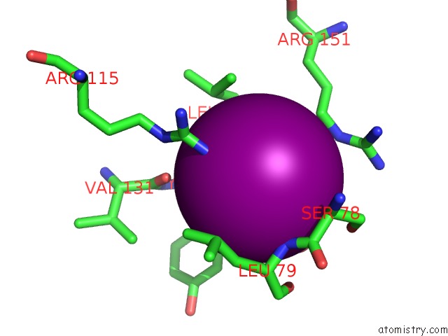
Mono view
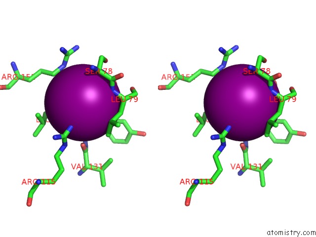
Stereo pair view
Mono view
Stereo pair view
A full contact list of Iodine with other atoms in the I binding
site number 1 of Structure of USP15 Dusp-Ubl Deletion Mutant within 5.0Å range:
|
Reference:
P.R.Elliott,
H.Liu,
M.W.Pastok,
G.J.Grossmann,
D.J.Rigden,
M.J.Clague,
S.Urbe,
I.L.Barsukov.
Structural Variability of the Ubiquitin Specific Protease Dusp-Ubl Double Domains. Febs Lett. V. 585 3385 2011.
ISSN: ISSN 0014-5793
PubMed: 22001210
DOI: 10.1016/J.FEBSLET.2011.09.040
Page generated: Sun Aug 11 17:20:29 2024
ISSN: ISSN 0014-5793
PubMed: 22001210
DOI: 10.1016/J.FEBSLET.2011.09.040
Last articles
Zn in 9J0NZn in 9J0O
Zn in 9J0P
Zn in 9FJX
Zn in 9EKB
Zn in 9C0F
Zn in 9CAH
Zn in 9CH0
Zn in 9CH3
Zn in 9CH1