Iodine »
PDB 442d-4dch »
4dch »
Iodine in PDB 4dch: Insights Into Glucokinase Activation Mechanism: Observation of Multiple Distinct Protein Conformations
Enzymatic activity of Insights Into Glucokinase Activation Mechanism: Observation of Multiple Distinct Protein Conformations
All present enzymatic activity of Insights Into Glucokinase Activation Mechanism: Observation of Multiple Distinct Protein Conformations:
2.7.1.2;
2.7.1.2;
Protein crystallography data
The structure of Insights Into Glucokinase Activation Mechanism: Observation of Multiple Distinct Protein Conformations, PDB code: 4dch
was solved by
S.E.Greasley,
M.Hickey,
J.Feng,
E.Garcia,
with X-Ray Crystallography technique. A brief refinement statistics is given in the table below:
| Resolution Low / High (Å) | 50.00 / 1.79 |
| Space group | P 1 21 1 |
| Cell size a, b, c (Å), α, β, γ (°) | 49.917, 85.794, 72.939, 90.00, 104.40, 90.00 |
| R / Rfree (%) | 19.8 / 23.1 |
Iodine Binding Sites:
The binding sites of Iodine atom in the Insights Into Glucokinase Activation Mechanism: Observation of Multiple Distinct Protein Conformations
(pdb code 4dch). This binding sites where shown within
5.0 Angstroms radius around Iodine atom.
In total 2 binding sites of Iodine where determined in the Insights Into Glucokinase Activation Mechanism: Observation of Multiple Distinct Protein Conformations, PDB code: 4dch:
Jump to Iodine binding site number: 1; 2;
In total 2 binding sites of Iodine where determined in the Insights Into Glucokinase Activation Mechanism: Observation of Multiple Distinct Protein Conformations, PDB code: 4dch:
Jump to Iodine binding site number: 1; 2;
Iodine binding site 1 out of 2 in 4dch
Go back to
Iodine binding site 1 out
of 2 in the Insights Into Glucokinase Activation Mechanism: Observation of Multiple Distinct Protein Conformations
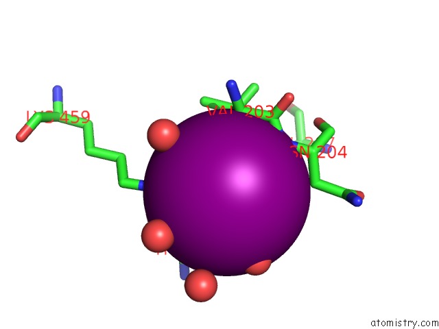
Mono view
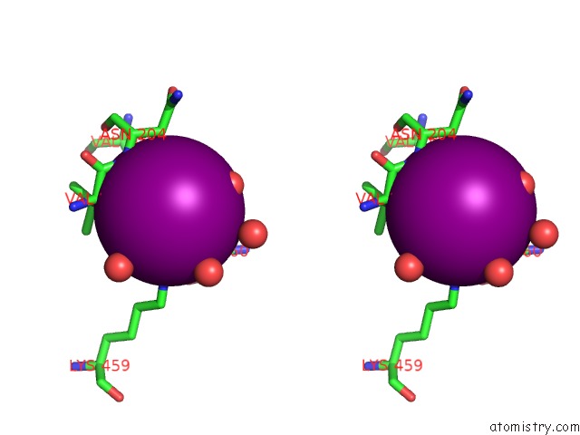
Stereo pair view
Mono view
Stereo pair view
A full contact list of Iodine with other atoms in the I binding
site number 1 of Insights Into Glucokinase Activation Mechanism: Observation of Multiple Distinct Protein Conformations within 5.0Å range:
|
Iodine binding site 2 out of 2 in 4dch
Go back to
Iodine binding site 2 out
of 2 in the Insights Into Glucokinase Activation Mechanism: Observation of Multiple Distinct Protein Conformations
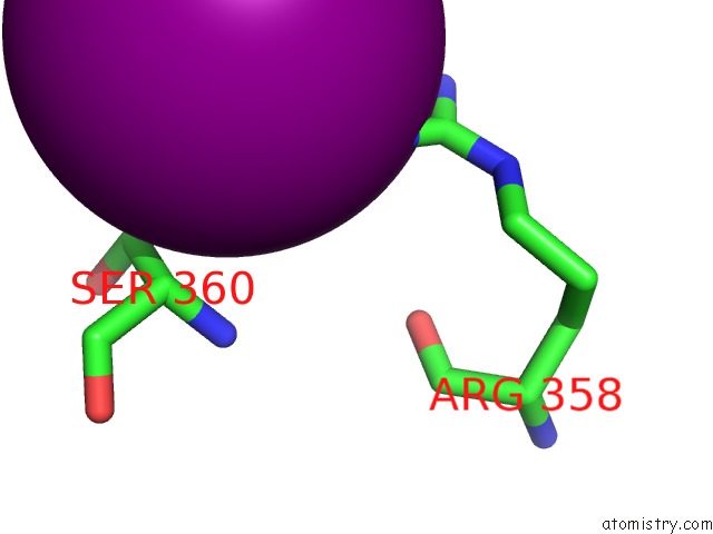
Mono view
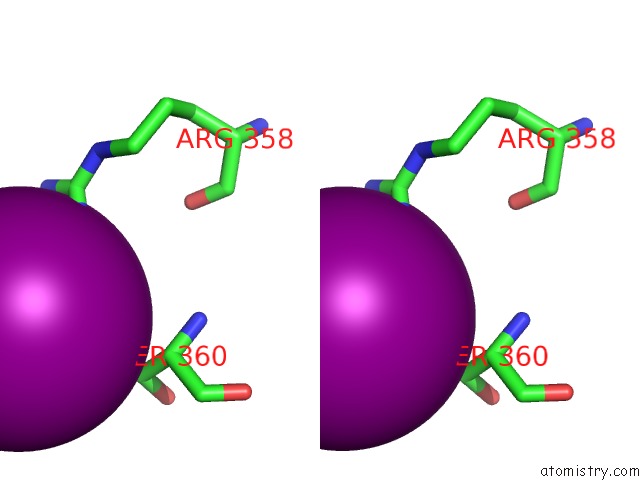
Stereo pair view
Mono view
Stereo pair view
A full contact list of Iodine with other atoms in the I binding
site number 2 of Insights Into Glucokinase Activation Mechanism: Observation of Multiple Distinct Protein Conformations within 5.0Å range:
|
Reference:
S.Liu,
M.J.Ammirati,
X.Song,
J.D.Knafels,
J.Zhang,
S.E.Greasley,
J.A.Pfefferkorn,
X.Qiu.
Insights Into Mechanism of Glucokinase Activation: Observation of Multiple Distinct Protein Conformations. J.Biol.Chem. V. 287 13598 2012.
ISSN: ISSN 0021-9258
PubMed: 22298776
DOI: 10.1074/JBC.M111.274126
Page generated: Fri Aug 8 16:53:54 2025
ISSN: ISSN 0021-9258
PubMed: 22298776
DOI: 10.1074/JBC.M111.274126
Last articles
K in 3S49K in 3S3X
K in 3RYD
K in 3RU3
K in 3RU2
K in 3RTG
K in 3RTC
K in 3RTB
K in 3RTD
K in 3RTE