Iodine »
PDB 4dgh-4hjh »
4dny »
Iodine in PDB 4dny: Crystal Structure of Enterohemorrhagic E. Coli Stce(132-251)
Protein crystallography data
The structure of Crystal Structure of Enterohemorrhagic E. Coli Stce(132-251), PDB code: 4dny
was solved by
A.C.Y.Yu,
N.C.J.Strynadka,
with X-Ray Crystallography technique. A brief refinement statistics is given in the table below:
| Resolution Low / High (Å) | 43.37 / 1.61 |
| Space group | H 3 |
| Cell size a, b, c (Å), α, β, γ (°) | 56.000, 56.000, 96.880, 90.00, 90.00, 120.00 |
| R / Rfree (%) | 18.2 / 21.3 |
Iodine Binding Sites:
The binding sites of Iodine atom in the Crystal Structure of Enterohemorrhagic E. Coli Stce(132-251)
(pdb code 4dny). This binding sites where shown within
5.0 Angstroms radius around Iodine atom.
In total 7 binding sites of Iodine where determined in the Crystal Structure of Enterohemorrhagic E. Coli Stce(132-251), PDB code: 4dny:
Jump to Iodine binding site number: 1; 2; 3; 4; 5; 6; 7;
In total 7 binding sites of Iodine where determined in the Crystal Structure of Enterohemorrhagic E. Coli Stce(132-251), PDB code: 4dny:
Jump to Iodine binding site number: 1; 2; 3; 4; 5; 6; 7;
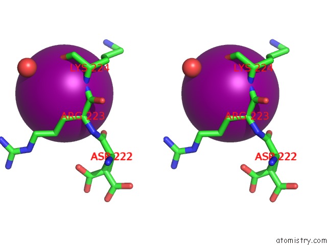
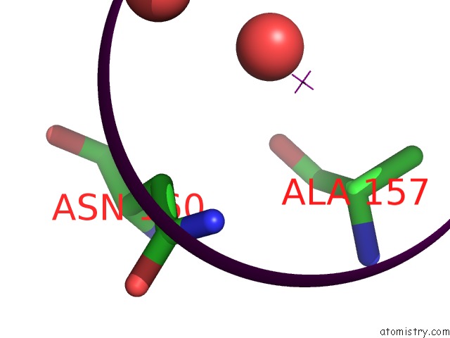
Iodine binding site 1 out of 7 in 4dny
Go back to
Iodine binding site 1 out
of 7 in the Crystal Structure of Enterohemorrhagic E. Coli Stce(132-251)

Mono view
Stereo pair view
Mono view
Stereo pair view
A full contact list of Iodine with other atoms in the I binding
site number 1 of Crystal Structure of Enterohemorrhagic E. Coli Stce(132-251) within 5.0Å range:
|
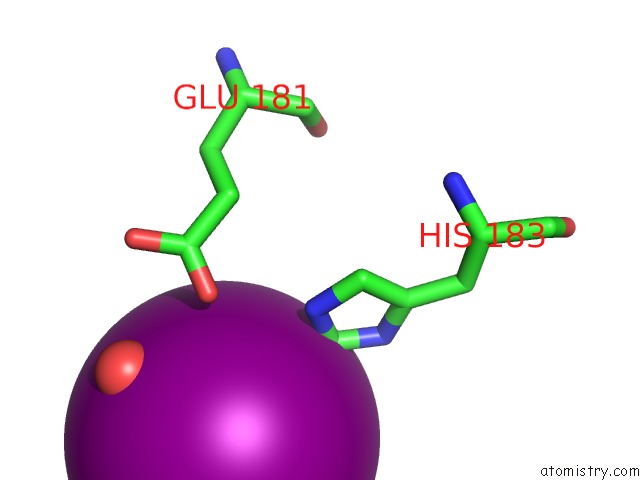
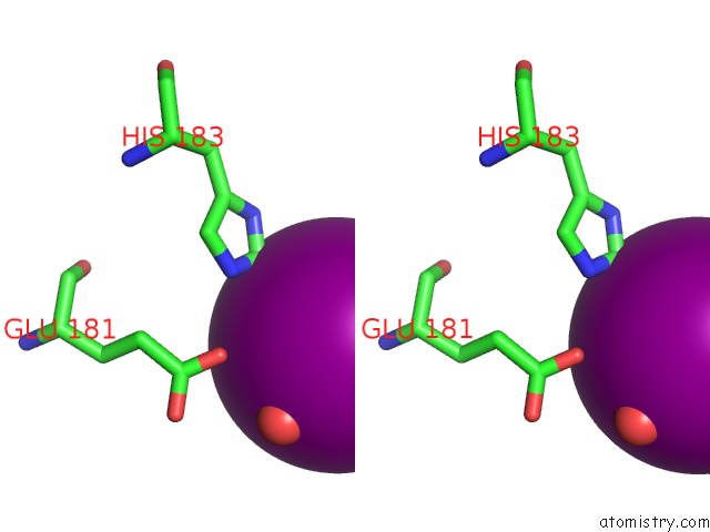
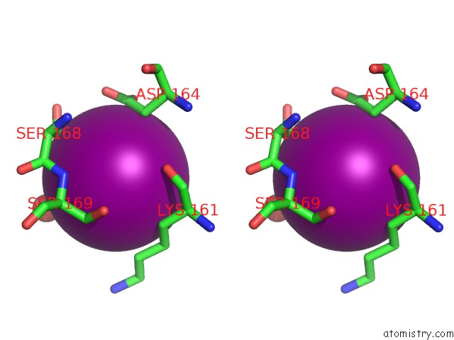
Iodine binding site 2 out of 7 in 4dny
Go back to
Iodine binding site 2 out
of 7 in the Crystal Structure of Enterohemorrhagic E. Coli Stce(132-251)
Mono view
Stereo pair view
Mono view
Stereo pair view
A full contact list of Iodine with other atoms in the I binding
site number 2 of Crystal Structure of Enterohemorrhagic E. Coli Stce(132-251) within 5.0Å range:
|
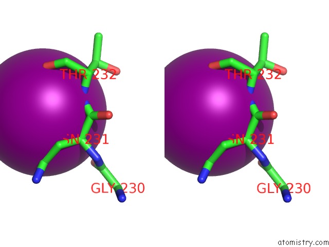
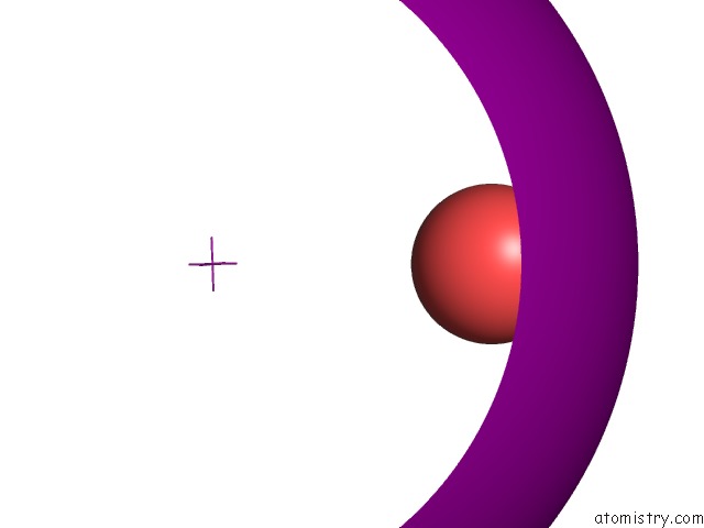
Iodine binding site 3 out of 7 in 4dny
Go back to
Iodine binding site 3 out
of 7 in the Crystal Structure of Enterohemorrhagic E. Coli Stce(132-251)

Mono view
Stereo pair view
Mono view
Stereo pair view
A full contact list of Iodine with other atoms in the I binding
site number 3 of Crystal Structure of Enterohemorrhagic E. Coli Stce(132-251) within 5.0Å range:
|
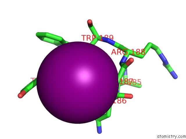
Iodine binding site 4 out of 7 in 4dny
Go back to
Iodine binding site 4 out
of 7 in the Crystal Structure of Enterohemorrhagic E. Coli Stce(132-251)
Mono view

Stereo pair view
Mono view
Stereo pair view
A full contact list of Iodine with other atoms in the I binding
site number 4 of Crystal Structure of Enterohemorrhagic E. Coli Stce(132-251) within 5.0Å range:
|
Iodine binding site 5 out of 7 in 4dny
Go back to
Iodine binding site 5 out
of 7 in the Crystal Structure of Enterohemorrhagic E. Coli Stce(132-251)

Mono view
Stereo pair view
Mono view
Stereo pair view
A full contact list of Iodine with other atoms in the I binding
site number 5 of Crystal Structure of Enterohemorrhagic E. Coli Stce(132-251) within 5.0Å range:
|
Iodine binding site 6 out of 7 in 4dny
Go back to
Iodine binding site 6 out
of 7 in the Crystal Structure of Enterohemorrhagic E. Coli Stce(132-251)
Mono view

Stereo pair view
Mono view
Stereo pair view
A full contact list of Iodine with other atoms in the I binding
site number 6 of Crystal Structure of Enterohemorrhagic E. Coli Stce(132-251) within 5.0Å range:
|
Iodine binding site 7 out of 7 in 4dny
Go back to
Iodine binding site 7 out
of 7 in the Crystal Structure of Enterohemorrhagic E. Coli Stce(132-251)
Mono view

Stereo pair view
Mono view
Stereo pair view
A full contact list of Iodine with other atoms in the I binding
site number 7 of Crystal Structure of Enterohemorrhagic E. Coli Stce(132-251) within 5.0Å range:
|
Reference:
A.C.Yu,
L.J.Worrall,
N.C.Strynadka.
Structural Insight Into the Bacterial Mucinase Stce Essential to Adhesion and Immune Evasion During Enterohemorrhagic E. Coli Infection. Structure V. 20 707 2012.
ISSN: ISSN 0969-2126
PubMed: 22483117
DOI: 10.1016/J.STR.2012.02.015
Page generated: Sun Aug 11 17:32:54 2024
ISSN: ISSN 0969-2126
PubMed: 22483117
DOI: 10.1016/J.STR.2012.02.015
Last articles
Zn in 9J0NZn in 9J0O
Zn in 9J0P
Zn in 9FJX
Zn in 9EKB
Zn in 9C0F
Zn in 9CAH
Zn in 9CH0
Zn in 9CH3
Zn in 9CH1