Iodine »
PDB 4dgh-4hjh »
4eye »
Iodine in PDB 4eye: Crystal Structure of A Probable Oxidoreductase From Mycobacterium Abscessus Solved By Iodide Ion Sad
Protein crystallography data
The structure of Crystal Structure of A Probable Oxidoreductase From Mycobacterium Abscessus Solved By Iodide Ion Sad, PDB code: 4eye
was solved by
T.E.Edwards,
J.Abendroth,
Seattle Structural Genomics Center Forinfectious Disease (Ssgcid),
with X-Ray Crystallography technique. A brief refinement statistics is given in the table below:
| Resolution Low / High (Å) | 50.00 / 2.10 |
| Space group | P 1 21 1 |
| Cell size a, b, c (Å), α, β, γ (°) | 78.640, 46.340, 84.130, 90.00, 93.14, 90.00 |
| R / Rfree (%) | 18.5 / 23.5 |
Iodine Binding Sites:
Pages:
>>> Page 1 <<< Page 2, Binding sites: 11 - 18;Binding sites:
The binding sites of Iodine atom in the Crystal Structure of A Probable Oxidoreductase From Mycobacterium Abscessus Solved By Iodide Ion Sad (pdb code 4eye). This binding sites where shown within 5.0 Angstroms radius around Iodine atom.In total 18 binding sites of Iodine where determined in the Crystal Structure of A Probable Oxidoreductase From Mycobacterium Abscessus Solved By Iodide Ion Sad, PDB code: 4eye:
Jump to Iodine binding site number: 1; 2; 3; 4; 5; 6; 7; 8; 9; 10;
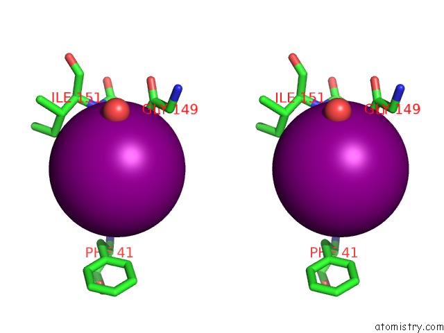
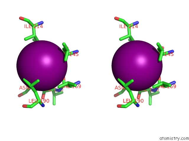
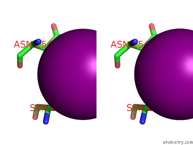
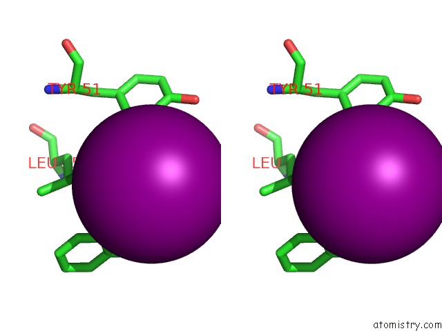
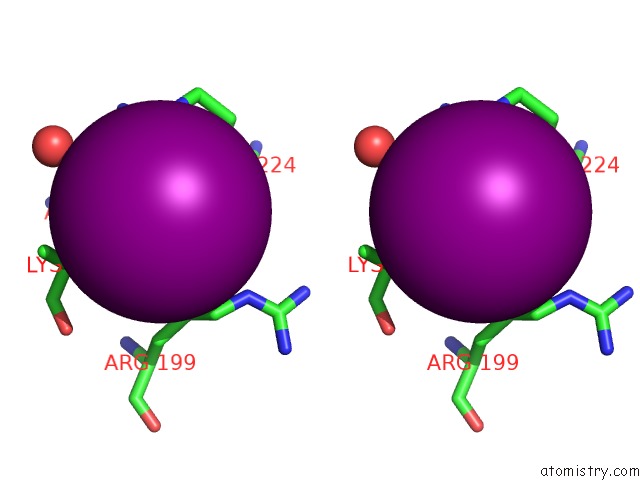
Iodine binding site 1 out of 18 in 4eye
Go back to
Iodine binding site 1 out
of 18 in the Crystal Structure of A Probable Oxidoreductase From Mycobacterium Abscessus Solved By Iodide Ion Sad

Mono view
Stereo pair view
Mono view
Stereo pair view
A full contact list of Iodine with other atoms in the I binding
site number 1 of Crystal Structure of A Probable Oxidoreductase From Mycobacterium Abscessus Solved By Iodide Ion Sad within 5.0Å range:
|
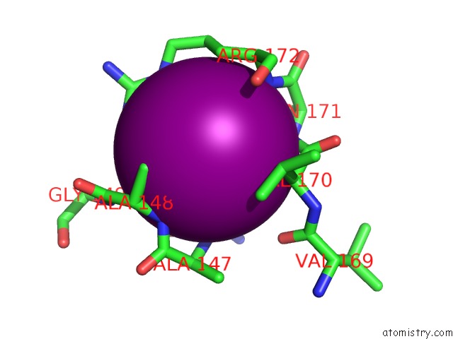
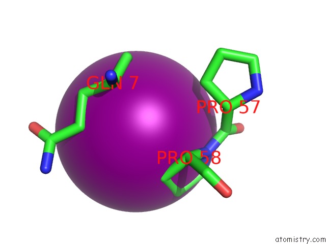
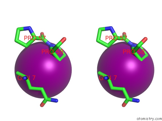
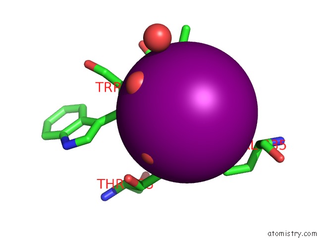
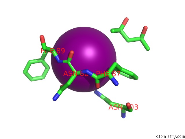
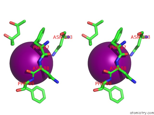
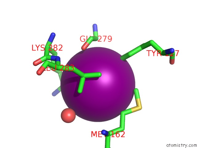
Iodine binding site 2 out of 18 in 4eye
Go back to
Iodine binding site 2 out
of 18 in the Crystal Structure of A Probable Oxidoreductase From Mycobacterium Abscessus Solved By Iodide Ion Sad
Mono view
Stereo pair view
Mono view
Stereo pair view
A full contact list of Iodine with other atoms in the I binding
site number 2 of Crystal Structure of A Probable Oxidoreductase From Mycobacterium Abscessus Solved By Iodide Ion Sad within 5.0Å range:
|
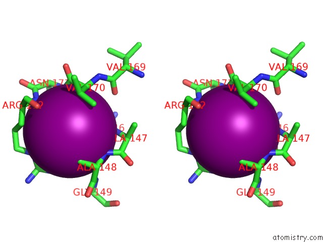
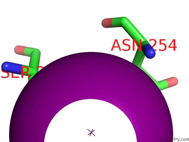
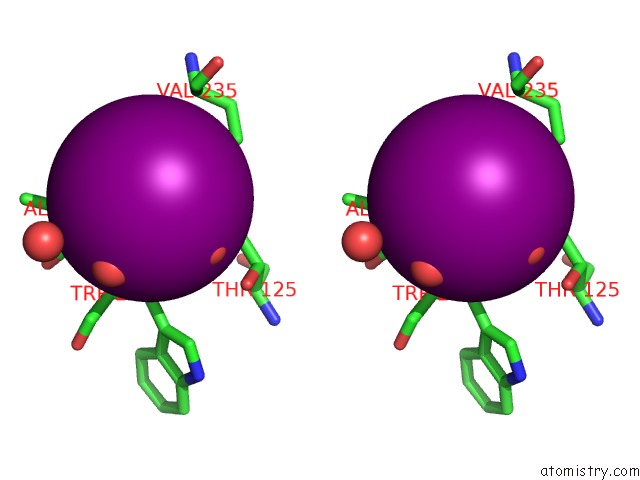
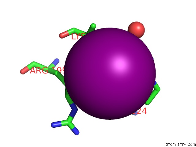
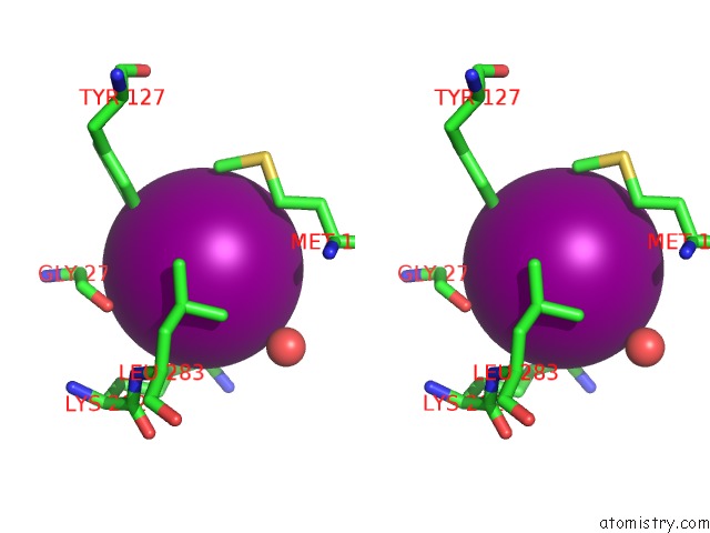
Iodine binding site 3 out of 18 in 4eye
Go back to
Iodine binding site 3 out
of 18 in the Crystal Structure of A Probable Oxidoreductase From Mycobacterium Abscessus Solved By Iodide Ion Sad
Mono view
Stereo pair view
Mono view
Stereo pair view
A full contact list of Iodine with other atoms in the I binding
site number 3 of Crystal Structure of A Probable Oxidoreductase From Mycobacterium Abscessus Solved By Iodide Ion Sad within 5.0Å range:
|
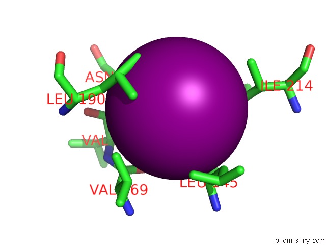
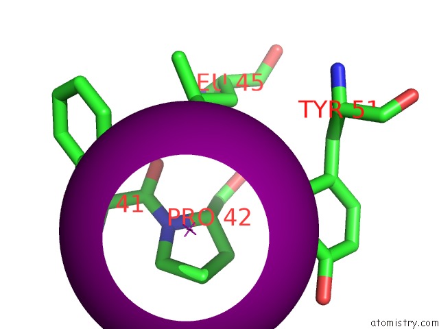
Iodine binding site 4 out of 18 in 4eye
Go back to
Iodine binding site 4 out
of 18 in the Crystal Structure of A Probable Oxidoreductase From Mycobacterium Abscessus Solved By Iodide Ion Sad
Mono view
Stereo pair view
Mono view
Stereo pair view
A full contact list of Iodine with other atoms in the I binding
site number 4 of Crystal Structure of A Probable Oxidoreductase From Mycobacterium Abscessus Solved By Iodide Ion Sad within 5.0Å range:
|
Iodine binding site 5 out of 18 in 4eye
Go back to
Iodine binding site 5 out
of 18 in the Crystal Structure of A Probable Oxidoreductase From Mycobacterium Abscessus Solved By Iodide Ion Sad
Mono view
Stereo pair view
Mono view
Stereo pair view
A full contact list of Iodine with other atoms in the I binding
site number 5 of Crystal Structure of A Probable Oxidoreductase From Mycobacterium Abscessus Solved By Iodide Ion Sad within 5.0Å range:
|
Iodine binding site 6 out of 18 in 4eye
Go back to
Iodine binding site 6 out
of 18 in the Crystal Structure of A Probable Oxidoreductase From Mycobacterium Abscessus Solved By Iodide Ion Sad
Mono view
Stereo pair view
Mono view
Stereo pair view
A full contact list of Iodine with other atoms in the I binding
site number 6 of Crystal Structure of A Probable Oxidoreductase From Mycobacterium Abscessus Solved By Iodide Ion Sad within 5.0Å range:
|
Iodine binding site 7 out of 18 in 4eye
Go back to
Iodine binding site 7 out
of 18 in the Crystal Structure of A Probable Oxidoreductase From Mycobacterium Abscessus Solved By Iodide Ion Sad
Mono view
Stereo pair view
Mono view
Stereo pair view
A full contact list of Iodine with other atoms in the I binding
site number 7 of Crystal Structure of A Probable Oxidoreductase From Mycobacterium Abscessus Solved By Iodide Ion Sad within 5.0Å range:
|
Iodine binding site 8 out of 18 in 4eye
Go back to
Iodine binding site 8 out
of 18 in the Crystal Structure of A Probable Oxidoreductase From Mycobacterium Abscessus Solved By Iodide Ion Sad
Mono view
Stereo pair view
Mono view
Stereo pair view
A full contact list of Iodine with other atoms in the I binding
site number 8 of Crystal Structure of A Probable Oxidoreductase From Mycobacterium Abscessus Solved By Iodide Ion Sad within 5.0Å range:
|
Iodine binding site 9 out of 18 in 4eye
Go back to
Iodine binding site 9 out
of 18 in the Crystal Structure of A Probable Oxidoreductase From Mycobacterium Abscessus Solved By Iodide Ion Sad
Mono view
Stereo pair view
Mono view
Stereo pair view
A full contact list of Iodine with other atoms in the I binding
site number 9 of Crystal Structure of A Probable Oxidoreductase From Mycobacterium Abscessus Solved By Iodide Ion Sad within 5.0Å range:
|
Iodine binding site 10 out of 18 in 4eye
Go back to
Iodine binding site 10 out
of 18 in the Crystal Structure of A Probable Oxidoreductase From Mycobacterium Abscessus Solved By Iodide Ion Sad
Mono view
Stereo pair view
Mono view
Stereo pair view
A full contact list of Iodine with other atoms in the I binding
site number 10 of Crystal Structure of A Probable Oxidoreductase From Mycobacterium Abscessus Solved By Iodide Ion Sad within 5.0Å range:
|
Reference:
L.Baugh,
I.Phan,
D.W.Begley,
M.C.Clifton,
B.Armour,
D.M.Dranow,
B.M.Taylor,
M.M.Muruthi,
J.Abendroth,
J.W.Fairman,
D.Fox,
S.H.Dieterich,
B.L.Staker,
A.S.Gardberg,
R.Choi,
S.N.Hewitt,
A.J.Napuli,
J.Myers,
L.K.Barrett,
Y.Zhang,
M.Ferrell,
E.Mundt,
K.Thompkins,
N.Tran,
S.Lyons-Abbott,
A.Abramov,
A.Sekar,
D.Serbzhinskiy,
D.Lorimer,
G.W.Buchko,
R.Stacy,
L.J.Stewart,
T.E.Edwards,
W.C.Van Voorhis,
P.J.Myler.
Increasing the Structural Coverage of Tuberculosis Drug Targets. Tuberculosis (Edinb) V. 95 142 2015.
ISSN: ISSN 1472-9792
PubMed: 25613812
DOI: 10.1016/J.TUBE.2014.12.003
Page generated: Sun Aug 11 17:39:27 2024
ISSN: ISSN 1472-9792
PubMed: 25613812
DOI: 10.1016/J.TUBE.2014.12.003
Last articles
Zn in 9J0NZn in 9J0O
Zn in 9J0P
Zn in 9FJX
Zn in 9EKB
Zn in 9C0F
Zn in 9CAH
Zn in 9CH0
Zn in 9CH3
Zn in 9CH1