Iodine in PDB 4hkl: Crystal Structures of Mutant Endo-Beta-1,4-Xylanase II Complexed with Substrate (1.15 A) and Products (1.6 A)
Enzymatic activity of Crystal Structures of Mutant Endo-Beta-1,4-Xylanase II Complexed with Substrate (1.15 A) and Products (1.6 A)
All present enzymatic activity of Crystal Structures of Mutant Endo-Beta-1,4-Xylanase II Complexed with Substrate (1.15 A) and Products (1.6 A):
3.2.1.8;
Protein crystallography data
The structure of Crystal Structures of Mutant Endo-Beta-1,4-Xylanase II Complexed with Substrate (1.15 A) and Products (1.6 A), PDB code: 4hkl
was solved by
P.Langan,
Q.Wan,
L.Coates,
A.Kovalevsky,
Oak Ridge National Lab,
with X-Ray Crystallography technique. A brief refinement statistics is given in the table below:
Resolution Low / High (Å)
|
15.00 /
1.10
|
Space group
|
P 21 21 21
|
Cell size a, b, c (Å), α, β, γ (°)
|
48.307,
59.032,
69.717,
90.00,
90.00,
90.00
|
R / Rfree (%)
|
11.9 /
12.9
|
Iodine Binding Sites:
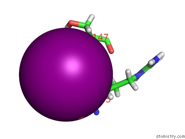
The binding sites of Iodine atom in the Crystal Structures of Mutant Endo-Beta-1,4-Xylanase II Complexed with Substrate (1.15 A) and Products (1.6 A)
(pdb code 4hkl). This binding sites where shown within
5.0 Angstroms radius around Iodine atom.
In total 4 binding sites of Iodine where determined in the
Crystal Structures of Mutant Endo-Beta-1,4-Xylanase II Complexed with Substrate (1.15 A) and Products (1.6 A), PDB code: 4hkl:
Jump to Iodine binding site number:
1;
2;
3;
4;
Iodine binding site 1 out
of 4 in 4hkl
Go back to
Iodine Binding Sites List in 4hkl
Iodine binding site 1 out
of 4 in the Crystal Structures of Mutant Endo-Beta-1,4-Xylanase II Complexed with Substrate (1.15 A) and Products (1.6 A)
Mono view
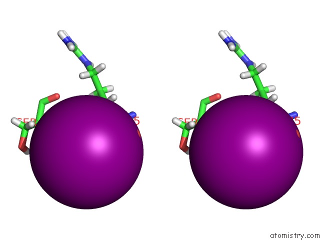
Stereo pair view
|
A full contact list of Iodine with other atoms in the I binding
site number 1 of Crystal Structures of Mutant Endo-Beta-1,4-Xylanase II Complexed with Substrate (1.15 A) and Products (1.6 A) within 5.0Å range:
probe
|
atom
|
residue
|
distance (Å)
|
B
|
Occ
|
A:I500
b:13.5
occ:0.33
|
H
|
A:SER146
|
2.4
|
11.9
|
0.2
|
HG
|
A:SER146
|
2.7
|
13.1
|
0.2
|
O
|
A:HOH604
|
3.1
|
9.7
|
0.9
|
O
|
A:HOH647
|
3.3
|
12.4
|
0.6
|
N
|
A:SER146
|
3.3
|
9.9
|
1.0
|
HA
|
A:ARG145
|
3.4
|
11.3
|
1.0
|
HB2
|
A:SER146
|
3.4
|
14.3
|
0.8
|
OG
|
A:SER146
|
3.4
|
10.9
|
0.2
|
O
|
A:HOH801
|
3.4
|
14.5
|
0.3
|
H
|
A:SER147
|
3.5
|
12.9
|
1.0
|
HB2
|
A:ARG145
|
3.5
|
11.1
|
1.0
|
CA
|
A:ARG145
|
4.0
|
9.4
|
1.0
|
CA
|
A:SER146
|
4.1
|
10.8
|
0.8
|
CB
|
A:SER146
|
4.1
|
12.0
|
0.8
|
N
|
A:SER147
|
4.1
|
10.8
|
1.0
|
CA
|
A:SER146
|
4.1
|
10.6
|
0.2
|
C
|
A:ARG145
|
4.1
|
9.4
|
1.0
|
CB
|
A:ARG145
|
4.2
|
9.2
|
1.0
|
CB
|
A:SER146
|
4.3
|
10.9
|
0.2
|
C
|
A:SER146
|
4.4
|
10.8
|
1.0
|
HB2
|
A:SER147
|
4.5
|
12.1
|
1.0
|
OG
|
A:SER146
|
4.5
|
12.6
|
0.8
|
HB3
|
A:ARG145
|
4.5
|
11.1
|
1.0
|
HG
|
A:SER146
|
4.6
|
15.1
|
0.8
|
HB3
|
A:SER146
|
4.6
|
13.1
|
0.2
|
HB3
|
A:SER146
|
4.9
|
14.3
|
0.8
|
HA
|
A:SER146
|
5.0
|
13.0
|
0.2
|
HG
|
A:SER147
|
5.0
|
12.3
|
1.0
|
|
Iodine binding site 2 out
of 4 in 4hkl
Go back to
Iodine Binding Sites List in 4hkl
Iodine binding site 2 out
of 4 in the Crystal Structures of Mutant Endo-Beta-1,4-Xylanase II Complexed with Substrate (1.15 A) and Products (1.6 A)
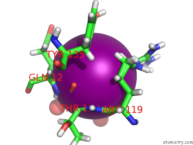
Mono view
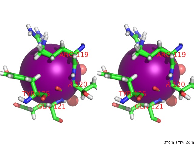
Stereo pair view
|
A full contact list of Iodine with other atoms in the I binding
site number 2 of Crystal Structures of Mutant Endo-Beta-1,4-Xylanase II Complexed with Substrate (1.15 A) and Products (1.6 A) within 5.0Å range:
probe
|
atom
|
residue
|
distance (Å)
|
B
|
Occ
|
A:I501
b:22.1
occ:0.14
|
HH21
|
A:ARG119
|
2.7
|
23.4
|
0.2
|
HH22
|
A:ARG119
|
2.9
|
23.4
|
0.2
|
NH2
|
A:ARG119
|
2.9
|
19.5
|
0.2
|
HB2
|
A:GLN121
|
3.2
|
14.7
|
1.0
|
HE1
|
A:TYR135
|
3.2
|
16.8
|
1.0
|
HG2
|
A:ARG119
|
3.2
|
16.3
|
0.2
|
HA
|
A:THR120
|
3.2
|
10.7
|
1.0
|
HG2
|
A:ARG119
|
3.3
|
14.8
|
0.8
|
HE
|
A:ARG119
|
3.3
|
22.8
|
0.8
|
O
|
A:HOH858
|
3.4
|
29.3
|
0.6
|
HD1
|
A:TYR135
|
3.5
|
15.0
|
1.0
|
C
|
A:THR120
|
3.6
|
8.9
|
1.0
|
CA
|
A:THR120
|
3.8
|
8.9
|
1.0
|
N
|
A:GLN121
|
3.8
|
10.1
|
1.0
|
CZ
|
A:ARG119
|
3.8
|
18.9
|
0.2
|
O
|
A:ARG119
|
3.9
|
10.1
|
1.0
|
CE1
|
A:TYR135
|
3.9
|
14.0
|
1.0
|
CB
|
A:GLN121
|
4.0
|
12.2
|
1.0
|
H
|
A:GLN121
|
4.0
|
12.1
|
1.0
|
O
|
A:THR120
|
4.0
|
9.3
|
1.0
|
NE
|
A:ARG119
|
4.0
|
19.0
|
0.8
|
HD2
|
A:ARG119
|
4.0
|
19.1
|
0.8
|
CD1
|
A:TYR135
|
4.0
|
12.5
|
1.0
|
HB3
|
A:GLN121
|
4.1
|
14.7
|
1.0
|
HD2
|
A:ARG119
|
4.1
|
19.1
|
0.2
|
CG
|
A:ARG119
|
4.1
|
13.6
|
0.2
|
CG
|
A:ARG119
|
4.1
|
12.3
|
0.8
|
C
|
A:ARG119
|
4.1
|
9.0
|
1.0
|
N
|
A:THR120
|
4.2
|
9.0
|
1.0
|
O
|
A:HOH622
|
4.2
|
11.4
|
0.7
|
CD
|
A:ARG119
|
4.3
|
15.9
|
0.8
|
HB3
|
A:ARG119
|
4.3
|
12.3
|
0.8
|
CD
|
A:ARG119
|
4.4
|
15.9
|
0.2
|
NE
|
A:ARG119
|
4.4
|
17.9
|
0.2
|
CA
|
A:GLN121
|
4.5
|
10.9
|
1.0
|
HB3
|
A:ARG119
|
4.5
|
13.8
|
0.2
|
O
|
A:HOH651
|
4.5
|
20.6
|
1.0
|
HH12
|
A:ARG119
|
4.6
|
23.0
|
0.2
|
NH1
|
A:ARG119
|
4.6
|
19.2
|
0.2
|
CB
|
A:ARG119
|
4.7
|
10.2
|
0.8
|
HG3
|
A:ARG119
|
4.7
|
16.3
|
0.2
|
H
|
A:THR120
|
4.7
|
10.8
|
1.0
|
CB
|
A:ARG119
|
4.8
|
11.5
|
0.2
|
HA
|
A:GLN121
|
4.8
|
13.1
|
1.0
|
HH21
|
A:ARG119
|
4.8
|
24.8
|
0.8
|
HG3
|
A:ARG119
|
4.9
|
14.8
|
0.8
|
|
Iodine binding site 3 out
of 4 in 4hkl
Go back to
Iodine Binding Sites List in 4hkl
Iodine binding site 3 out
of 4 in the Crystal Structures of Mutant Endo-Beta-1,4-Xylanase II Complexed with Substrate (1.15 A) and Products (1.6 A)
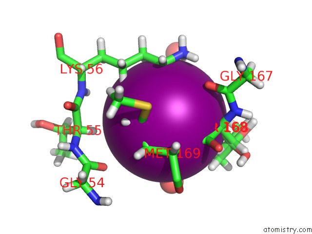
Mono view
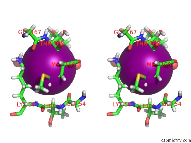
Stereo pair view
|
A full contact list of Iodine with other atoms in the I binding
site number 3 of Crystal Structures of Mutant Endo-Beta-1,4-Xylanase II Complexed with Substrate (1.15 A) and Products (1.6 A) within 5.0Å range:
probe
|
atom
|
residue
|
distance (Å)
|
B
|
Occ
|
A:I502
b:20.7
occ:0.25
|
H
|
A:MET169
|
2.3
|
11.6
|
1.0
|
HG2
|
A:MET169
|
2.7
|
15.5
|
1.0
|
HA
|
A:THR168
|
3.0
|
12.5
|
0.3
|
HA
|
A:THR168
|
3.0
|
13.2
|
0.7
|
HE3
|
A:LYS56
|
3.1
|
16.2
|
1.0
|
O
|
A:HOH789
|
3.1
|
20.1
|
0.5
|
N
|
A:MET169
|
3.2
|
9.7
|
1.0
|
O
|
A:HOH655
|
3.2
|
14.5
|
0.7
|
HB2
|
A:MET169
|
3.5
|
13.4
|
1.0
|
CG
|
A:MET169
|
3.5
|
12.9
|
1.0
|
HG23
|
A:THR168
|
3.6
|
17.2
|
0.7
|
CA
|
A:THR168
|
3.8
|
10.4
|
0.3
|
HG3
|
A:LYS56
|
3.8
|
13.8
|
1.0
|
CA
|
A:THR168
|
3.9
|
11.0
|
0.7
|
CB
|
A:MET169
|
3.9
|
11.2
|
1.0
|
HG3
|
A:MET169
|
3.9
|
15.5
|
1.0
|
O
|
A:HOH660
|
3.9
|
19.7
|
0.9
|
HB
|
A:THR168
|
4.0
|
13.7
|
0.3
|
CE
|
A:LYS56
|
4.0
|
13.5
|
1.0
|
C
|
A:THR168
|
4.0
|
9.9
|
1.0
|
HD2
|
A:LYS56
|
4.1
|
15.2
|
1.0
|
CA
|
A:MET169
|
4.1
|
9.8
|
1.0
|
HZ2
|
A:LYS56
|
4.2
|
17.2
|
1.0
|
HG22
|
A:THR168
|
4.2
|
17.2
|
0.7
|
CG2
|
A:THR168
|
4.3
|
14.4
|
0.7
|
HG22
|
A:THR168
|
4.3
|
14.1
|
0.3
|
O
|
A:GLY167
|
4.3
|
12.8
|
1.0
|
CB
|
A:THR168
|
4.4
|
11.4
|
0.3
|
CD
|
A:LYS56
|
4.5
|
12.7
|
1.0
|
NZ
|
A:LYS56
|
4.5
|
14.3
|
1.0
|
HZ1
|
A:LYS56
|
4.6
|
17.2
|
1.0
|
CG
|
A:LYS56
|
4.6
|
11.5
|
1.0
|
HA
|
A:THR55
|
4.6
|
11.3
|
1.0
|
HE2
|
A:LYS56
|
4.6
|
16.2
|
1.0
|
CB
|
A:THR168
|
4.6
|
13.2
|
0.7
|
HA
|
A:MET169
|
4.8
|
11.8
|
1.0
|
O
|
A:HOH776
|
4.8
|
27.0
|
0.5
|
HB3
|
A:MET169
|
4.8
|
13.4
|
1.0
|
CG2
|
A:THR168
|
4.9
|
11.7
|
0.3
|
SD
|
A:MET169
|
4.9
|
13.3
|
1.0
|
N
|
A:THR168
|
4.9
|
10.1
|
1.0
|
O
|
A:GLY54
|
5.0
|
10.4
|
1.0
|
|
Iodine binding site 4 out
of 4 in 4hkl
Go back to
Iodine Binding Sites List in 4hkl
Iodine binding site 4 out
of 4 in the Crystal Structures of Mutant Endo-Beta-1,4-Xylanase II Complexed with Substrate (1.15 A) and Products (1.6 A)
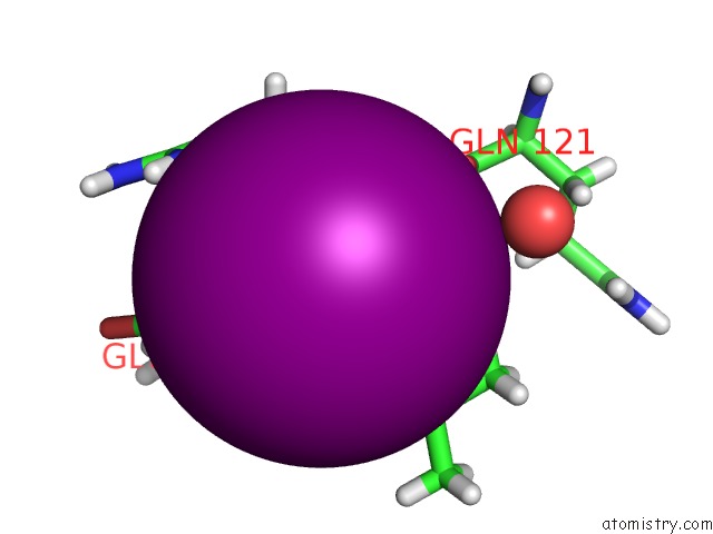
Mono view
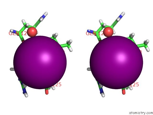
Stereo pair view
|
A full contact list of Iodine with other atoms in the I binding
site number 4 of Crystal Structures of Mutant Endo-Beta-1,4-Xylanase II Complexed with Substrate (1.15 A) and Products (1.6 A) within 5.0Å range:
probe
|
atom
|
residue
|
distance (Å)
|
B
|
Occ
|
A:I503
b:22.4
occ:0.19
|
H
|
A:VAL123
|
2.3
|
14.7
|
1.0
|
HE21
|
A:GLN125
|
2.4
|
25.4
|
1.0
|
HA
|
A:ARG122
|
2.9
|
13.1
|
1.0
|
N
|
A:VAL123
|
3.2
|
12.2
|
1.0
|
NE2
|
A:GLN125
|
3.3
|
21.2
|
1.0
|
HG3
|
A:GLN125
|
3.3
|
19.3
|
1.0
|
HD2
|
A:ARG122
|
3.4
|
16.8
|
1.0
|
HH11
|
A:ARG122
|
3.5
|
21.6
|
1.0
|
HB
|
A:VAL123
|
3.5
|
18.0
|
1.0
|
HG2
|
A:GLN125
|
3.5
|
19.3
|
1.0
|
CA
|
A:ARG122
|
3.7
|
10.9
|
1.0
|
CG
|
A:GLN125
|
3.8
|
16.1
|
1.0
|
HE22
|
A:GLN125
|
3.8
|
25.4
|
1.0
|
NH1
|
A:ARG122
|
3.9
|
18.0
|
1.0
|
C
|
A:ARG122
|
3.9
|
11.1
|
1.0
|
O
|
A:VAL123
|
4.0
|
14.3
|
1.0
|
CD
|
A:GLN125
|
4.0
|
19.1
|
1.0
|
HG23
|
A:VAL123
|
4.0
|
19.2
|
1.0
|
CA
|
A:VAL123
|
4.1
|
13.3
|
1.0
|
HH12
|
A:ARG122
|
4.2
|
21.6
|
1.0
|
HB3
|
A:ARG122
|
4.2
|
14.0
|
1.0
|
O
|
A:GLN121
|
4.2
|
12.6
|
1.0
|
CB
|
A:VAL123
|
4.2
|
15.0
|
1.0
|
CD
|
A:ARG122
|
4.4
|
14.0
|
1.0
|
C
|
A:VAL123
|
4.5
|
13.7
|
1.0
|
CB
|
A:ARG122
|
4.5
|
11.7
|
1.0
|
CG2
|
A:VAL123
|
4.6
|
16.0
|
1.0
|
CZ
|
A:ARG122
|
4.6
|
16.9
|
1.0
|
O
|
A:HOH844
|
4.7
|
29.8
|
0.7
|
NE
|
A:ARG122
|
4.8
|
15.2
|
1.0
|
N
|
A:ARG122
|
4.8
|
10.4
|
1.0
|
C
|
A:GLN121
|
4.9
|
11.0
|
1.0
|
HD3
|
A:ARG122
|
5.0
|
16.8
|
1.0
|
HA
|
A:VAL123
|
5.0
|
15.9
|
1.0
|
|
Reference:
Q.Wan,
Q.Zhang,
S.Hamilton-Brehm,
K.Weiss,
M.Mustyakimov,
L.Coates,
P.Langan,
D.Graham,
A.Kovalevsky.
X-Ray Crystallographic Studies of Family 11 Xylanase Michaelis and Product Complexes: Implications For the Catalytic Mechanism. Acta Crystallogr.,Sect.D V. 70 11 2014.
ISSN: ISSN 0907-4449
PubMed: 24419374
DOI: 10.1107/S1399004713023626
Page generated: Sun Aug 11 18:04:21 2024
|