Iodine »
PDB 4hkl-4jxj »
4hko »
Iodine in PDB 4hko: Crystal Structures of Mutant Endo-Beta-1,4-Xylanase II (E177Q) in the Apo Form
Enzymatic activity of Crystal Structures of Mutant Endo-Beta-1,4-Xylanase II (E177Q) in the Apo Form
All present enzymatic activity of Crystal Structures of Mutant Endo-Beta-1,4-Xylanase II (E177Q) in the Apo Form:
3.2.1.8;
3.2.1.8;
Protein crystallography data
The structure of Crystal Structures of Mutant Endo-Beta-1,4-Xylanase II (E177Q) in the Apo Form, PDB code: 4hko
was solved by
P.Langan,
Q.Wan,
L.Coates,
A.Kovalevsky,
with X-Ray Crystallography technique. A brief refinement statistics is given in the table below:
| Resolution Low / High (Å) | 19.83 / 1.50 |
| Space group | P 21 21 21 |
| Cell size a, b, c (Å), α, β, γ (°) | 48.216, 59.209, 69.718, 90.00, 90.00, 90.00 |
| R / Rfree (%) | 17.1 / 18.6 |
Iodine Binding Sites:
The binding sites of Iodine atom in the Crystal Structures of Mutant Endo-Beta-1,4-Xylanase II (E177Q) in the Apo Form
(pdb code 4hko). This binding sites where shown within
5.0 Angstroms radius around Iodine atom.
In total 6 binding sites of Iodine where determined in the Crystal Structures of Mutant Endo-Beta-1,4-Xylanase II (E177Q) in the Apo Form, PDB code: 4hko:
Jump to Iodine binding site number: 1; 2; 3; 4; 5; 6;
In total 6 binding sites of Iodine where determined in the Crystal Structures of Mutant Endo-Beta-1,4-Xylanase II (E177Q) in the Apo Form, PDB code: 4hko:
Jump to Iodine binding site number: 1; 2; 3; 4; 5; 6;
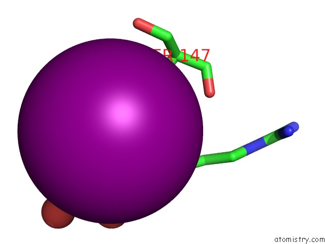
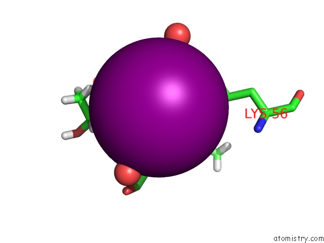
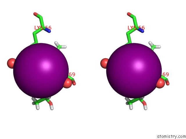
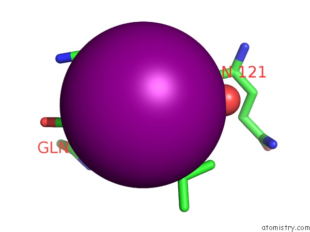
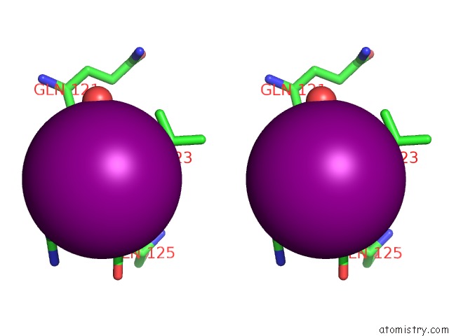
Iodine binding site 1 out of 6 in 4hko
Go back to
Iodine binding site 1 out
of 6 in the Crystal Structures of Mutant Endo-Beta-1,4-Xylanase II (E177Q) in the Apo Form
Mono view
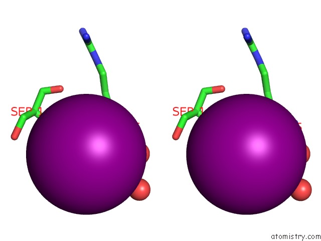
Stereo pair view
Mono view
Stereo pair view
A full contact list of Iodine with other atoms in the I binding
site number 1 of Crystal Structures of Mutant Endo-Beta-1,4-Xylanase II (E177Q) in the Apo Form within 5.0Å range:
|
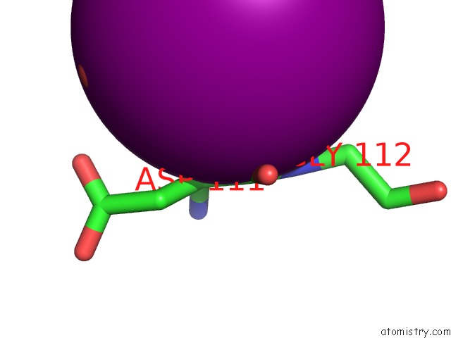
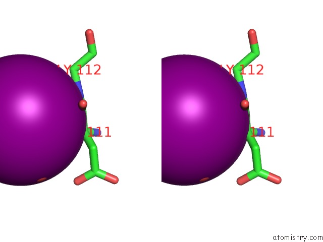
Iodine binding site 2 out of 6 in 4hko
Go back to
Iodine binding site 2 out
of 6 in the Crystal Structures of Mutant Endo-Beta-1,4-Xylanase II (E177Q) in the Apo Form
Mono view
Stereo pair view
Mono view
Stereo pair view
A full contact list of Iodine with other atoms in the I binding
site number 2 of Crystal Structures of Mutant Endo-Beta-1,4-Xylanase II (E177Q) in the Apo Form within 5.0Å range:
|
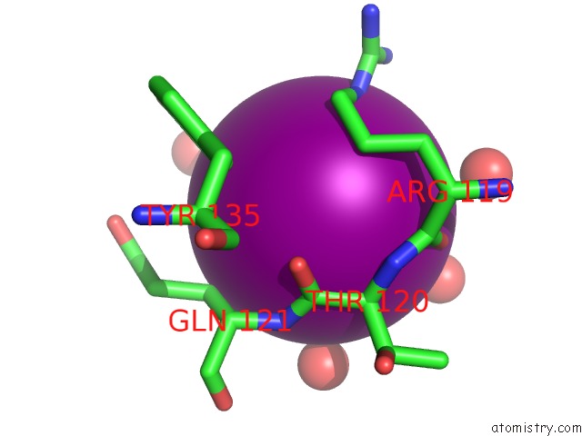
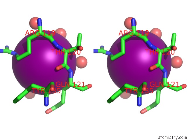
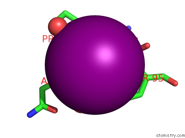
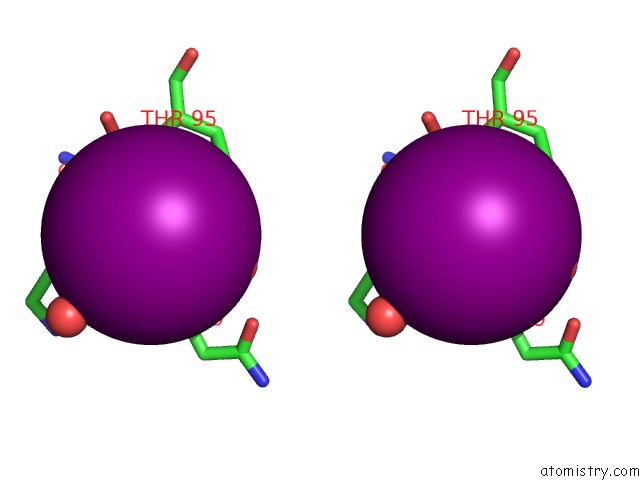
Iodine binding site 3 out of 6 in 4hko
Go back to
Iodine binding site 3 out
of 6 in the Crystal Structures of Mutant Endo-Beta-1,4-Xylanase II (E177Q) in the Apo Form
Mono view
Stereo pair view
Mono view
Stereo pair view
A full contact list of Iodine with other atoms in the I binding
site number 3 of Crystal Structures of Mutant Endo-Beta-1,4-Xylanase II (E177Q) in the Apo Form within 5.0Å range:
|
Iodine binding site 4 out of 6 in 4hko
Go back to
Iodine binding site 4 out
of 6 in the Crystal Structures of Mutant Endo-Beta-1,4-Xylanase II (E177Q) in the Apo Form
Mono view
Stereo pair view
Mono view
Stereo pair view
A full contact list of Iodine with other atoms in the I binding
site number 4 of Crystal Structures of Mutant Endo-Beta-1,4-Xylanase II (E177Q) in the Apo Form within 5.0Å range:
|
Iodine binding site 5 out of 6 in 4hko
Go back to
Iodine binding site 5 out
of 6 in the Crystal Structures of Mutant Endo-Beta-1,4-Xylanase II (E177Q) in the Apo Form
Mono view
Stereo pair view
Mono view
Stereo pair view
A full contact list of Iodine with other atoms in the I binding
site number 5 of Crystal Structures of Mutant Endo-Beta-1,4-Xylanase II (E177Q) in the Apo Form within 5.0Å range:
|
Iodine binding site 6 out of 6 in 4hko
Go back to
Iodine binding site 6 out
of 6 in the Crystal Structures of Mutant Endo-Beta-1,4-Xylanase II (E177Q) in the Apo Form
Mono view
Stereo pair view
Mono view
Stereo pair view
A full contact list of Iodine with other atoms in the I binding
site number 6 of Crystal Structures of Mutant Endo-Beta-1,4-Xylanase II (E177Q) in the Apo Form within 5.0Å range:
|
Reference:
Q.Wan,
Q.Zhang,
S.Hamilton-Brehm,
K.Weiss,
M.Mustyakimov,
L.Coates,
P.Langan,
D.Graham,
A.Kovalevsky.
X-Ray Crystallographic Studies of Family 11 Xylanase Michaelis and Product Complexes: Implications For the Catalytic Mechanism. Acta Crystallogr.,Sect.D V. 70 11 2014.
ISSN: ISSN 0907-4449
PubMed: 24419374
DOI: 10.1107/S1399004713023626
Page generated: Sun Aug 11 18:04:21 2024
ISSN: ISSN 0907-4449
PubMed: 24419374
DOI: 10.1107/S1399004713023626
Last articles
Zn in 9J0NZn in 9J0O
Zn in 9J0P
Zn in 9FJX
Zn in 9EKB
Zn in 9C0F
Zn in 9CAH
Zn in 9CH0
Zn in 9CH3
Zn in 9CH1