Iodine »
PDB 4hkl-4jxj »
4iw0 »
Iodine in PDB 4iw0: Crystal Structure and Mechanism of Activation of TBK1
Enzymatic activity of Crystal Structure and Mechanism of Activation of TBK1
All present enzymatic activity of Crystal Structure and Mechanism of Activation of TBK1:
2.7.11.1;
2.7.11.1;
Protein crystallography data
The structure of Crystal Structure and Mechanism of Activation of TBK1, PDB code: 4iw0
was solved by
A.Larabi,
J.M.Devos,
S.-L.Ng,
M.H.Nanao,
A.Round,
T.Maniatis,
D.Panne,
with X-Ray Crystallography technique. A brief refinement statistics is given in the table below:
| Resolution Low / High (Å) | 47.56 / 4.00 |
| Space group | P 65 2 2 |
| Cell size a, b, c (Å), α, β, γ (°) | 228.910, 228.910, 97.980, 90.00, 90.00, 120.00 |
| R / Rfree (%) | 23.7 / 29.1 |
Iodine Binding Sites:
The binding sites of Iodine atom in the Crystal Structure and Mechanism of Activation of TBK1
(pdb code 4iw0). This binding sites where shown within
5.0 Angstroms radius around Iodine atom.
In total only one binding site of Iodine was determined in the Crystal Structure and Mechanism of Activation of TBK1, PDB code: 4iw0:
In total only one binding site of Iodine was determined in the Crystal Structure and Mechanism of Activation of TBK1, PDB code: 4iw0:
Iodine binding site 1 out of 1 in 4iw0
Go back to
Iodine binding site 1 out
of 1 in the Crystal Structure and Mechanism of Activation of TBK1
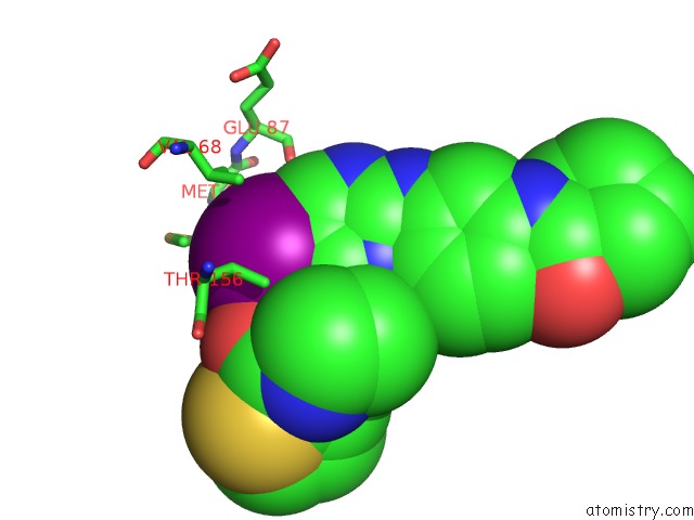
Mono view
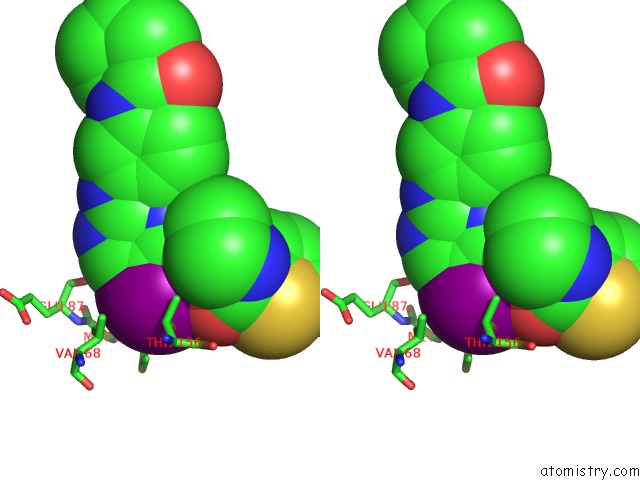
Stereo pair view
Mono view
Stereo pair view
A full contact list of Iodine with other atoms in the I binding
site number 1 of Crystal Structure and Mechanism of Activation of TBK1 within 5.0Å range:
|
Reference:
A.Larabi,
J.M.Devos,
S.L.Ng,
M.H.Nanao,
A.Round,
T.Maniatis,
D.Panne.
Crystal Structure and Mechanism of Activation of Tank-Binding Kinase 1. Cell Rep V. 3 734 2013.
ISSN: ESSN 2211-1247
PubMed: 23453971
DOI: 10.1016/J.CELREP.2013.01.034
Page generated: Sun Aug 11 18:12:15 2024
ISSN: ESSN 2211-1247
PubMed: 23453971
DOI: 10.1016/J.CELREP.2013.01.034
Last articles
Zn in 9J0NZn in 9J0O
Zn in 9J0P
Zn in 9FJX
Zn in 9EKB
Zn in 9C0F
Zn in 9CAH
Zn in 9CH0
Zn in 9CH3
Zn in 9CH1