Iodine »
PDB 4hkl-4jxj »
4j2v »
Iodine in PDB 4j2v: Crystal Structure of Equine Serum Albumin in Complex with 3,5- Diiodosalicylic Acid
Protein crystallography data
The structure of Crystal Structure of Equine Serum Albumin in Complex with 3,5- Diiodosalicylic Acid, PDB code: 4j2v
was solved by
B.Sekula,
A.Bujacz,
K.Zielinski,
G.Bujacz,
with X-Ray Crystallography technique. A brief refinement statistics is given in the table below:
| Resolution Low / High (Å) | 33.39 / 2.12 |
| Space group | P 61 |
| Cell size a, b, c (Å), α, β, γ (°) | 88.880, 88.880, 134.340, 90.00, 90.00, 120.00 |
| R / Rfree (%) | 18.1 / 23.8 |
Iodine Binding Sites:
The binding sites of Iodine atom in the Crystal Structure of Equine Serum Albumin in Complex with 3,5- Diiodosalicylic Acid
(pdb code 4j2v). This binding sites where shown within
5.0 Angstroms radius around Iodine atom.
In total 8 binding sites of Iodine where determined in the Crystal Structure of Equine Serum Albumin in Complex with 3,5- Diiodosalicylic Acid, PDB code: 4j2v:
Jump to Iodine binding site number: 1; 2; 3; 4; 5; 6; 7; 8;
In total 8 binding sites of Iodine where determined in the Crystal Structure of Equine Serum Albumin in Complex with 3,5- Diiodosalicylic Acid, PDB code: 4j2v:
Jump to Iodine binding site number: 1; 2; 3; 4; 5; 6; 7; 8;
Iodine binding site 1 out of 8 in 4j2v
Go back to
Iodine binding site 1 out
of 8 in the Crystal Structure of Equine Serum Albumin in Complex with 3,5- Diiodosalicylic Acid

Mono view
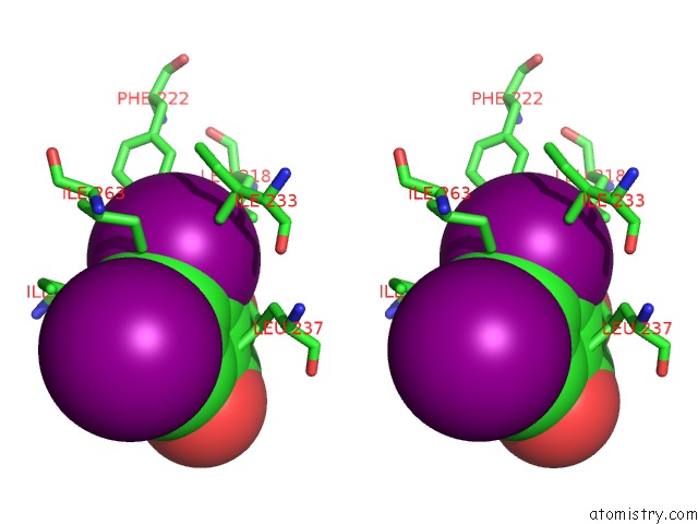
Stereo pair view
Mono view
Stereo pair view
A full contact list of Iodine with other atoms in the I binding
site number 1 of Crystal Structure of Equine Serum Albumin in Complex with 3,5- Diiodosalicylic Acid within 5.0Å range:
|
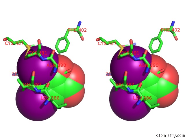
Iodine binding site 2 out of 8 in 4j2v
Go back to
Iodine binding site 2 out
of 8 in the Crystal Structure of Equine Serum Albumin in Complex with 3,5- Diiodosalicylic Acid

Mono view

Stereo pair view
Mono view
Stereo pair view
A full contact list of Iodine with other atoms in the I binding
site number 2 of Crystal Structure of Equine Serum Albumin in Complex with 3,5- Diiodosalicylic Acid within 5.0Å range:
|
Iodine binding site 3 out of 8 in 4j2v
Go back to
Iodine binding site 3 out
of 8 in the Crystal Structure of Equine Serum Albumin in Complex with 3,5- Diiodosalicylic Acid

Mono view
Stereo pair view
Mono view
Stereo pair view
A full contact list of Iodine with other atoms in the I binding
site number 3 of Crystal Structure of Equine Serum Albumin in Complex with 3,5- Diiodosalicylic Acid within 5.0Å range:
|
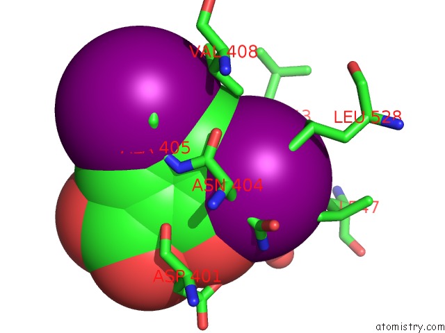
Iodine binding site 4 out of 8 in 4j2v
Go back to
Iodine binding site 4 out
of 8 in the Crystal Structure of Equine Serum Albumin in Complex with 3,5- Diiodosalicylic Acid

Mono view

Stereo pair view
Mono view
Stereo pair view
A full contact list of Iodine with other atoms in the I binding
site number 4 of Crystal Structure of Equine Serum Albumin in Complex with 3,5- Diiodosalicylic Acid within 5.0Å range:
|
Iodine binding site 5 out of 8 in 4j2v
Go back to
Iodine binding site 5 out
of 8 in the Crystal Structure of Equine Serum Albumin in Complex with 3,5- Diiodosalicylic Acid

Mono view

Stereo pair view
Mono view
Stereo pair view
A full contact list of Iodine with other atoms in the I binding
site number 5 of Crystal Structure of Equine Serum Albumin in Complex with 3,5- Diiodosalicylic Acid within 5.0Å range:
|
Iodine binding site 6 out of 8 in 4j2v
Go back to
Iodine binding site 6 out
of 8 in the Crystal Structure of Equine Serum Albumin in Complex with 3,5- Diiodosalicylic Acid

Mono view

Stereo pair view
Mono view
Stereo pair view
A full contact list of Iodine with other atoms in the I binding
site number 6 of Crystal Structure of Equine Serum Albumin in Complex with 3,5- Diiodosalicylic Acid within 5.0Å range:
|
Iodine binding site 7 out of 8 in 4j2v
Go back to
Iodine binding site 7 out
of 8 in the Crystal Structure of Equine Serum Albumin in Complex with 3,5- Diiodosalicylic Acid
Mono view

Stereo pair view
Mono view
Stereo pair view
A full contact list of Iodine with other atoms in the I binding
site number 7 of Crystal Structure of Equine Serum Albumin in Complex with 3,5- Diiodosalicylic Acid within 5.0Å range:
|
Iodine binding site 8 out of 8 in 4j2v
Go back to
Iodine binding site 8 out
of 8 in the Crystal Structure of Equine Serum Albumin in Complex with 3,5- Diiodosalicylic Acid

Mono view

Stereo pair view
Mono view
Stereo pair view
A full contact list of Iodine with other atoms in the I binding
site number 8 of Crystal Structure of Equine Serum Albumin in Complex with 3,5- Diiodosalicylic Acid within 5.0Å range:
|
Reference:
B.Sekula,
K.Zielinski,
A.Bujacz.
Crystallographic Studies of the Complexes of Bovine and Equine Serum Albumin with 3,5-Diiodosalicylic Acid. Int.J.Biol.Macromol. V. 60C 316 2013.
ISSN: ISSN 0141-8130
PubMed: 23769932
DOI: 10.1016/J.IJBIOMAC.2013.06.004
Page generated: Sun Aug 11 18:13:23 2024
ISSN: ISSN 0141-8130
PubMed: 23769932
DOI: 10.1016/J.IJBIOMAC.2013.06.004
Last articles
Cl in 8AEUCl in 8AEP
Cl in 8AEM
Cl in 8AEC
Cl in 8ADK
Cl in 8ADJ
Cl in 8ADM
Cl in 8ACV
Cl in 8ACL
Cl in 8AD2