Iodine »
PDB 4k1c-4mw7 »
4l85 »
Iodine in PDB 4l85: Crystal Structure of Receiver Domain of Kdpe D52A Mutant From E. Coli
Protein crystallography data
The structure of Crystal Structure of Receiver Domain of Kdpe D52A Mutant From E. Coli, PDB code: 4l85
was solved by
S.Kumar,
D.A.Yernool,
with X-Ray Crystallography technique. A brief refinement statistics is given in the table below:
| Resolution Low / High (Å) | 29.21 / 2.20 |
| Space group | P 31 2 1 |
| Cell size a, b, c (Å), α, β, γ (°) | 108.918, 108.918, 74.408, 90.00, 90.00, 120.00 |
| R / Rfree (%) | 17.4 / 21.9 |
Iodine Binding Sites:
Pages:
>>> Page 1 <<< Page 2, Binding sites: 11 - 20; Page 3, Binding sites: 21 - 30; Page 4, Binding sites: 31 - 35;Binding sites:
The binding sites of Iodine atom in the Crystal Structure of Receiver Domain of Kdpe D52A Mutant From E. Coli (pdb code 4l85). This binding sites where shown within 5.0 Angstroms radius around Iodine atom.In total 35 binding sites of Iodine where determined in the Crystal Structure of Receiver Domain of Kdpe D52A Mutant From E. Coli, PDB code: 4l85:
Jump to Iodine binding site number: 1; 2; 3; 4; 5; 6; 7; 8; 9; 10;
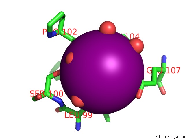
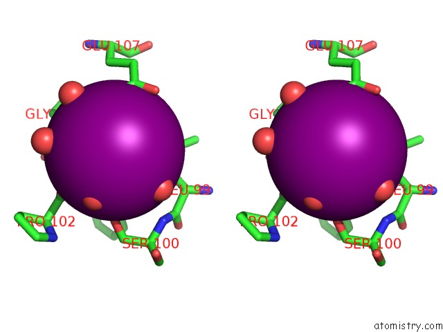
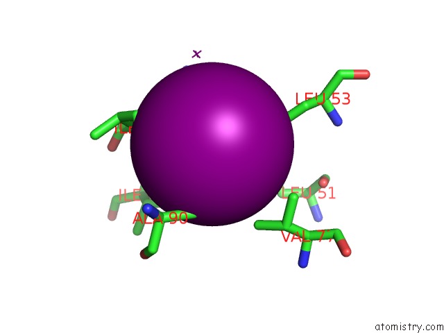
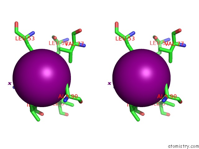
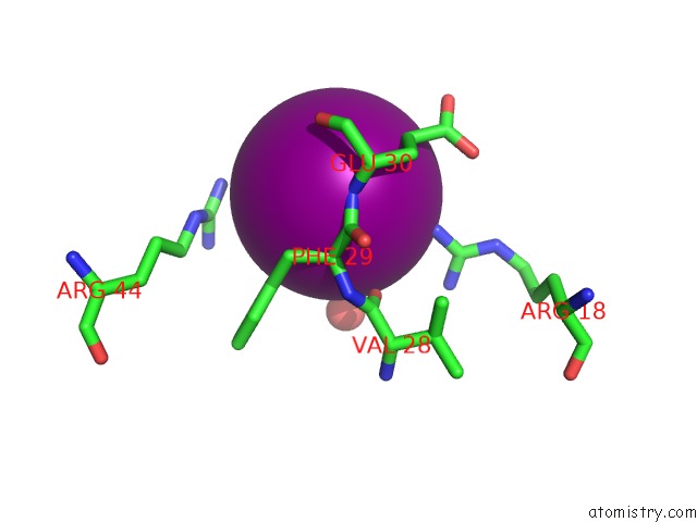
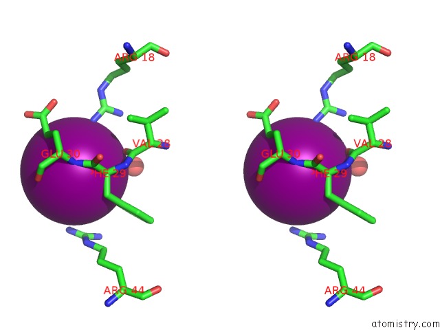
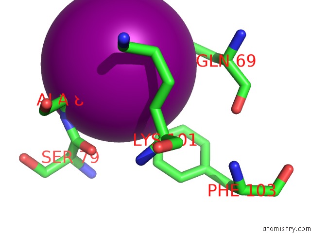
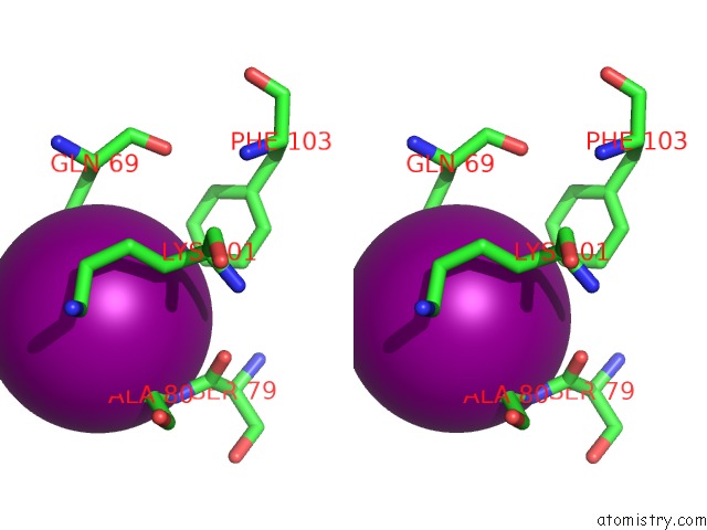
Iodine binding site 1 out of 35 in 4l85
Go back to
Iodine binding site 1 out
of 35 in the Crystal Structure of Receiver Domain of Kdpe D52A Mutant From E. Coli
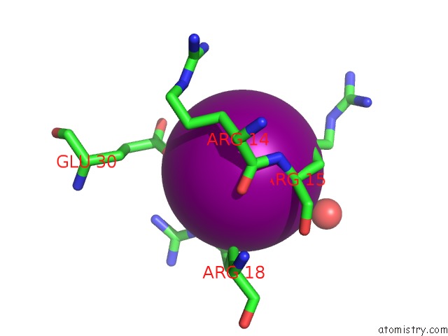
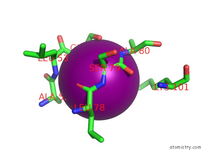
Mono view
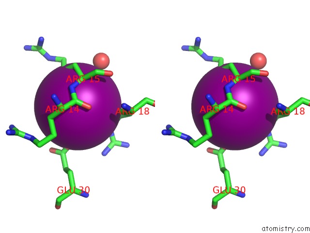
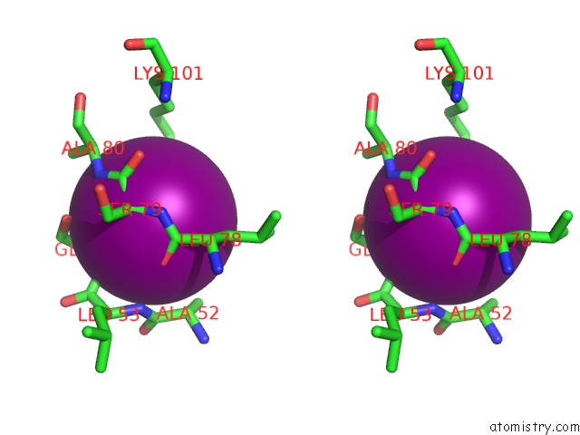
Stereo pair view
Mono view
Stereo pair view
A full contact list of Iodine with other atoms in the I binding
site number 1 of Crystal Structure of Receiver Domain of Kdpe D52A Mutant From E. Coli within 5.0Å range:
|
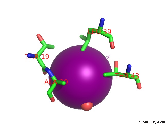
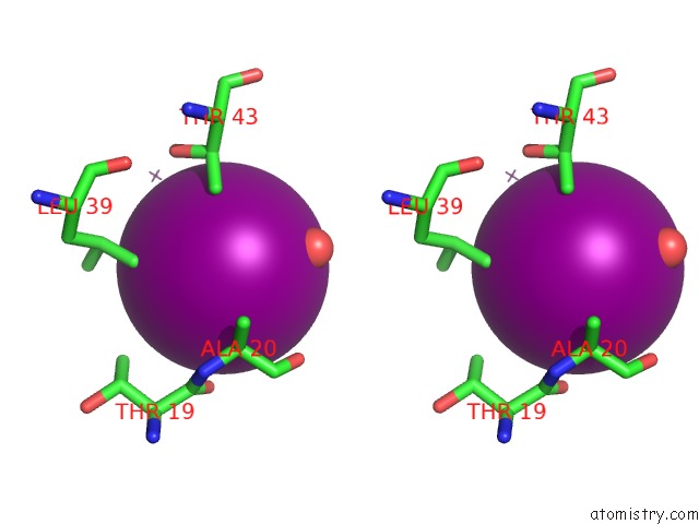
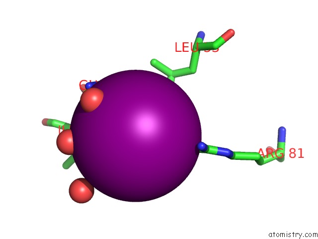
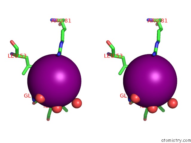
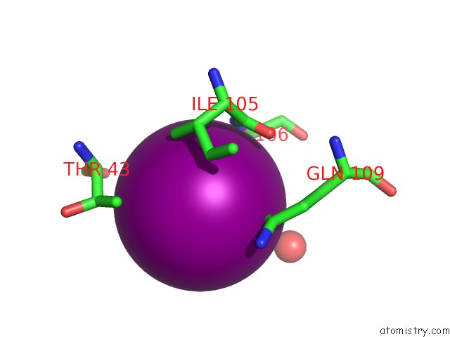
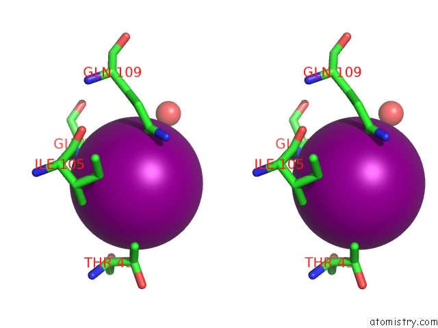
Iodine binding site 2 out of 35 in 4l85
Go back to
Iodine binding site 2 out
of 35 in the Crystal Structure of Receiver Domain of Kdpe D52A Mutant From E. Coli
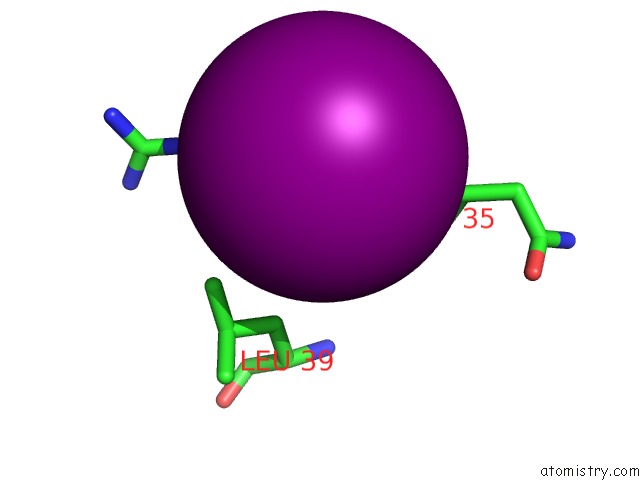
Mono view
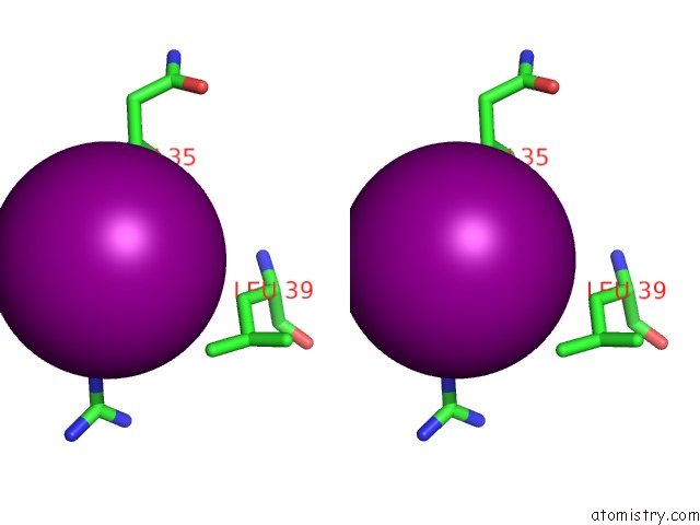
Stereo pair view
Mono view
Stereo pair view
A full contact list of Iodine with other atoms in the I binding
site number 2 of Crystal Structure of Receiver Domain of Kdpe D52A Mutant From E. Coli within 5.0Å range:
|
Iodine binding site 3 out of 35 in 4l85
Go back to
Iodine binding site 3 out
of 35 in the Crystal Structure of Receiver Domain of Kdpe D52A Mutant From E. Coli
Mono view
Stereo pair view
Mono view
Stereo pair view
A full contact list of Iodine with other atoms in the I binding
site number 3 of Crystal Structure of Receiver Domain of Kdpe D52A Mutant From E. Coli within 5.0Å range:
|
Iodine binding site 4 out of 35 in 4l85
Go back to
Iodine binding site 4 out
of 35 in the Crystal Structure of Receiver Domain of Kdpe D52A Mutant From E. Coli
Mono view
Stereo pair view
Mono view
Stereo pair view
A full contact list of Iodine with other atoms in the I binding
site number 4 of Crystal Structure of Receiver Domain of Kdpe D52A Mutant From E. Coli within 5.0Å range:
|
Iodine binding site 5 out of 35 in 4l85
Go back to
Iodine binding site 5 out
of 35 in the Crystal Structure of Receiver Domain of Kdpe D52A Mutant From E. Coli
Mono view
Stereo pair view
Mono view
Stereo pair view
A full contact list of Iodine with other atoms in the I binding
site number 5 of Crystal Structure of Receiver Domain of Kdpe D52A Mutant From E. Coli within 5.0Å range:
|
Iodine binding site 6 out of 35 in 4l85
Go back to
Iodine binding site 6 out
of 35 in the Crystal Structure of Receiver Domain of Kdpe D52A Mutant From E. Coli
Mono view
Stereo pair view
Mono view
Stereo pair view
A full contact list of Iodine with other atoms in the I binding
site number 6 of Crystal Structure of Receiver Domain of Kdpe D52A Mutant From E. Coli within 5.0Å range:
|
Iodine binding site 7 out of 35 in 4l85
Go back to
Iodine binding site 7 out
of 35 in the Crystal Structure of Receiver Domain of Kdpe D52A Mutant From E. Coli
Mono view
Stereo pair view
Mono view
Stereo pair view
A full contact list of Iodine with other atoms in the I binding
site number 7 of Crystal Structure of Receiver Domain of Kdpe D52A Mutant From E. Coli within 5.0Å range:
|
Iodine binding site 8 out of 35 in 4l85
Go back to
Iodine binding site 8 out
of 35 in the Crystal Structure of Receiver Domain of Kdpe D52A Mutant From E. Coli
Mono view
Stereo pair view
Mono view
Stereo pair view
A full contact list of Iodine with other atoms in the I binding
site number 8 of Crystal Structure of Receiver Domain of Kdpe D52A Mutant From E. Coli within 5.0Å range:
|
Iodine binding site 9 out of 35 in 4l85
Go back to
Iodine binding site 9 out
of 35 in the Crystal Structure of Receiver Domain of Kdpe D52A Mutant From E. Coli
Mono view
Stereo pair view
Mono view
Stereo pair view
A full contact list of Iodine with other atoms in the I binding
site number 9 of Crystal Structure of Receiver Domain of Kdpe D52A Mutant From E. Coli within 5.0Å range:
|
Iodine binding site 10 out of 35 in 4l85
Go back to
Iodine binding site 10 out
of 35 in the Crystal Structure of Receiver Domain of Kdpe D52A Mutant From E. Coli
Mono view
Stereo pair view
Mono view
Stereo pair view
A full contact list of Iodine with other atoms in the I binding
site number 10 of Crystal Structure of Receiver Domain of Kdpe D52A Mutant From E. Coli within 5.0Å range:
|
Reference:
A.Narayanan,
S.Kumar,
A.N.Evrard,
L.N.Paul,
D.A.Yernool.
An Asymmetric Heterodomain Interface Stabilizes A Response Regulator-Dna Complex. Nat Commun V. 5 3282 2014.
ISSN: ESSN 2041-1723
PubMed: 24526190
DOI: 10.1038/NCOMMS4282
Page generated: Sun Aug 11 18:31:00 2024
ISSN: ESSN 2041-1723
PubMed: 24526190
DOI: 10.1038/NCOMMS4282
Last articles
Zn in 9J0NZn in 9J0O
Zn in 9J0P
Zn in 9FJX
Zn in 9EKB
Zn in 9C0F
Zn in 9CAH
Zn in 9CH0
Zn in 9CH3
Zn in 9CH1