Iodine »
PDB 4k1c-4mw7 »
4m71 »
Iodine in PDB 4m71: Mutant Structure of Methyltransferase From Streptomyces Hygroscopicus Complexed with S-Adenosyl-L-Homocysteine and Methylphenylpyruvic Acid
Protein crystallography data
The structure of Mutant Structure of Methyltransferase From Streptomyces Hygroscopicus Complexed with S-Adenosyl-L-Homocysteine and Methylphenylpyruvic Acid, PDB code: 4m71
was solved by
Y.C.Liu,
X.W.Zou,
H.C.Chan,
C.J.Huang,
T.L.Li,
with X-Ray Crystallography technique. A brief refinement statistics is given in the table below:
| Resolution Low / High (Å) | 28.95 / 2.21 |
| Space group | P 21 21 21 |
| Cell size a, b, c (Å), α, β, γ (°) | 60.833, 93.693, 137.375, 90.00, 90.00, 90.00 |
| R / Rfree (%) | 15.8 / 22.9 |
Other elements in 4m71:
The structure of Mutant Structure of Methyltransferase From Streptomyces Hygroscopicus Complexed with S-Adenosyl-L-Homocysteine and Methylphenylpyruvic Acid also contains other interesting chemical elements:
| Iron | (Fe) | 2 atoms |
| Calcium | (Ca) | 14 atoms |
Iodine Binding Sites:
The binding sites of Iodine atom in the Mutant Structure of Methyltransferase From Streptomyces Hygroscopicus Complexed with S-Adenosyl-L-Homocysteine and Methylphenylpyruvic Acid
(pdb code 4m71). This binding sites where shown within
5.0 Angstroms radius around Iodine atom.
In total only one binding site of Iodine was determined in the Mutant Structure of Methyltransferase From Streptomyces Hygroscopicus Complexed with S-Adenosyl-L-Homocysteine and Methylphenylpyruvic Acid, PDB code: 4m71:
In total only one binding site of Iodine was determined in the Mutant Structure of Methyltransferase From Streptomyces Hygroscopicus Complexed with S-Adenosyl-L-Homocysteine and Methylphenylpyruvic Acid, PDB code: 4m71:
Iodine binding site 1 out of 1 in 4m71
Go back to
Iodine binding site 1 out
of 1 in the Mutant Structure of Methyltransferase From Streptomyces Hygroscopicus Complexed with S-Adenosyl-L-Homocysteine and Methylphenylpyruvic Acid
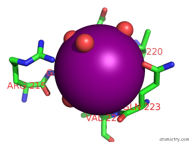
Mono view
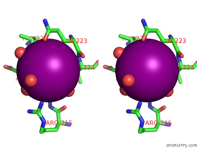
Stereo pair view
Mono view
Stereo pair view
A full contact list of Iodine with other atoms in the I binding
site number 1 of Mutant Structure of Methyltransferase From Streptomyces Hygroscopicus Complexed with S-Adenosyl-L-Homocysteine and Methylphenylpyruvic Acid within 5.0Å range:
|
Reference:
X.W.Zou,
Y.C.Liu,
N.S.Hsu,
C.J.Huang,
S.Y.Lyu,
H.C.Chan,
C.Y.Chang,
H.W.Yeh,
K.H.Lin,
C.J.Wu,
M.D.Tsai,
T.L.Li.
Structure and Mechanism of A Nonhaem-Iron Sam-Dependent C-Methyltransferase and Its Engineering to A Hydratase and An O-Methyltransferase Acta Crystallogr.,Sect.D V. 70 1549 2014.
ISSN: ISSN 0907-4449
PubMed: 24914966
DOI: 10.1107/S1399004714005239
Page generated: Sun Aug 11 18:41:26 2024
ISSN: ISSN 0907-4449
PubMed: 24914966
DOI: 10.1107/S1399004714005239
Last articles
Zn in 9MJ5Zn in 9HNW
Zn in 9G0L
Zn in 9FNE
Zn in 9DZN
Zn in 9E0I
Zn in 9D32
Zn in 9DAK
Zn in 8ZXC
Zn in 8ZUF