Iodine in PDB 4nhb: Crystal Structure of A Trap Periplasmic Solute Binding Protein From Desulfovibrio Desulfuricans (DDES_1525), Target Efi-510107, with Bound Sn-Glycerol-3-Phosphate
Protein crystallography data
The structure of Crystal Structure of A Trap Periplasmic Solute Binding Protein From Desulfovibrio Desulfuricans (DDES_1525), Target Efi-510107, with Bound Sn-Glycerol-3-Phosphate, PDB code: 4nhb
was solved by
M.W.Vetting,
N.F.Al Obaidi,
L.L.Morisco,
S.R.Wasserman,
S.Sojitra,
M.Stead,
J.D.Attonito,
A.Scott Glenn,
S.Chowdhury,
B.Evans,
B.Hillerich,
J.Love,
R.D.Seidel,
H.J.Imker,
J.A.Gerlt,
S.C.Almo,
Enzyme Functioninitiative (Efi),
with X-Ray Crystallography technique. A brief refinement statistics is given in the table below:
Resolution Low / High (Å)
|
25.64 /
1.90
|
Space group
|
P 1 21 1
|
Cell size a, b, c (Å), α, β, γ (°)
|
53.090,
83.407,
65.043,
90.00,
91.83,
90.00
|
R / Rfree (%)
|
18.5 /
24.8
|
Iodine Binding Sites:
Pages:
>>> Page 1 <<<
Page 2, Binding sites: 11 -
20;
Page 3, Binding sites: 21 -
23;
Binding sites:
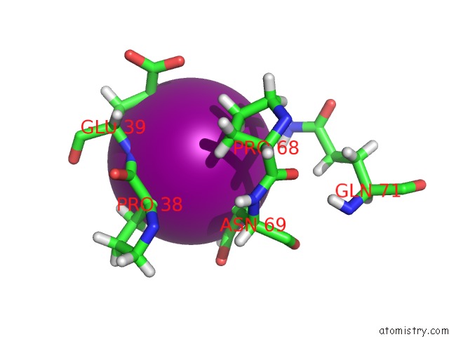
The binding sites of Iodine atom in the Crystal Structure of A Trap Periplasmic Solute Binding Protein From Desulfovibrio Desulfuricans (DDES_1525), Target Efi-510107, with Bound Sn-Glycerol-3-Phosphate
(pdb code 4nhb). This binding sites where shown within
5.0 Angstroms radius around Iodine atom.
In total 23 binding sites of Iodine where determined in the
Crystal Structure of A Trap Periplasmic Solute Binding Protein From Desulfovibrio Desulfuricans (DDES_1525), Target Efi-510107, with Bound Sn-Glycerol-3-Phosphate, PDB code: 4nhb:
Jump to Iodine binding site number:
1;
2;
3;
4;
5;
6;
7;
8;
9;
10;
Iodine binding site 1 out
of 23 in 4nhb
Go back to
Iodine Binding Sites List in 4nhb
Iodine binding site 1 out
of 23 in the Crystal Structure of A Trap Periplasmic Solute Binding Protein From Desulfovibrio Desulfuricans (DDES_1525), Target Efi-510107, with Bound Sn-Glycerol-3-Phosphate
Mono view
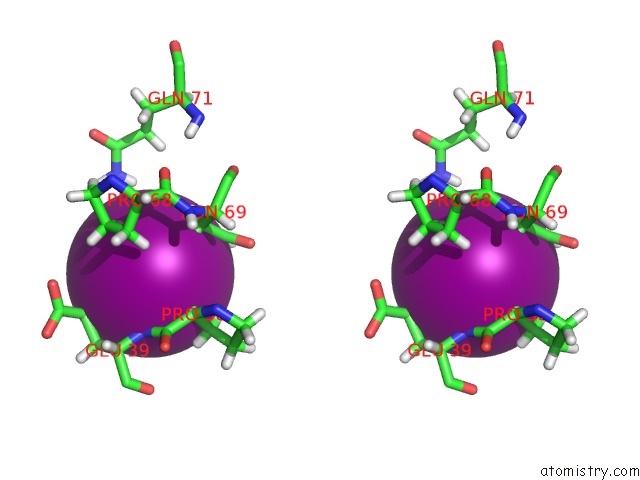
Stereo pair view
|
A full contact list of Iodine with other atoms in the I binding
site number 1 of Crystal Structure of A Trap Periplasmic Solute Binding Protein From Desulfovibrio Desulfuricans (DDES_1525), Target Efi-510107, with Bound Sn-Glycerol-3-Phosphate within 5.0Å range:
probe
|
atom
|
residue
|
distance (Å)
|
B
|
Occ
|
A:I401
b:39.2
occ:0.61
|
H
|
A:GLU39
|
2.9
|
20.0
|
1.0
|
O
|
A:HOH635
|
3.1
|
29.1
|
1.0
|
HG2
|
A:GLU39
|
3.2
|
23.8
|
1.0
|
HD21
|
A:ASN69
|
3.2
|
26.0
|
1.0
|
HA
|
A:PRO38
|
3.3
|
7.8
|
1.0
|
HB2
|
A:ASN69
|
3.5
|
15.9
|
1.0
|
ND2
|
A:ASN69
|
3.7
|
21.7
|
1.0
|
N
|
A:GLU39
|
3.7
|
16.7
|
1.0
|
CG
|
A:GLU39
|
3.9
|
19.9
|
1.0
|
HB3
|
A:PRO38
|
3.9
|
17.1
|
1.0
|
HG3
|
A:GLU39
|
3.9
|
23.8
|
1.0
|
HB3
|
A:ASN69
|
4.0
|
15.9
|
1.0
|
HB2
|
A:GLU39
|
4.0
|
16.9
|
1.0
|
CA
|
A:PRO38
|
4.0
|
6.5
|
1.0
|
CB
|
A:ASN69
|
4.0
|
13.2
|
1.0
|
HD22
|
A:ASN69
|
4.1
|
26.0
|
1.0
|
HB2
|
A:PRO38
|
4.2
|
17.1
|
1.0
|
HG2
|
A:PRO68
|
4.2
|
20.0
|
1.0
|
CG
|
A:ASN69
|
4.2
|
14.8
|
1.0
|
CB
|
A:PRO38
|
4.2
|
14.2
|
1.0
|
CB
|
A:GLU39
|
4.4
|
14.1
|
1.0
|
C
|
A:PRO38
|
4.4
|
7.1
|
1.0
|
HE21
|
A:GLN71
|
4.4
|
53.8
|
1.0
|
CA
|
A:GLU39
|
4.7
|
18.8
|
1.0
|
HE22
|
A:GLN71
|
4.9
|
53.8
|
1.0
|
NE2
|
A:GLN71
|
5.0
|
44.8
|
1.0
|
CG
|
A:PRO68
|
5.0
|
16.7
|
1.0
|
|
Iodine binding site 2 out
of 23 in 4nhb
Go back to
Iodine Binding Sites List in 4nhb
Iodine binding site 2 out
of 23 in the Crystal Structure of A Trap Periplasmic Solute Binding Protein From Desulfovibrio Desulfuricans (DDES_1525), Target Efi-510107, with Bound Sn-Glycerol-3-Phosphate
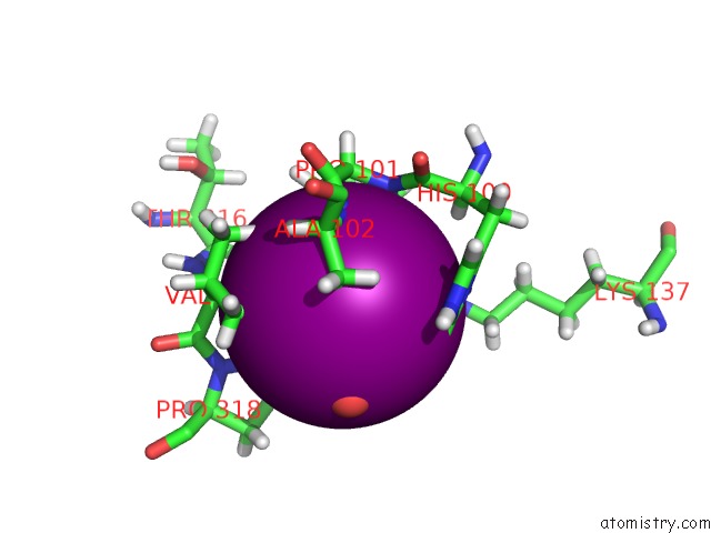
Mono view
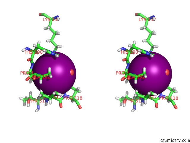
Stereo pair view
|
A full contact list of Iodine with other atoms in the I binding
site number 2 of Crystal Structure of A Trap Periplasmic Solute Binding Protein From Desulfovibrio Desulfuricans (DDES_1525), Target Efi-510107, with Bound Sn-Glycerol-3-Phosphate within 5.0Å range:
probe
|
atom
|
residue
|
distance (Å)
|
B
|
Occ
|
A:I402
b:30.2
occ:0.48
|
HD2
|
A:PRO318
|
2.6
|
43.7
|
1.0
|
HB2
|
A:PRO101
|
3.0
|
28.7
|
1.0
|
H
|
A:ALA102
|
3.1
|
15.4
|
1.0
|
HA
|
A:VAL317
|
3.1
|
21.9
|
1.0
|
HG13
|
A:VAL317
|
3.2
|
22.0
|
1.0
|
HD3
|
A:PRO318
|
3.2
|
43.7
|
1.0
|
CD
|
A:PRO318
|
3.3
|
36.4
|
1.0
|
HB2
|
A:ALA102
|
3.3
|
25.2
|
1.0
|
O
|
A:HOH595
|
3.4
|
39.0
|
1.0
|
HD2
|
A:PRO101
|
3.4
|
22.8
|
1.0
|
HZ1
|
A:LYS137
|
3.6
|
35.1
|
1.0
|
HE1
|
A:HIS100
|
3.6
|
26.8
|
1.0
|
N
|
A:ALA102
|
3.7
|
12.9
|
1.0
|
HG22
|
A:VAL317
|
3.8
|
14.3
|
1.0
|
CB
|
A:PRO101
|
3.9
|
23.9
|
1.0
|
CE1
|
A:HIS100
|
3.9
|
22.4
|
1.0
|
HZ2
|
A:LYS137
|
3.9
|
35.1
|
1.0
|
HG2
|
A:PRO101
|
3.9
|
30.9
|
1.0
|
HD1
|
A:HIS100
|
3.9
|
27.6
|
1.0
|
HG2
|
A:PRO318
|
4.0
|
45.5
|
1.0
|
ND1
|
A:HIS100
|
4.0
|
23.0
|
1.0
|
O
|
A:THR316
|
4.0
|
27.4
|
1.0
|
CD
|
A:PRO101
|
4.1
|
19.0
|
1.0
|
CG1
|
A:VAL317
|
4.1
|
18.4
|
1.0
|
CA
|
A:VAL317
|
4.1
|
18.3
|
1.0
|
CB
|
A:ALA102
|
4.1
|
21.0
|
1.0
|
N
|
A:PRO101
|
4.1
|
17.2
|
1.0
|
NZ
|
A:LYS137
|
4.1
|
29.2
|
1.0
|
CG
|
A:PRO101
|
4.2
|
25.8
|
1.0
|
HB3
|
A:ALA102
|
4.2
|
25.2
|
1.0
|
CG
|
A:PRO318
|
4.3
|
37.9
|
1.0
|
CA
|
A:PRO101
|
4.4
|
19.5
|
1.0
|
CA
|
A:ALA102
|
4.4
|
12.3
|
1.0
|
C
|
A:PRO101
|
4.4
|
13.6
|
1.0
|
HG12
|
A:VAL317
|
4.4
|
22.0
|
1.0
|
N
|
A:PRO318
|
4.4
|
27.7
|
1.0
|
CB
|
A:VAL317
|
4.5
|
15.9
|
1.0
|
HA
|
A:ALA102
|
4.6
|
14.8
|
1.0
|
CG2
|
A:VAL317
|
4.6
|
11.9
|
1.0
|
HB3
|
A:PRO101
|
4.6
|
28.7
|
1.0
|
NE2
|
A:HIS100
|
4.6
|
22.5
|
1.0
|
HZ3
|
A:LYS137
|
4.6
|
35.1
|
1.0
|
HG11
|
A:VAL317
|
4.7
|
22.0
|
1.0
|
C
|
A:VAL317
|
4.8
|
23.3
|
1.0
|
HA
|
A:HIS100
|
4.8
|
15.3
|
1.0
|
HG3
|
A:PRO318
|
4.8
|
45.5
|
1.0
|
C
|
A:HIS100
|
4.8
|
19.0
|
1.0
|
CG
|
A:HIS100
|
4.9
|
19.0
|
1.0
|
HB1
|
A:ALA102
|
4.9
|
25.2
|
1.0
|
C
|
A:THR316
|
4.9
|
26.1
|
1.0
|
HE2
|
A:HIS100
|
5.0
|
27.0
|
1.0
|
HD3
|
A:PRO101
|
5.0
|
22.8
|
1.0
|
|
Iodine binding site 3 out
of 23 in 4nhb
Go back to
Iodine Binding Sites List in 4nhb
Iodine binding site 3 out
of 23 in the Crystal Structure of A Trap Periplasmic Solute Binding Protein From Desulfovibrio Desulfuricans (DDES_1525), Target Efi-510107, with Bound Sn-Glycerol-3-Phosphate
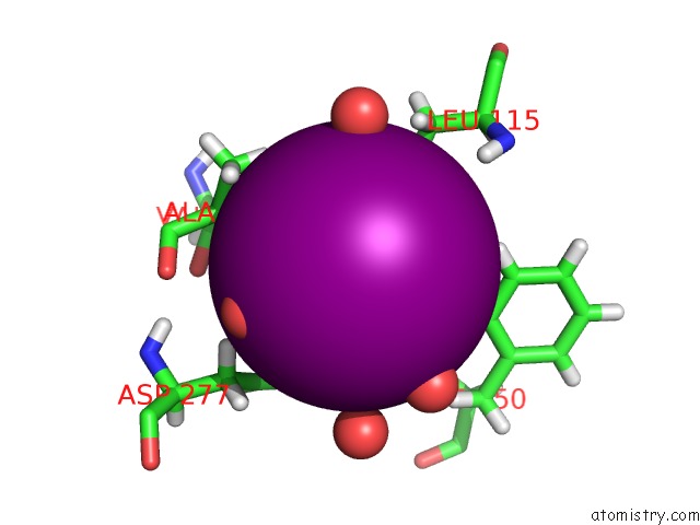
Mono view
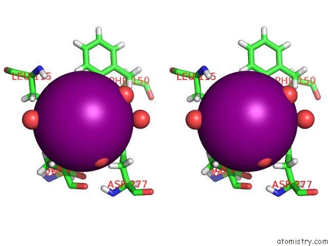
Stereo pair view
|
A full contact list of Iodine with other atoms in the I binding
site number 3 of Crystal Structure of A Trap Periplasmic Solute Binding Protein From Desulfovibrio Desulfuricans (DDES_1525), Target Efi-510107, with Bound Sn-Glycerol-3-Phosphate within 5.0Å range:
probe
|
atom
|
residue
|
distance (Å)
|
B
|
Occ
|
A:I403
b:22.3
occ:0.68
|
O
|
A:HOH524
|
3.2
|
14.1
|
1.0
|
HD12
|
A:LEU115
|
3.2
|
21.6
|
1.0
|
HA
|
A:ALA274
|
3.2
|
28.9
|
1.0
|
HG
|
A:LEU115
|
3.3
|
19.7
|
1.0
|
O
|
A:HOH507
|
3.3
|
6.9
|
1.0
|
HD11
|
A:LEU115
|
3.4
|
21.6
|
1.0
|
O
|
A:HOH686
|
3.4
|
22.8
|
1.0
|
CD1
|
A:LEU115
|
3.6
|
18.0
|
1.0
|
HD2
|
A:PHE150
|
3.8
|
17.1
|
1.0
|
O
|
A:HOH624
|
3.9
|
30.7
|
1.0
|
HB2
|
A:ALA274
|
3.9
|
33.5
|
1.0
|
CG
|
A:LEU115
|
3.9
|
16.4
|
1.0
|
CA
|
A:ALA274
|
4.1
|
24.0
|
1.0
|
O
|
A:HOH698
|
4.2
|
20.1
|
1.0
|
HB1
|
A:ALA274
|
4.2
|
33.5
|
1.0
|
HE2
|
A:PHE150
|
4.3
|
18.9
|
1.0
|
HB2
|
A:ASP277
|
4.3
|
12.3
|
1.0
|
CB
|
A:ALA274
|
4.3
|
27.9
|
1.0
|
HG12
|
A:VAL273
|
4.3
|
16.2
|
1.0
|
OD2
|
A:ASP277
|
4.3
|
16.8
|
1.0
|
HB2
|
A:LEU115
|
4.5
|
24.1
|
1.0
|
HD13
|
A:LEU115
|
4.5
|
21.6
|
1.0
|
CD2
|
A:PHE150
|
4.5
|
14.3
|
1.0
|
O
|
A:HOH541
|
4.6
|
21.1
|
1.0
|
H
|
A:LEU115
|
4.7
|
12.9
|
1.0
|
N
|
A:ALA274
|
4.7
|
20.6
|
1.0
|
CE2
|
A:PHE150
|
4.8
|
15.8
|
1.0
|
CB
|
A:LEU115
|
4.8
|
20.1
|
1.0
|
HD21
|
A:LEU115
|
4.9
|
19.1
|
1.0
|
CB
|
A:ASP277
|
4.9
|
10.2
|
1.0
|
HB3
|
A:ASP277
|
4.9
|
12.3
|
1.0
|
CG
|
A:ASP277
|
5.0
|
10.5
|
1.0
|
|
Iodine binding site 4 out
of 23 in 4nhb
Go back to
Iodine Binding Sites List in 4nhb
Iodine binding site 4 out
of 23 in the Crystal Structure of A Trap Periplasmic Solute Binding Protein From Desulfovibrio Desulfuricans (DDES_1525), Target Efi-510107, with Bound Sn-Glycerol-3-Phosphate
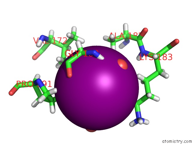
Mono view
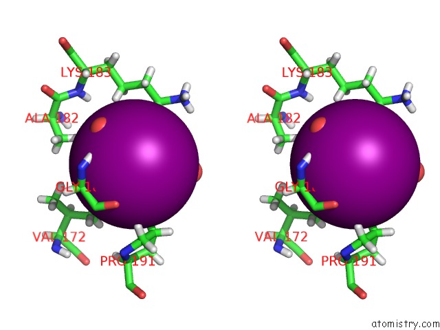
Stereo pair view
|
A full contact list of Iodine with other atoms in the I binding
site number 4 of Crystal Structure of A Trap Periplasmic Solute Binding Protein From Desulfovibrio Desulfuricans (DDES_1525), Target Efi-510107, with Bound Sn-Glycerol-3-Phosphate within 5.0Å range:
probe
|
atom
|
residue
|
distance (Å)
|
B
|
Occ
|
A:I404
b:39.9
occ:0.51
|
HB
|
A:THR179
|
2.9
|
19.8
|
1.0
|
HG22
|
A:THR179
|
3.2
|
24.1
|
1.0
|
HA3
|
A:GLY189
|
3.2
|
28.6
|
1.0
|
HE2
|
A:LYS183
|
3.3
|
35.9
|
1.0
|
HA
|
A:THR179
|
3.3
|
18.1
|
1.0
|
O
|
A:HOH654
|
3.4
|
33.1
|
1.0
|
HG3
|
A:PRO191
|
3.5
|
32.2
|
1.0
|
CB
|
A:THR179
|
3.5
|
16.5
|
1.0
|
HG21
|
A:VAL172
|
3.6
|
22.4
|
1.0
|
CG2
|
A:THR179
|
3.7
|
20.1
|
1.0
|
O
|
A:HOH705
|
3.8
|
23.3
|
1.0
|
H
|
A:GLY189
|
3.8
|
24.6
|
1.0
|
CA
|
A:THR179
|
3.9
|
15.1
|
1.0
|
O
|
A:GLY189
|
3.9
|
27.1
|
1.0
|
HG21
|
A:THR179
|
4.0
|
24.1
|
1.0
|
CA
|
A:GLY189
|
4.0
|
23.9
|
1.0
|
O
|
A:THR179
|
4.0
|
16.9
|
1.0
|
HD3
|
A:PRO191
|
4.0
|
25.5
|
1.0
|
HB3
|
A:ALA182
|
4.1
|
19.8
|
1.0
|
HG2
|
A:LYS183
|
4.2
|
41.6
|
1.0
|
C
|
A:GLY189
|
4.2
|
19.1
|
1.0
|
CE
|
A:LYS183
|
4.3
|
29.9
|
1.0
|
N
|
A:GLY189
|
4.3
|
20.5
|
1.0
|
C
|
A:THR179
|
4.3
|
14.6
|
1.0
|
CG
|
A:PRO191
|
4.4
|
26.8
|
1.0
|
HE3
|
A:LYS183
|
4.5
|
35.9
|
1.0
|
CG2
|
A:VAL172
|
4.5
|
18.7
|
1.0
|
HG22
|
A:VAL172
|
4.6
|
22.4
|
1.0
|
HG23
|
A:THR179
|
4.6
|
24.1
|
1.0
|
HG2
|
A:PRO191
|
4.7
|
32.2
|
1.0
|
CD
|
A:PRO191
|
4.7
|
21.3
|
1.0
|
HA2
|
A:GLY189
|
4.8
|
28.6
|
1.0
|
OG1
|
A:THR179
|
4.8
|
18.4
|
1.0
|
HG23
|
A:VAL172
|
4.8
|
22.4
|
1.0
|
HG11
|
A:VAL172
|
4.9
|
14.5
|
1.0
|
CG
|
A:LYS183
|
4.9
|
34.6
|
1.0
|
HZ2
|
A:LYS183
|
5.0
|
38.0
|
1.0
|
HD3
|
A:LYS183
|
5.0
|
32.9
|
1.0
|
CB
|
A:ALA182
|
5.0
|
16.5
|
1.0
|
HG3
|
A:LYS183
|
5.0
|
41.6
|
1.0
|
|
Iodine binding site 5 out
of 23 in 4nhb
Go back to
Iodine Binding Sites List in 4nhb
Iodine binding site 5 out
of 23 in the Crystal Structure of A Trap Periplasmic Solute Binding Protein From Desulfovibrio Desulfuricans (DDES_1525), Target Efi-510107, with Bound Sn-Glycerol-3-Phosphate
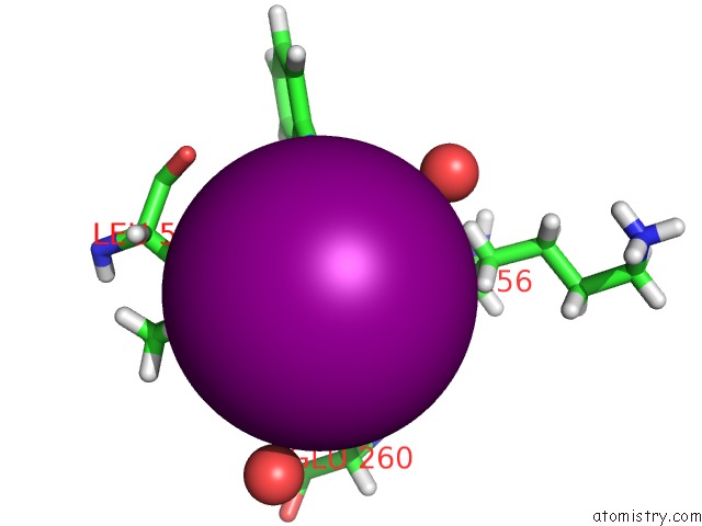
Mono view
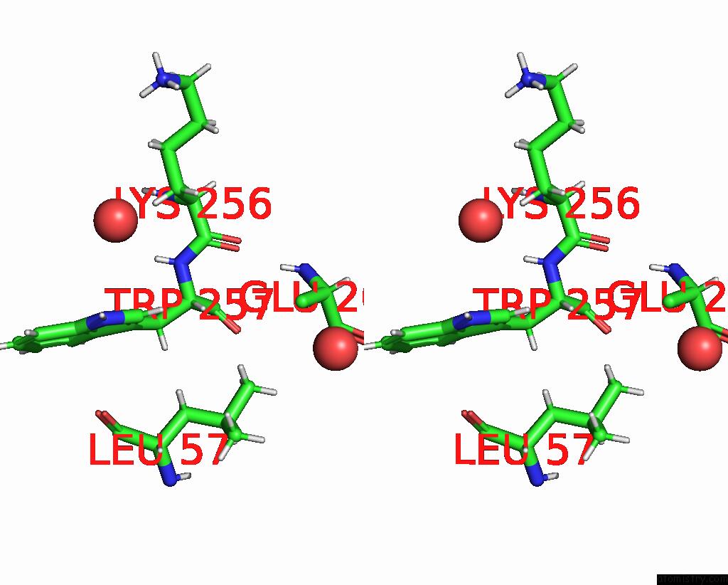
Stereo pair view
|
A full contact list of Iodine with other atoms in the I binding
site number 5 of Crystal Structure of A Trap Periplasmic Solute Binding Protein From Desulfovibrio Desulfuricans (DDES_1525), Target Efi-510107, with Bound Sn-Glycerol-3-Phosphate within 5.0Å range:
probe
|
atom
|
residue
|
distance (Å)
|
B
|
Occ
|
A:I405
b:27.0
occ:0.61
|
HD1
|
A:TRP257
|
2.4
|
48.5
|
1.0
|
HD13
|
A:LEU57
|
3.1
|
36.9
|
1.0
|
CD1
|
A:TRP257
|
3.2
|
40.4
|
1.0
|
HB3
|
A:LEU57
|
3.5
|
32.7
|
1.0
|
HD22
|
A:LEU57
|
3.6
|
38.0
|
1.0
|
HE1
|
A:TRP257
|
3.6
|
53.4
|
1.0
|
HA
|
A:TRP257
|
3.7
|
18.4
|
1.0
|
NE1
|
A:TRP257
|
3.8
|
44.5
|
1.0
|
CD1
|
A:LEU57
|
4.1
|
30.7
|
1.0
|
CB
|
A:LEU57
|
4.4
|
27.3
|
1.0
|
CG
|
A:TRP257
|
4.4
|
36.0
|
1.0
|
CD2
|
A:LEU57
|
4.4
|
31.6
|
1.0
|
CB
|
A:GLU260
|
4.5
|
26.2
|
1.0
|
CA
|
A:TRP257
|
4.5
|
15.3
|
1.0
|
CG
|
A:LEU57
|
4.5
|
28.5
|
1.0
|
HD11
|
A:LEU57
|
4.5
|
36.9
|
1.0
|
HD12
|
A:LEU57
|
4.5
|
36.9
|
1.0
|
O
|
A:HOH594
|
4.6
|
37.2
|
1.0
|
O
|
A:HOH707
|
4.7
|
36.0
|
1.0
|
N
|
A:TRP257
|
4.8
|
16.5
|
1.0
|
HB3
|
A:LYS256
|
4.9
|
35.4
|
1.0
|
HB2
|
A:LEU57
|
4.9
|
32.7
|
1.0
|
HD21
|
A:LEU57
|
4.9
|
38.0
|
1.0
|
CB
|
A:TRP257
|
4.9
|
16.1
|
1.0
|
|
Iodine binding site 6 out
of 23 in 4nhb
Go back to
Iodine Binding Sites List in 4nhb
Iodine binding site 6 out
of 23 in the Crystal Structure of A Trap Periplasmic Solute Binding Protein From Desulfovibrio Desulfuricans (DDES_1525), Target Efi-510107, with Bound Sn-Glycerol-3-Phosphate
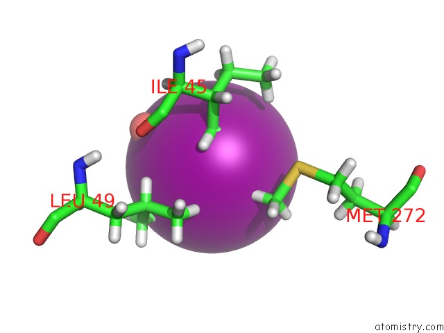
Mono view
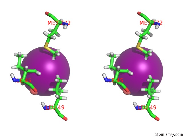
Stereo pair view
|
A full contact list of Iodine with other atoms in the I binding
site number 6 of Crystal Structure of A Trap Periplasmic Solute Binding Protein From Desulfovibrio Desulfuricans (DDES_1525), Target Efi-510107, with Bound Sn-Glycerol-3-Phosphate within 5.0Å range:
probe
|
atom
|
residue
|
distance (Å)
|
B
|
Occ
|
A:I406
b:48.8
occ:0.43
|
O
|
A:HOH545
|
2.6
|
30.7
|
1.0
|
HG23
|
A:ILE45
|
3.2
|
29.1
|
1.0
|
HD21
|
A:LEU49
|
3.5
|
26.4
|
1.0
|
HG21
|
A:ILE45
|
3.6
|
29.1
|
1.0
|
O
|
A:HOH563
|
3.7
|
26.0
|
1.0
|
CG2
|
A:ILE45
|
3.8
|
24.2
|
1.0
|
HE1
|
A:MET272
|
3.9
|
42.4
|
1.0
|
HD11
|
A:LEU49
|
4.0
|
13.6
|
1.0
|
HG22
|
A:ILE45
|
4.2
|
29.1
|
1.0
|
CD2
|
A:LEU49
|
4.4
|
22.0
|
1.0
|
HG
|
A:LEU49
|
4.5
|
20.0
|
1.0
|
CE
|
A:MET272
|
4.7
|
35.3
|
1.0
|
HG12
|
A:ILE45
|
4.7
|
28.5
|
1.0
|
HD23
|
A:LEU49
|
4.7
|
26.4
|
1.0
|
HD13
|
A:ILE45
|
4.8
|
18.7
|
1.0
|
HE2
|
A:MET272
|
4.8
|
42.4
|
1.0
|
CG
|
A:LEU49
|
4.8
|
16.7
|
1.0
|
CD1
|
A:LEU49
|
4.9
|
11.3
|
1.0
|
|
Iodine binding site 7 out
of 23 in 4nhb
Go back to
Iodine Binding Sites List in 4nhb
Iodine binding site 7 out
of 23 in the Crystal Structure of A Trap Periplasmic Solute Binding Protein From Desulfovibrio Desulfuricans (DDES_1525), Target Efi-510107, with Bound Sn-Glycerol-3-Phosphate
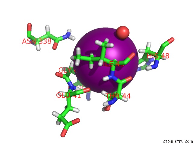
Mono view
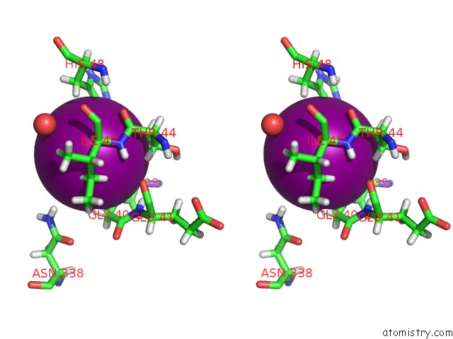
Stereo pair view
|
A full contact list of Iodine with other atoms in the I binding
site number 7 of Crystal Structure of A Trap Periplasmic Solute Binding Protein From Desulfovibrio Desulfuricans (DDES_1525), Target Efi-510107, with Bound Sn-Glycerol-3-Phosphate within 5.0Å range:
probe
|
atom
|
residue
|
distance (Å)
|
B
|
Occ
|
A:I407
b:31.4
occ:0.37
|
OE1
|
A:GLU39
|
2.4
|
24.2
|
1.0
|
H
|
A:GLY40
|
2.7
|
17.3
|
1.0
|
HD2
|
A:HIS48
|
2.8
|
17.3
|
1.0
|
HG22
|
A:THR44
|
3.3
|
19.1
|
1.0
|
HA
|
A:GLU39
|
3.4
|
22.6
|
1.0
|
CD2
|
A:HIS48
|
3.5
|
14.4
|
1.0
|
N
|
A:GLY40
|
3.5
|
14.4
|
1.0
|
HA
|
A:ILE45
|
3.5
|
20.3
|
1.0
|
CD
|
A:GLU39
|
3.6
|
21.2
|
1.0
|
HB3
|
A:GLU39
|
3.6
|
16.9
|
1.0
|
HG12
|
A:ILE45
|
3.7
|
28.5
|
1.0
|
HE2
|
A:HIS48
|
3.8
|
24.5
|
1.0
|
HG13
|
A:ILE45
|
3.9
|
28.5
|
1.0
|
HG21
|
A:THR44
|
4.0
|
19.1
|
1.0
|
NE2
|
A:HIS48
|
4.0
|
20.4
|
1.0
|
HA2
|
A:GLY40
|
4.0
|
16.0
|
1.0
|
CG2
|
A:THR44
|
4.0
|
15.9
|
1.0
|
CA
|
A:GLU39
|
4.1
|
18.8
|
1.0
|
HB
|
A:THR44
|
4.2
|
17.3
|
1.0
|
CB
|
A:GLU39
|
4.2
|
14.1
|
1.0
|
CG1
|
A:ILE45
|
4.2
|
23.8
|
1.0
|
N
|
A:ILE45
|
4.2
|
13.5
|
1.0
|
CA
|
A:ILE45
|
4.3
|
16.9
|
1.0
|
C
|
A:GLU39
|
4.3
|
17.5
|
1.0
|
OE2
|
A:GLU39
|
4.3
|
19.5
|
1.0
|
CA
|
A:GLY40
|
4.3
|
13.3
|
1.0
|
H
|
A:ILE45
|
4.4
|
16.2
|
1.0
|
C
|
A:THR44
|
4.5
|
10.6
|
1.0
|
O
|
A:THR44
|
4.5
|
9.1
|
1.0
|
CG
|
A:GLU39
|
4.5
|
19.9
|
1.0
|
CG
|
A:HIS48
|
4.6
|
18.0
|
1.0
|
CB
|
A:THR44
|
4.6
|
14.4
|
1.0
|
O
|
A:HOH563
|
4.7
|
26.0
|
1.0
|
HB2
|
A:HIS48
|
4.7
|
21.9
|
1.0
|
HD22
|
B:ASN338
|
4.8
|
51.9
|
1.0
|
HG23
|
A:THR44
|
4.8
|
19.1
|
1.0
|
CB
|
A:ILE45
|
4.9
|
23.9
|
1.0
|
H
|
A:GLU41
|
5.0
|
13.2
|
1.0
|
|
Iodine binding site 8 out
of 23 in 4nhb
Go back to
Iodine Binding Sites List in 4nhb
Iodine binding site 8 out
of 23 in the Crystal Structure of A Trap Periplasmic Solute Binding Protein From Desulfovibrio Desulfuricans (DDES_1525), Target Efi-510107, with Bound Sn-Glycerol-3-Phosphate
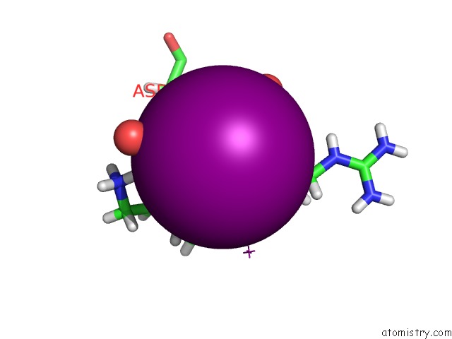
Mono view
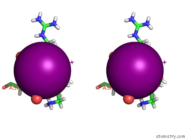
Stereo pair view
|
A full contact list of Iodine with other atoms in the I binding
site number 8 of Crystal Structure of A Trap Periplasmic Solute Binding Protein From Desulfovibrio Desulfuricans (DDES_1525), Target Efi-510107, with Bound Sn-Glycerol-3-Phosphate within 5.0Å range:
probe
|
atom
|
residue
|
distance (Å)
|
B
|
Occ
|
A:I408
b:29.6
occ:0.35
|
OD2
|
A:ASP267
|
2.5
|
27.0
|
1.0
|
HA
|
A:ARG264
|
3.3
|
16.7
|
1.0
|
HG3
|
A:LYS263
|
3.3
|
20.8
|
1.0
|
HD2
|
A:ARG264
|
3.4
|
52.5
|
1.0
|
CG
|
A:ASP267
|
3.4
|
31.6
|
1.0
|
HB2
|
A:ASP267
|
3.5
|
22.0
|
1.0
|
CB
|
A:ASP267
|
3.8
|
18.3
|
1.0
|
HB3
|
A:ASP267
|
3.8
|
22.0
|
1.0
|
HZ2
|
A:LYS263
|
3.9
|
38.0
|
1.0
|
HG2
|
A:ARG264
|
4.0
|
32.3
|
1.0
|
O
|
A:HOH588
|
4.0
|
23.9
|
1.0
|
O
|
A:HOH667
|
4.1
|
42.0
|
1.0
|
CA
|
A:ARG264
|
4.2
|
13.9
|
1.0
|
I
|
A:IOD409
|
4.2
|
54.6
|
0.5
|
CD
|
A:ARG264
|
4.2
|
43.7
|
1.0
|
CG
|
A:LYS263
|
4.3
|
17.4
|
1.0
|
HD2
|
A:LYS263
|
4.4
|
38.0
|
1.0
|
O
|
A:LYS263
|
4.5
|
18.4
|
1.0
|
CG
|
A:ARG264
|
4.5
|
26.9
|
1.0
|
OD1
|
A:ASP267
|
4.5
|
28.6
|
1.0
|
N
|
A:ARG264
|
4.5
|
11.2
|
1.0
|
HD3
|
A:ARG264
|
4.6
|
52.5
|
1.0
|
HG2
|
A:LYS263
|
4.6
|
20.8
|
1.0
|
C
|
A:LYS263
|
4.7
|
12.7
|
1.0
|
CB
|
A:ARG264
|
4.7
|
14.6
|
1.0
|
HB3
|
A:ARG264
|
4.8
|
17.5
|
1.0
|
NZ
|
A:LYS263
|
4.8
|
31.7
|
1.0
|
CD
|
A:LYS263
|
4.8
|
31.7
|
1.0
|
H
|
A:ARG264
|
5.0
|
13.4
|
1.0
|
|
Iodine binding site 9 out
of 23 in 4nhb
Go back to
Iodine Binding Sites List in 4nhb
Iodine binding site 9 out
of 23 in the Crystal Structure of A Trap Periplasmic Solute Binding Protein From Desulfovibrio Desulfuricans (DDES_1525), Target Efi-510107, with Bound Sn-Glycerol-3-Phosphate
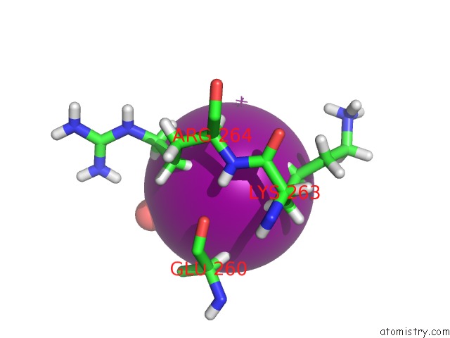
Mono view
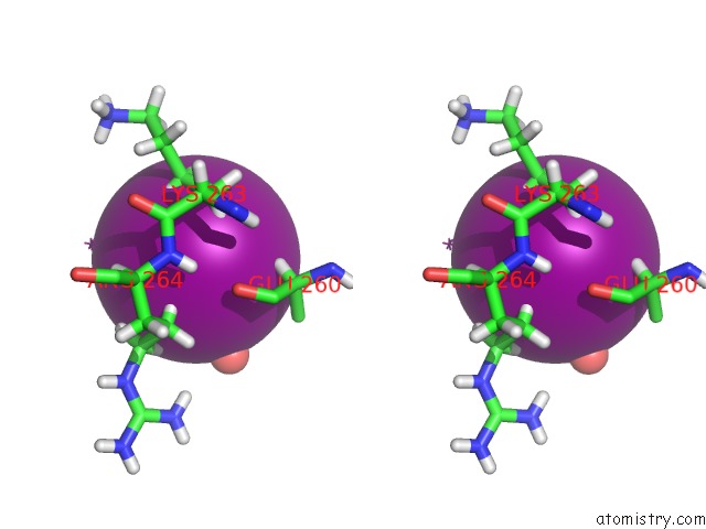
Stereo pair view
|
A full contact list of Iodine with other atoms in the I binding
site number 9 of Crystal Structure of A Trap Periplasmic Solute Binding Protein From Desulfovibrio Desulfuricans (DDES_1525), Target Efi-510107, with Bound Sn-Glycerol-3-Phosphate within 5.0Å range:
probe
|
atom
|
residue
|
distance (Å)
|
B
|
Occ
|
A:I409
b:54.6
occ:0.49
|
HG3
|
A:LYS263
|
3.6
|
20.8
|
1.0
|
HG2
|
A:ARG264
|
3.7
|
32.3
|
1.0
|
HG2
|
A:LYS263
|
3.7
|
20.8
|
1.0
|
HB3
|
A:LYS263
|
3.7
|
19.4
|
1.0
|
O
|
A:HOH707
|
3.8
|
36.0
|
1.0
|
HA
|
A:GLU260
|
4.0
|
24.8
|
1.0
|
HD3
|
A:ARG264
|
4.0
|
52.5
|
1.0
|
CG
|
A:LYS263
|
4.0
|
17.4
|
1.0
|
I
|
A:IOD408
|
4.2
|
29.6
|
0.3
|
CB
|
A:LYS263
|
4.4
|
16.2
|
1.0
|
HD2
|
A:ARG264
|
4.4
|
52.5
|
1.0
|
CG
|
A:ARG264
|
4.5
|
26.9
|
1.0
|
CD
|
A:ARG264
|
4.5
|
43.7
|
1.0
|
O
|
A:GLU260
|
4.7
|
17.5
|
1.0
|
CA
|
A:GLU260
|
4.8
|
20.6
|
1.0
|
CB
|
A:GLU260
|
4.8
|
26.2
|
1.0
|
HG3
|
A:ARG264
|
4.8
|
32.3
|
1.0
|
HB2
|
A:LYS263
|
4.8
|
19.4
|
1.0
|
H
|
A:ARG264
|
5.0
|
13.4
|
1.0
|
|
Iodine binding site 10 out
of 23 in 4nhb
Go back to
Iodine Binding Sites List in 4nhb
Iodine binding site 10 out
of 23 in the Crystal Structure of A Trap Periplasmic Solute Binding Protein From Desulfovibrio Desulfuricans (DDES_1525), Target Efi-510107, with Bound Sn-Glycerol-3-Phosphate
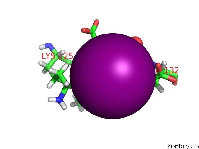
Mono view
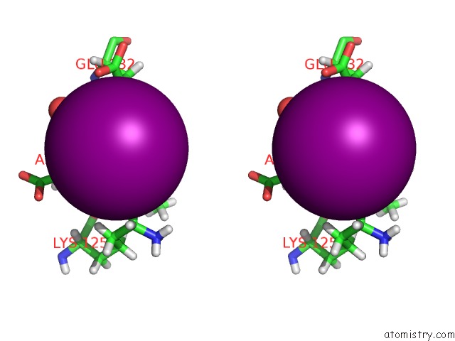
Stereo pair view
|
A full contact list of Iodine with other atoms in the I binding
site number 10 of Crystal Structure of A Trap Periplasmic Solute Binding Protein From Desulfovibrio Desulfuricans (DDES_1525), Target Efi-510107, with Bound Sn-Glycerol-3-Phosphate within 5.0Å range:
probe
|
atom
|
residue
|
distance (Å)
|
B
|
Occ
|
A:I410
b:47.8
occ:0.40
|
HB3
|
A:ASP128
|
3.0
|
31.2
|
1.0
|
HG2
|
A:GLU132
|
3.0
|
50.5
|
1.0
|
HG3
|
A:GLU132
|
3.3
|
50.5
|
1.0
|
HE2
|
A:LYS125
|
3.5
|
52.0
|
1.0
|
HD23
|
A:LEU129
|
3.5
|
24.4
|
1.0
|
CG
|
A:GLU132
|
3.6
|
42.1
|
1.0
|
HA
|
A:LEU129
|
3.7
|
21.7
|
1.0
|
CB
|
A:ASP128
|
3.9
|
26.0
|
1.0
|
O
|
A:ASP128
|
3.9
|
21.1
|
1.0
|
C
|
A:ASP128
|
4.0
|
17.9
|
1.0
|
HB2
|
A:ASP128
|
4.0
|
31.2
|
1.0
|
O
|
A:HOH617
|
4.1
|
28.4
|
1.0
|
HG
|
A:LEU129
|
4.2
|
17.9
|
1.0
|
N
|
A:LEU129
|
4.2
|
16.9
|
1.0
|
CD2
|
A:LEU129
|
4.3
|
20.3
|
1.0
|
CE
|
A:LYS125
|
4.3
|
43.3
|
1.0
|
HD21
|
A:LEU129
|
4.3
|
24.4
|
1.0
|
OE2
|
A:GLU132
|
4.3
|
52.8
|
1.0
|
HE3
|
A:LYS125
|
4.3
|
52.0
|
1.0
|
CA
|
A:LEU129
|
4.4
|
18.1
|
1.0
|
CD
|
A:GLU132
|
4.5
|
50.2
|
1.0
|
HD2
|
A:LYS125
|
4.5
|
54.4
|
1.0
|
HB2
|
A:GLU132
|
4.5
|
31.9
|
1.0
|
CA
|
A:ASP128
|
4.5
|
21.0
|
1.0
|
H
|
A:LEU129
|
4.6
|
20.3
|
1.0
|
CG
|
A:LEU129
|
4.7
|
15.0
|
1.0
|
CB
|
A:GLU132
|
4.7
|
26.6
|
1.0
|
HA
|
A:ASP128
|
4.9
|
25.2
|
1.0
|
CG
|
A:ASP128
|
4.9
|
23.5
|
1.0
|
|
Reference:
M.W.Vetting,
N.F.Al Obaidi,
L.L.Morisco,
S.R.Wasserman,
S.Sojitra,
M.Stead,
J.D.Attonito,
A.Scott Glenn,
S.Chowdhury,
B.Evans,
B.Hillerich,
J.Love,
R.D.Seidel,
H.J.Imker,
J.A.Gerlt,
S.C.Almo.
Crystal Structure of A Trap Periplasmic Solute Binding Protein From Desulfovibrio Desulfuricans (DDES_1525), Target Efi-510107, with Bound Sn-Glycerol-3-Phosphate To Be Published.
Page generated: Sun Aug 11 18:55:07 2024
|