Iodine »
PDB 4p9t-4ttc »
4pvq »
Iodine in PDB 4pvq: Crystal Structure of Sulfate-Bound Human L-Asparaginase Protein
Enzymatic activity of Crystal Structure of Sulfate-Bound Human L-Asparaginase Protein
All present enzymatic activity of Crystal Structure of Sulfate-Bound Human L-Asparaginase Protein:
3.4.19.5; 3.5.1.1;
3.4.19.5; 3.5.1.1;
Protein crystallography data
The structure of Crystal Structure of Sulfate-Bound Human L-Asparaginase Protein, PDB code: 4pvq
was solved by
J.Nomme,
A.Lavie,
with X-Ray Crystallography technique. A brief refinement statistics is given in the table below:
| Resolution Low / High (Å) | 30.00 / 2.13 |
| Space group | P 65 |
| Cell size a, b, c (Å), α, β, γ (°) | 59.220, 59.220, 298.500, 90.00, 90.00, 120.00 |
| R / Rfree (%) | 16.6 / 22.2 |
Other elements in 4pvq:
The structure of Crystal Structure of Sulfate-Bound Human L-Asparaginase Protein also contains other interesting chemical elements:
| Sodium | (Na) | 2 atoms |
Iodine Binding Sites:
The binding sites of Iodine atom in the Crystal Structure of Sulfate-Bound Human L-Asparaginase Protein
(pdb code 4pvq). This binding sites where shown within
5.0 Angstroms radius around Iodine atom.
In total 4 binding sites of Iodine where determined in the Crystal Structure of Sulfate-Bound Human L-Asparaginase Protein, PDB code: 4pvq:
Jump to Iodine binding site number: 1; 2; 3; 4;
In total 4 binding sites of Iodine where determined in the Crystal Structure of Sulfate-Bound Human L-Asparaginase Protein, PDB code: 4pvq:
Jump to Iodine binding site number: 1; 2; 3; 4;
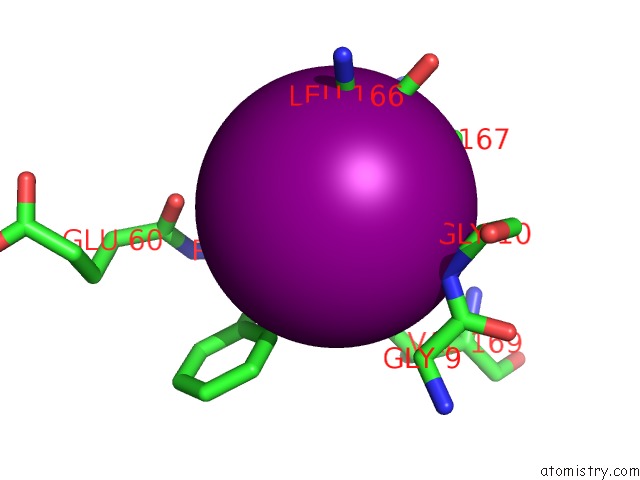
Iodine binding site 1 out of 4 in 4pvq
Go back to
Iodine binding site 1 out
of 4 in the Crystal Structure of Sulfate-Bound Human L-Asparaginase Protein
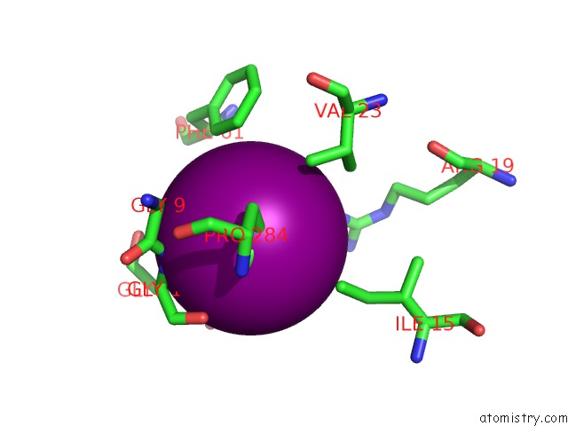
Mono view
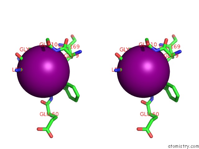
Stereo pair view
Mono view
Stereo pair view
A full contact list of Iodine with other atoms in the I binding
site number 1 of Crystal Structure of Sulfate-Bound Human L-Asparaginase Protein within 5.0Å range:
|
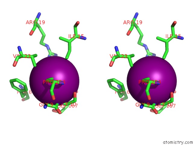
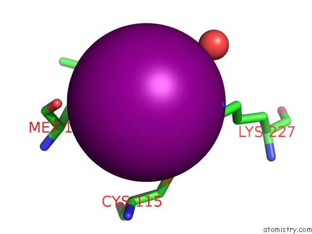
Iodine binding site 2 out of 4 in 4pvq
Go back to
Iodine binding site 2 out
of 4 in the Crystal Structure of Sulfate-Bound Human L-Asparaginase Protein
Mono view
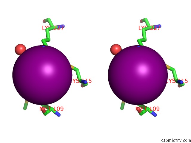
Stereo pair view
Mono view
Stereo pair view
A full contact list of Iodine with other atoms in the I binding
site number 2 of Crystal Structure of Sulfate-Bound Human L-Asparaginase Protein within 5.0Å range:
|
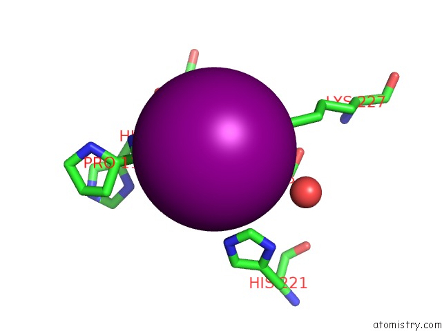
Iodine binding site 3 out of 4 in 4pvq
Go back to
Iodine binding site 3 out
of 4 in the Crystal Structure of Sulfate-Bound Human L-Asparaginase Protein
Mono view
Stereo pair view
Mono view
Stereo pair view
A full contact list of Iodine with other atoms in the I binding
site number 3 of Crystal Structure of Sulfate-Bound Human L-Asparaginase Protein within 5.0Å range:
|
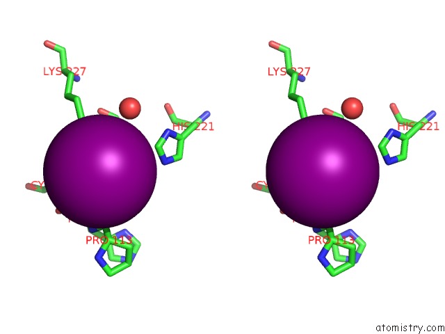
Iodine binding site 4 out of 4 in 4pvq
Go back to
Iodine binding site 4 out
of 4 in the Crystal Structure of Sulfate-Bound Human L-Asparaginase Protein
Mono view
Stereo pair view
Mono view
Stereo pair view
A full contact list of Iodine with other atoms in the I binding
site number 4 of Crystal Structure of Sulfate-Bound Human L-Asparaginase Protein within 5.0Å range:
|
Reference:
J.Nomme,
Y.Su,
M.Konrad,
A.Lavie.
Structures of Apo and Product-Bound Human L-Asparaginase: Insights Into the Mechanism of Autoproteolysis and Substrate Hydrolysis. Biochemistry V. 51 6816 2012.
ISSN: ISSN 0006-2960
PubMed: 22861376
DOI: 10.1021/BI300870G
Page generated: Sun Aug 11 19:30:31 2024
ISSN: ISSN 0006-2960
PubMed: 22861376
DOI: 10.1021/BI300870G
Last articles
Zn in 9J0NZn in 9J0O
Zn in 9J0P
Zn in 9FJX
Zn in 9EKB
Zn in 9C0F
Zn in 9CAH
Zn in 9CH0
Zn in 9CH3
Zn in 9CH1