Iodine in PDB 4q53: Crystal Structure of A Hypothetical Protein (BACUNI_04292) From Bacteroides Uniformis Atcc 8492 at 1.27 A Resolution
Protein crystallography data
The structure of Crystal Structure of A Hypothetical Protein (BACUNI_04292) From Bacteroides Uniformis Atcc 8492 at 1.27 A Resolution, PDB code: 4q53
was solved by
Joint Center For Structural Genomics (Jcsg),
with X-Ray Crystallography technique. A brief refinement statistics is given in the table below:
Resolution Low / High (Å)
|
28.00 /
1.27
|
Space group
|
C 1 2 1
|
Cell size a, b, c (Å), α, β, γ (°)
|
108.136,
37.109,
61.376,
90.00,
121.66,
90.00
|
R / Rfree (%)
|
15.4 /
17.6
|
Other elements in 4q53:
The structure of Crystal Structure of A Hypothetical Protein (BACUNI_04292) From Bacteroides Uniformis Atcc 8492 at 1.27 A Resolution also contains other interesting chemical elements:
Iodine Binding Sites:
Pages:
>>> Page 1 <<<
Page 2, Binding sites: 11 -
11;
Binding sites:
The binding sites of Iodine atom in the Crystal Structure of A Hypothetical Protein (BACUNI_04292) From Bacteroides Uniformis Atcc 8492 at 1.27 A Resolution
(pdb code 4q53). This binding sites where shown within
5.0 Angstroms radius around Iodine atom.
In total 11 binding sites of Iodine where determined in the
Crystal Structure of A Hypothetical Protein (BACUNI_04292) From Bacteroides Uniformis Atcc 8492 at 1.27 A Resolution, PDB code: 4q53:
Jump to Iodine binding site number:
1;
2;
3;
4;
5;
6;
7;
8;
9;
10;
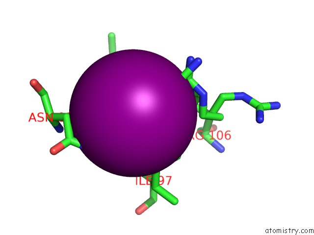
Iodine binding site 1 out
of 11 in 4q53
Go back to
Iodine Binding Sites List in 4q53
Iodine binding site 1 out
of 11 in the Crystal Structure of A Hypothetical Protein (BACUNI_04292) From Bacteroides Uniformis Atcc 8492 at 1.27 A Resolution
 Mono view
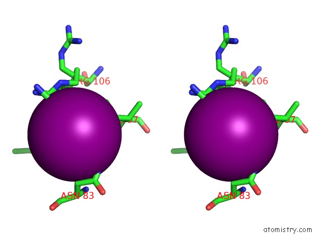
 Stereo pair view
|
A full contact list of Iodine with other atoms in the I binding
site number 1 of Crystal Structure of A Hypothetical Protein (BACUNI_04292) From Bacteroides Uniformis Atcc 8492 at 1.27 A Resolution within 5.0Å range:
probe
|
atom
|
residue
|
distance (Å)
|
B
|
Occ
|
A:I201
b:20.9
occ:0.60
|
NH1
|
A:ARG106
|
3.2
|
11.3
|
0.3
|
NE
|
A:ARG106
|
3.4
|
22.5
|
0.3
|
NH2
|
A:ARG106
|
3.4
|
25.4
|
0.3
|
ND2
|
A:ASN83
|
3.5
|
21.2
|
1.0
|
CG1
|
A:ILE97
|
3.6
|
21.6
|
1.0
|
CD1
|
A:ILE97
|
3.8
|
26.4
|
1.0
|
CZ
|
A:ARG106
|
3.8
|
23.9
|
0.3
|
C
|
A:GLY96
|
4.0
|
12.0
|
1.0
|
CA
|
A:GLY96
|
4.0
|
12.7
|
1.0
|
CZ
|
A:ARG106
|
4.0
|
18.7
|
0.3
|
CG2
|
A:ILE95
|
4.1
|
15.4
|
1.0
|
CB
|
A:ASN83
|
4.1
|
15.4
|
1.0
|
O
|
A:GLY96
|
4.3
|
12.9
|
1.0
|
N
|
A:GLY96
|
4.3
|
11.6
|
1.0
|
CG
|
A:ASN83
|
4.3
|
18.5
|
1.0
|
N
|
A:ILE97
|
4.4
|
12.2
|
1.0
|
CD
|
A:ARG106
|
4.4
|
21.4
|
0.3
|
CG
|
A:ARG106
|
4.5
|
19.4
|
0.3
|
CD
|
A:ARG106
|
4.5
|
21.8
|
0.3
|
NE
|
A:ARG106
|
4.5
|
21.7
|
0.3
|
C
|
A:ILE95
|
4.6
|
11.6
|
1.0
|
O
|
A:ILE95
|
4.6
|
12.0
|
1.0
|
NH2
|
A:ARG106
|
4.8
|
22.4
|
0.3
|
CB
|
A:ILE97
|
4.9
|
15.5
|
1.0
|
|
Iodine binding site 2 out
of 11 in 4q53
Go back to
Iodine Binding Sites List in 4q53
Iodine binding site 2 out
of 11 in the Crystal Structure of A Hypothetical Protein (BACUNI_04292) From Bacteroides Uniformis Atcc 8492 at 1.27 A Resolution
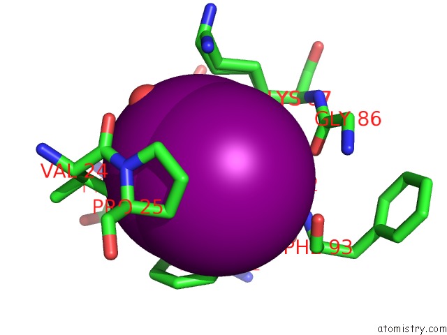
 Mono view
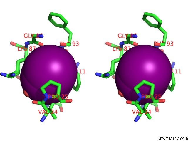
 Stereo pair view
|
A full contact list of Iodine with other atoms in the I binding
site number 2 of Crystal Structure of A Hypothetical Protein (BACUNI_04292) From Bacteroides Uniformis Atcc 8492 at 1.27 A Resolution within 5.0Å range:
probe
|
atom
|
residue
|
distance (Å)
|
B
|
Occ
|
A:I202
b:22.4
occ:0.35
|
I
|
A:IOD202
|
0.0
|
22.4
|
0.3
|
I
|
A:IOD202
|
1.1
|
22.4
|
0.3
|
O
|
A:HOH365
|
3.5
|
39.9
|
1.0
|
CG1
|
A:VAL24
|
3.6
|
31.7
|
1.0
|
I
|
A:IOD205
|
3.6
|
10.9
|
0.2
|
CG
|
A:TYR118
|
3.9
|
19.1
|
1.0
|
CG
|
A:PHE111
|
3.9
|
15.0
|
1.0
|
CB
|
A:TYR118
|
4.0
|
19.3
|
1.0
|
CD1
|
A:TYR118
|
4.0
|
20.7
|
1.0
|
CD1
|
A:PHE111
|
4.0
|
16.7
|
1.0
|
CB
|
A:PHE111
|
4.2
|
15.1
|
1.0
|
CB
|
A:LYS87
|
4.2
|
19.3
|
0.5
|
CD2
|
A:PHE111
|
4.2
|
16.6
|
1.0
|
CB
|
A:LYS87
|
4.3
|
19.5
|
0.5
|
CD
|
A:LYS87
|
4.3
|
24.6
|
0.5
|
CA
|
A:GLY92
|
4.3
|
14.3
|
1.0
|
CD2
|
A:TYR118
|
4.4
|
23.1
|
1.0
|
CE1
|
A:PHE111
|
4.5
|
16.7
|
1.0
|
CD
|
A:LYS87
|
4.5
|
24.2
|
0.5
|
CE1
|
A:TYR118
|
4.6
|
22.0
|
1.0
|
CE2
|
A:PHE111
|
4.6
|
17.6
|
1.0
|
CB
|
A:PRO25
|
4.7
|
24.5
|
1.0
|
C
|
A:GLY92
|
4.8
|
12.3
|
1.0
|
CB
|
A:VAL24
|
4.8
|
29.8
|
1.0
|
CZ
|
A:PHE111
|
4.8
|
18.4
|
1.0
|
CD
|
A:PRO25
|
4.8
|
27.3
|
1.0
|
CG
|
A:LYS87
|
4.8
|
21.2
|
0.5
|
N
|
A:PRO25
|
4.8
|
26.1
|
1.0
|
C
|
A:VAL24
|
4.9
|
27.7
|
1.0
|
CG
|
A:LYS87
|
4.9
|
20.9
|
0.5
|
O
|
A:GLY86
|
4.9
|
14.8
|
1.0
|
O
|
A:VAL24
|
4.9
|
29.9
|
1.0
|
N
|
A:PHE93
|
4.9
|
11.9
|
1.0
|
CE2
|
A:TYR118
|
4.9
|
24.8
|
1.0
|
CG
|
A:PRO25
|
5.0
|
25.2
|
1.0
|
N
|
A:GLY92
|
5.0
|
14.5
|
1.0
|
|
Iodine binding site 3 out
of 11 in 4q53
Go back to
Iodine Binding Sites List in 4q53
Iodine binding site 3 out
of 11 in the Crystal Structure of A Hypothetical Protein (BACUNI_04292) From Bacteroides Uniformis Atcc 8492 at 1.27 A Resolution
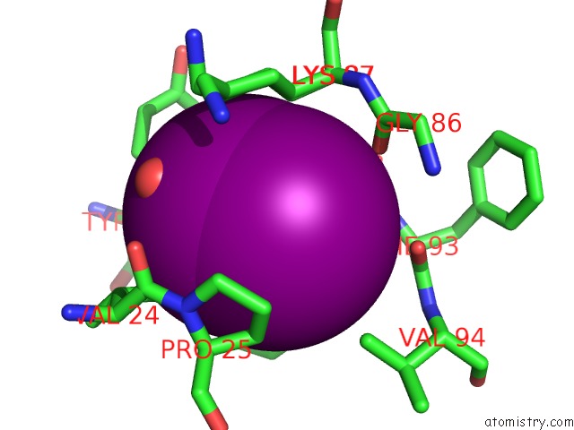
 Mono view
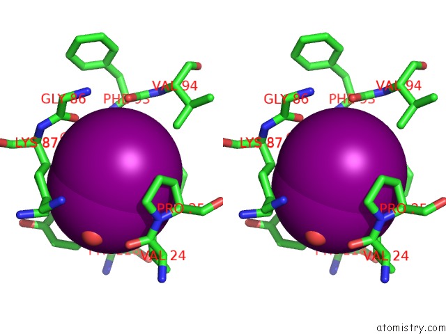
 Stereo pair view
|
A full contact list of Iodine with other atoms in the I binding
site number 3 of Crystal Structure of A Hypothetical Protein (BACUNI_04292) From Bacteroides Uniformis Atcc 8492 at 1.27 A Resolution within 5.0Å range:
probe
|
atom
|
residue
|
distance (Å)
|
B
|
Occ
|
A:I202
b:22.4
occ:0.35
|
I
|
A:IOD202
|
0.0
|
22.4
|
0.3
|
I
|
A:IOD202
|
1.1
|
22.4
|
0.3
|
I
|
A:IOD205
|
2.9
|
10.9
|
0.2
|
CD1
|
A:PHE111
|
3.7
|
16.7
|
1.0
|
CG
|
A:PHE111
|
4.0
|
15.0
|
1.0
|
CE1
|
A:PHE111
|
4.0
|
16.7
|
1.0
|
CA
|
A:GLY92
|
4.1
|
14.3
|
1.0
|
CG1
|
A:VAL24
|
4.1
|
31.7
|
1.0
|
N
|
A:PHE93
|
4.1
|
11.9
|
1.0
|
O
|
A:HOH365
|
4.2
|
39.9
|
1.0
|
O
|
A:GLY86
|
4.2
|
14.8
|
1.0
|
CB
|
A:PRO25
|
4.2
|
24.5
|
1.0
|
CB
|
A:LYS87
|
4.3
|
19.3
|
0.5
|
C
|
A:GLY92
|
4.3
|
12.3
|
1.0
|
CB
|
A:LYS87
|
4.3
|
19.5
|
0.5
|
CG
|
A:PRO25
|
4.4
|
25.2
|
1.0
|
CB
|
A:PHE111
|
4.5
|
15.1
|
1.0
|
CD2
|
A:PHE111
|
4.5
|
16.6
|
1.0
|
CD
|
A:PRO25
|
4.5
|
27.3
|
1.0
|
CZ
|
A:PHE111
|
4.5
|
18.4
|
1.0
|
O
|
A:PHE93
|
4.6
|
12.0
|
1.0
|
CD
|
A:LYS87
|
4.7
|
24.6
|
0.5
|
CE2
|
A:PHE111
|
4.7
|
17.6
|
1.0
|
CG2
|
A:VAL94
|
4.7
|
15.4
|
1.0
|
N
|
A:PRO25
|
4.7
|
26.1
|
1.0
|
CD
|
A:LYS87
|
4.8
|
24.2
|
0.5
|
C
|
A:PHE93
|
4.9
|
10.9
|
1.0
|
CG
|
A:LYS87
|
4.9
|
21.2
|
0.5
|
CG
|
A:LYS87
|
4.9
|
20.9
|
0.5
|
C
|
A:GLY86
|
4.9
|
13.4
|
1.0
|
CD1
|
A:TYR118
|
5.0
|
20.7
|
1.0
|
CG
|
A:TYR118
|
5.0
|
19.1
|
1.0
|
CA
|
A:PRO25
|
5.0
|
23.6
|
1.0
|
CA
|
A:PHE93
|
5.0
|
12.0
|
1.0
|
|
Iodine binding site 4 out
of 11 in 4q53
Go back to
Iodine Binding Sites List in 4q53
Iodine binding site 4 out
of 11 in the Crystal Structure of A Hypothetical Protein (BACUNI_04292) From Bacteroides Uniformis Atcc 8492 at 1.27 A Resolution
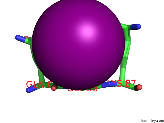
 Mono view
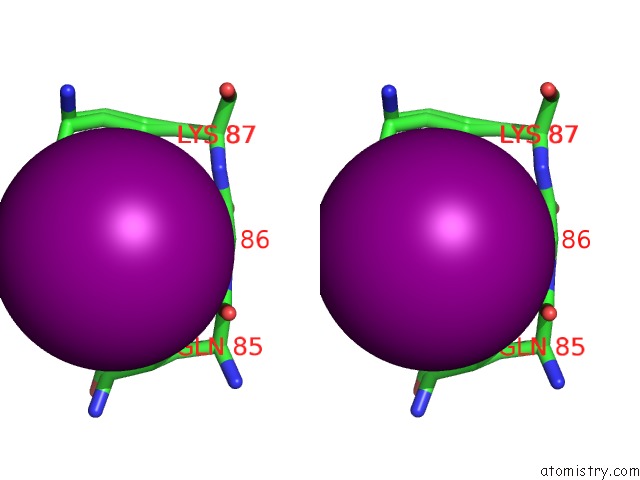
 Stereo pair view
|
A full contact list of Iodine with other atoms in the I binding
site number 4 of Crystal Structure of A Hypothetical Protein (BACUNI_04292) From Bacteroides Uniformis Atcc 8492 at 1.27 A Resolution within 5.0Å range:
probe
|
atom
|
residue
|
distance (Å)
|
B
|
Occ
|
A:I203
b:19.0
occ:0.80
|
I
|
A:IOD205
|
3.0
|
10.9
|
0.2
|
NZ
|
A:LYS87
|
3.4
|
27.7
|
0.5
|
NE2
|
A:GLN85
|
3.5
|
14.9
|
0.5
|
CA
|
A:GLY86
|
3.9
|
13.0
|
1.0
|
CG
|
A:GLN85
|
4.0
|
13.3
|
0.5
|
CG
|
A:GLN85
|
4.2
|
15.9
|
0.5
|
N
|
A:GLY86
|
4.2
|
10.7
|
1.0
|
OE1
|
A:GLN85
|
4.2
|
20.7
|
0.5
|
CD
|
A:GLN85
|
4.2
|
14.1
|
0.5
|
O
|
A:GLN85
|
4.3
|
11.4
|
1.0
|
C
|
A:GLN85
|
4.3
|
10.5
|
1.0
|
CG
|
A:LYS87
|
4.4
|
20.9
|
0.5
|
C
|
A:GLY86
|
4.4
|
13.4
|
1.0
|
CD
|
A:GLN85
|
4.5
|
17.8
|
0.5
|
CE
|
A:LYS87
|
4.5
|
27.0
|
0.5
|
N
|
A:LYS87
|
4.6
|
14.6
|
1.0
|
CG
|
A:LYS87
|
4.6
|
21.2
|
0.5
|
CB
|
A:GLN85
|
4.7
|
11.5
|
0.5
|
CB
|
A:GLN85
|
4.7
|
13.2
|
0.5
|
CE
|
A:LYS87
|
4.8
|
26.8
|
0.5
|
CD
|
A:LYS87
|
4.9
|
24.2
|
0.5
|
|
Iodine binding site 5 out
of 11 in 4q53
Go back to
Iodine Binding Sites List in 4q53
Iodine binding site 5 out
of 11 in the Crystal Structure of A Hypothetical Protein (BACUNI_04292) From Bacteroides Uniformis Atcc 8492 at 1.27 A Resolution
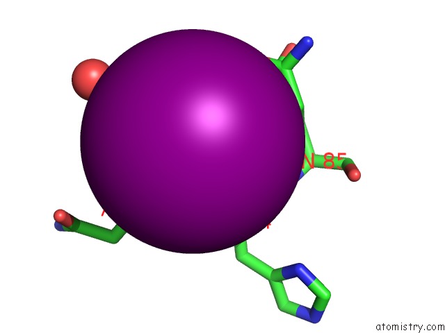
 Mono view
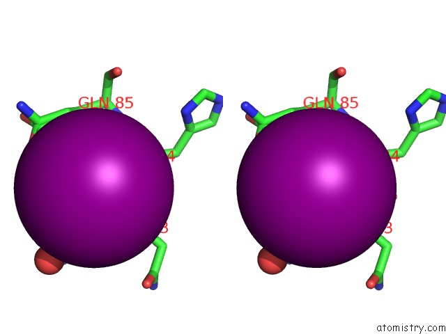
 Stereo pair view
|
A full contact list of Iodine with other atoms in the I binding
site number 5 of Crystal Structure of A Hypothetical Protein (BACUNI_04292) From Bacteroides Uniformis Atcc 8492 at 1.27 A Resolution within 5.0Å range:
probe
|
atom
|
residue
|
distance (Å)
|
B
|
Occ
|
A:I204
b:15.4
occ:0.70
|
O
|
A:VAL82
|
3.8
|
13.8
|
1.0
|
C
|
A:ASN83
|
3.8
|
10.8
|
1.0
|
NE2
|
A:GLN85
|
3.8
|
18.7
|
0.5
|
N
|
A:HIS84
|
4.0
|
10.9
|
1.0
|
CA
|
A:ASN83
|
4.0
|
11.0
|
1.0
|
CG1
|
A:VAL82
|
4.0
|
14.2
|
1.0
|
O
|
A:ASN83
|
4.0
|
11.1
|
1.0
|
C
|
A:VAL82
|
4.1
|
12.3
|
1.0
|
O
|
A:HOH353
|
4.2
|
29.6
|
1.0
|
N
|
A:ASN83
|
4.3
|
11.2
|
1.0
|
CA
|
A:HIS84
|
4.4
|
10.4
|
1.0
|
CG
|
A:GLN85
|
4.5
|
15.9
|
0.5
|
CB
|
A:VAL82
|
4.6
|
13.9
|
1.0
|
CG
|
A:GLN85
|
4.7
|
13.3
|
0.5
|
CD
|
A:GLN85
|
4.7
|
17.8
|
0.5
|
OE1
|
A:GLN85
|
4.8
|
15.7
|
0.5
|
C
|
A:HIS84
|
4.8
|
10.0
|
1.0
|
N
|
A:GLN85
|
4.8
|
10.5
|
1.0
|
CD
|
A:GLN85
|
4.9
|
14.1
|
0.5
|
|
Iodine binding site 6 out
of 11 in 4q53
Go back to
Iodine Binding Sites List in 4q53
Iodine binding site 6 out
of 11 in the Crystal Structure of A Hypothetical Protein (BACUNI_04292) From Bacteroides Uniformis Atcc 8492 at 1.27 A Resolution
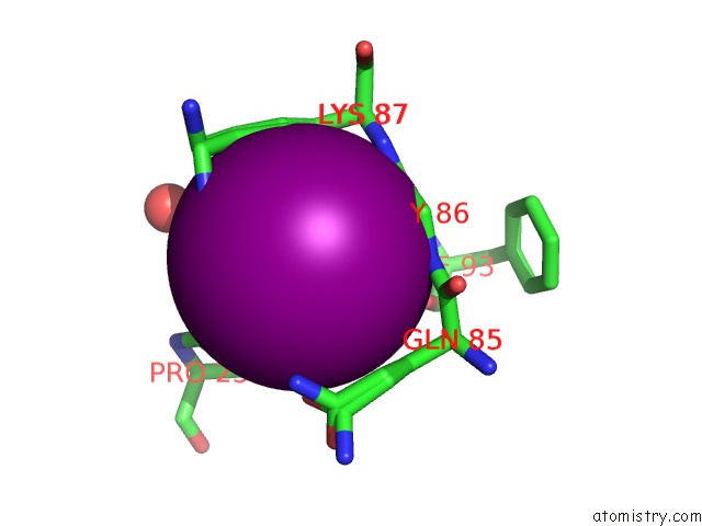
 Mono view
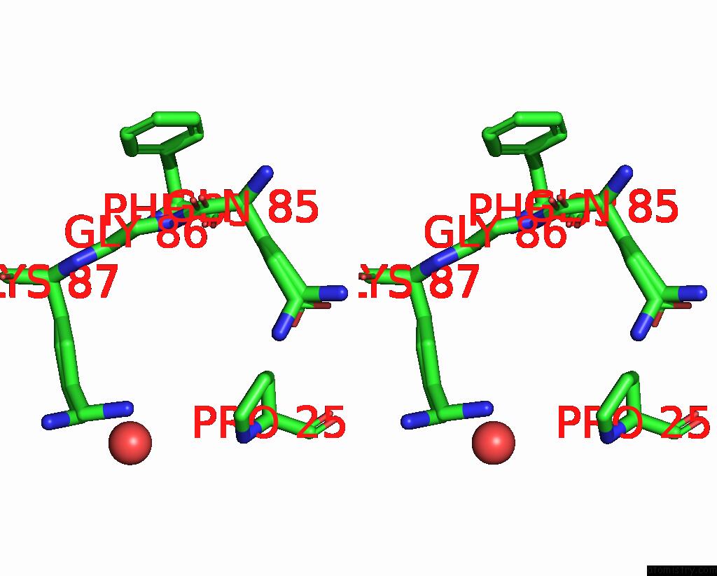
 Stereo pair view
|
A full contact list of Iodine with other atoms in the I binding
site number 6 of Crystal Structure of A Hypothetical Protein (BACUNI_04292) From Bacteroides Uniformis Atcc 8492 at 1.27 A Resolution within 5.0Å range:
probe
|
atom
|
residue
|
distance (Å)
|
B
|
Occ
|
A:I205
b:10.9
occ:0.20
|
I
|
A:IOD202
|
2.9
|
22.4
|
0.3
|
I
|
A:IOD203
|
3.0
|
19.0
|
0.8
|
I
|
A:IOD202
|
3.6
|
22.4
|
0.3
|
O
|
A:GLY86
|
3.6
|
14.8
|
1.0
|
NZ
|
A:LYS87
|
3.6
|
27.7
|
0.5
|
C
|
A:GLY86
|
3.7
|
13.4
|
1.0
|
CG
|
A:LYS87
|
3.8
|
20.9
|
0.5
|
CG
|
A:PRO25
|
3.8
|
25.2
|
1.0
|
N
|
A:GLY86
|
3.9
|
10.7
|
1.0
|
CD
|
A:PRO25
|
3.9
|
27.3
|
1.0
|
CG
|
A:LYS87
|
3.9
|
21.2
|
0.5
|
CB
|
A:LYS87
|
3.9
|
19.5
|
0.5
|
CB
|
A:LYS87
|
4.0
|
19.3
|
0.5
|
CA
|
A:GLY86
|
4.0
|
13.0
|
1.0
|
CD
|
A:LYS87
|
4.0
|
24.2
|
0.5
|
CD
|
A:LYS87
|
4.1
|
24.6
|
0.5
|
N
|
A:LYS87
|
4.2
|
14.6
|
1.0
|
OE1
|
A:GLN85
|
4.2
|
20.7
|
0.5
|
O
|
A:PHE93
|
4.4
|
12.0
|
1.0
|
NE2
|
A:GLN85
|
4.4
|
14.9
|
0.5
|
CE
|
A:LYS87
|
4.5
|
27.0
|
0.5
|
CE
|
A:LYS87
|
4.5
|
26.8
|
0.5
|
CB
|
A:GLN85
|
4.5
|
13.2
|
0.5
|
CB
|
A:GLN85
|
4.5
|
11.5
|
0.5
|
O
|
A:HOH365
|
4.5
|
39.9
|
1.0
|
CB
|
A:PRO25
|
4.6
|
24.5
|
1.0
|
C
|
A:GLN85
|
4.6
|
10.5
|
1.0
|
CA
|
A:LYS87
|
4.6
|
17.4
|
0.5
|
CA
|
A:LYS87
|
4.7
|
17.3
|
0.5
|
CG
|
A:GLN85
|
4.7
|
13.3
|
0.5
|
CD
|
A:GLN85
|
4.8
|
14.1
|
0.5
|
CG
|
A:GLN85
|
4.8
|
15.9
|
0.5
|
N
|
A:PRO25
|
5.0
|
26.1
|
1.0
|
CD
|
A:GLN85
|
5.0
|
17.8
|
0.5
|
|
Iodine binding site 7 out
of 11 in 4q53
Go back to
Iodine Binding Sites List in 4q53
Iodine binding site 7 out
of 11 in the Crystal Structure of A Hypothetical Protein (BACUNI_04292) From Bacteroides Uniformis Atcc 8492 at 1.27 A Resolution
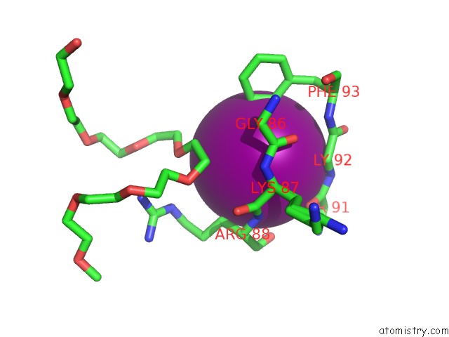
 Mono view
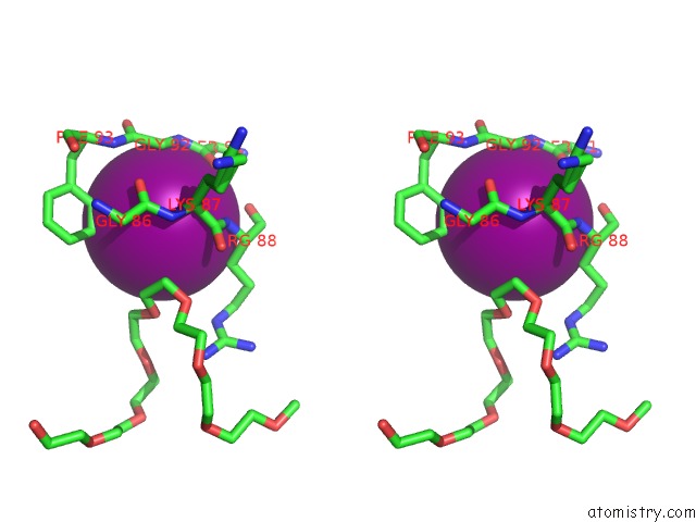
 Stereo pair view
|
A full contact list of Iodine with other atoms in the I binding
site number 7 of Crystal Structure of A Hypothetical Protein (BACUNI_04292) From Bacteroides Uniformis Atcc 8492 at 1.27 A Resolution within 5.0Å range:
probe
|
atom
|
residue
|
distance (Å)
|
B
|
Occ
|
B:I201
b:14.5
occ:0.90
|
I
|
B:IOD204
|
3.0
|
14.8
|
0.5
|
CD1
|
B:PHE93
|
3.8
|
12.1
|
1.0
|
CE1
|
B:PHE93
|
4.0
|
14.6
|
1.0
|
N
|
B:ARG88
|
4.0
|
12.3
|
1.0
|
CB
|
B:SER91
|
4.1
|
13.2
|
1.0
|
O
|
B:SER91
|
4.1
|
11.5
|
1.0
|
C
|
B:SER91
|
4.2
|
10.9
|
1.0
|
CB
|
B:ARG88
|
4.2
|
17.6
|
1.0
|
CA
|
B:GLY92
|
4.2
|
11.4
|
1.0
|
N
|
B:GLY92
|
4.2
|
10.7
|
1.0
|
CA
|
B:LYS87
|
4.4
|
12.2
|
0.5
|
CA
|
B:LYS87
|
4.4
|
11.2
|
0.5
|
C
|
B:LYS87
|
4.4
|
11.5
|
1.0
|
O
|
B:ARG88
|
4.5
|
13.2
|
1.0
|
O
|
B:GLY86
|
4.6
|
11.6
|
1.0
|
CA
|
B:ARG88
|
4.7
|
13.8
|
1.0
|
C9
|
B:7PG205
|
4.7
|
23.3
|
1.0
|
C
|
B:GLY92
|
4.7
|
10.6
|
1.0
|
CA
|
B:SER91
|
4.8
|
11.8
|
1.0
|
N
|
B:LYS87
|
4.8
|
10.5
|
1.0
|
C10
|
B:7PG205
|
4.8
|
26.8
|
1.0
|
C
|
B:GLY86
|
4.9
|
10.4
|
1.0
|
O4
|
B:7PG205
|
4.9
|
20.0
|
1.0
|
C8
|
B:7PG205
|
4.9
|
21.7
|
1.0
|
|
Iodine binding site 8 out
of 11 in 4q53
Go back to
Iodine Binding Sites List in 4q53
Iodine binding site 8 out
of 11 in the Crystal Structure of A Hypothetical Protein (BACUNI_04292) From Bacteroides Uniformis Atcc 8492 at 1.27 A Resolution
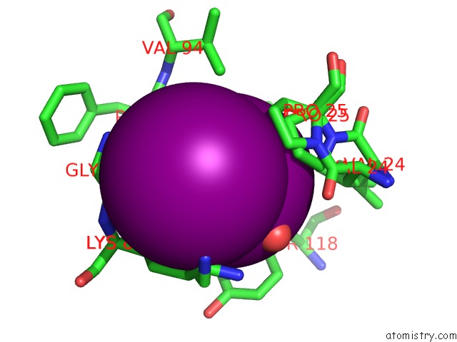
 Mono view
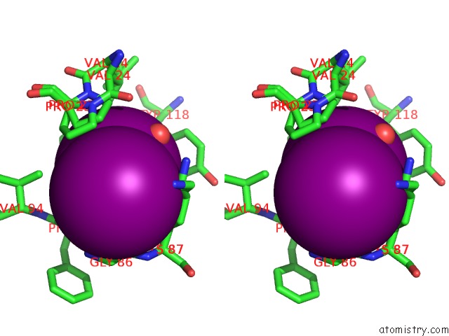
 Stereo pair view
|
A full contact list of Iodine with other atoms in the I binding
site number 8 of Crystal Structure of A Hypothetical Protein (BACUNI_04292) From Bacteroides Uniformis Atcc 8492 at 1.27 A Resolution within 5.0Å range:
probe
|
atom
|
residue
|
distance (Å)
|
B
|
Occ
|
B:I202
b:16.2
occ:0.30
|
I
|
B:IOD202
|
0.0
|
16.2
|
0.3
|
I
|
B:IOD202
|
3.1
|
21.4
|
0.7
|
O
|
B:HOH327
|
3.7
|
29.2
|
1.0
|
CD1
|
B:PHE111
|
3.9
|
14.2
|
1.0
|
CG
|
B:PHE111
|
4.1
|
12.0
|
1.0
|
CA
|
B:GLY92
|
4.2
|
11.4
|
1.0
|
N
|
B:PHE93
|
4.3
|
10.9
|
1.0
|
CE1
|
B:PHE111
|
4.3
|
13.5
|
1.0
|
CB
|
B:PRO25
|
4.4
|
21.3
|
0.5
|
CG
|
B:PRO25
|
4.4
|
21.6
|
0.5
|
CD2
|
B:TYR118
|
4.4
|
15.7
|
1.0
|
CB
|
B:PHE111
|
4.4
|
11.6
|
1.0
|
CG1
|
B:VAL24
|
4.4
|
21.3
|
0.5
|
CD
|
B:PRO25
|
4.4
|
18.0
|
0.5
|
C
|
B:GLY92
|
4.5
|
10.6
|
1.0
|
O
|
B:GLY86
|
4.5
|
11.6
|
1.0
|
CD
|
B:PRO25
|
4.5
|
20.8
|
0.5
|
CB
|
B:PRO25
|
4.5
|
17.8
|
0.5
|
CG
|
B:TYR118
|
4.6
|
15.4
|
1.0
|
CD
|
B:LYS87
|
4.6
|
20.1
|
0.5
|
CD2
|
B:PHE111
|
4.6
|
12.7
|
1.0
|
CG
|
B:PRO25
|
4.6
|
16.9
|
0.5
|
CB
|
B:LYS87
|
4.7
|
15.5
|
0.5
|
O
|
B:PHE93
|
4.7
|
10.8
|
1.0
|
CE2
|
B:TYR118
|
4.8
|
15.6
|
1.0
|
CB
|
B:LYS87
|
4.8
|
12.4
|
0.5
|
CZ
|
B:PHE111
|
4.8
|
13.7
|
1.0
|
N
|
B:PRO25
|
4.8
|
15.9
|
0.5
|
CB
|
B:TYR118
|
4.9
|
15.2
|
1.0
|
CE2
|
B:PHE111
|
4.9
|
13.5
|
1.0
|
CG2
|
B:VAL94
|
5.0
|
16.2
|
1.0
|
|
Iodine binding site 9 out
of 11 in 4q53
Go back to
Iodine Binding Sites List in 4q53
Iodine binding site 9 out
of 11 in the Crystal Structure of A Hypothetical Protein (BACUNI_04292) From Bacteroides Uniformis Atcc 8492 at 1.27 A Resolution
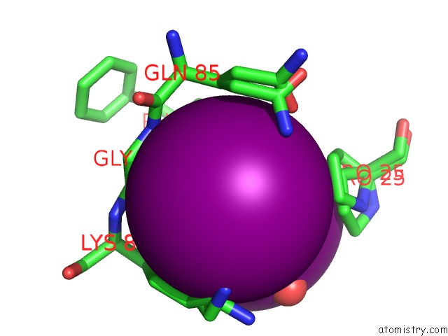
 Mono view
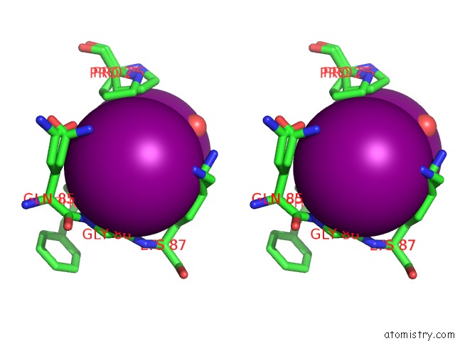
 Stereo pair view
|
A full contact list of Iodine with other atoms in the I binding
site number 9 of Crystal Structure of A Hypothetical Protein (BACUNI_04292) From Bacteroides Uniformis Atcc 8492 at 1.27 A Resolution within 5.0Å range:
probe
|
atom
|
residue
|
distance (Å)
|
B
|
Occ
|
B:I202
b:21.4
occ:0.70
|
I
|
B:IOD202
|
0.0
|
21.4
|
0.7
|
I
|
B:IOD202
|
3.1
|
16.2
|
0.3
|
O
|
B:GLY86
|
3.7
|
11.6
|
1.0
|
C
|
B:GLY86
|
3.8
|
10.4
|
1.0
|
CD
|
B:LYS87
|
3.8
|
20.1
|
0.5
|
NZ
|
B:LYS87
|
3.9
|
25.1
|
0.5
|
CE
|
B:LYS87
|
3.9
|
19.5
|
0.5
|
N
|
B:GLY86
|
4.0
|
11.4
|
1.0
|
CG
|
B:LYS87
|
4.0
|
16.8
|
0.5
|
CA
|
B:GLY86
|
4.1
|
10.6
|
1.0
|
NE2
|
B:GLN85
|
4.2
|
18.0
|
0.5
|
CD
|
B:LYS87
|
4.2
|
17.7
|
0.5
|
CG
|
B:LYS87
|
4.2
|
15.0
|
0.5
|
CB
|
B:LYS87
|
4.2
|
15.5
|
0.5
|
CB
|
B:LYS87
|
4.2
|
12.4
|
0.5
|
CD
|
B:PRO25
|
4.2
|
18.0
|
0.5
|
O
|
B:HOH327
|
4.3
|
29.2
|
1.0
|
O
|
B:PHE93
|
4.3
|
10.8
|
1.0
|
CG
|
B:PRO25
|
4.3
|
16.9
|
0.5
|
N
|
B:LYS87
|
4.4
|
10.5
|
1.0
|
OE1
|
B:GLN85
|
4.4
|
22.3
|
0.5
|
CE
|
B:LYS87
|
4.5
|
22.3
|
0.5
|
CB
|
B:GLN85
|
4.5
|
17.5
|
0.5
|
CB
|
B:GLN85
|
4.5
|
15.4
|
0.5
|
CG
|
B:PRO25
|
4.5
|
21.6
|
0.5
|
NZ
|
B:LYS87
|
4.6
|
22.9
|
0.5
|
CB
|
B:PRO25
|
4.6
|
21.3
|
0.5
|
C
|
B:GLN85
|
4.7
|
10.8
|
1.0
|
CG
|
B:GLN85
|
4.7
|
14.1
|
0.5
|
CD
|
B:GLN85
|
4.7
|
15.5
|
0.5
|
CA
|
B:LYS87
|
4.9
|
11.2
|
0.5
|
CA
|
B:LYS87
|
4.9
|
12.2
|
0.5
|
|
Iodine binding site 10 out
of 11 in 4q53
Go back to
Iodine Binding Sites List in 4q53
Iodine binding site 10 out
of 11 in the Crystal Structure of A Hypothetical Protein (BACUNI_04292) From Bacteroides Uniformis Atcc 8492 at 1.27 A Resolution
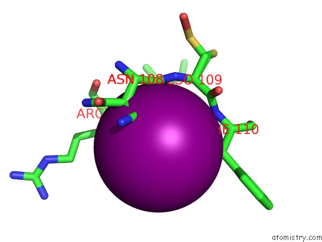
 Mono view
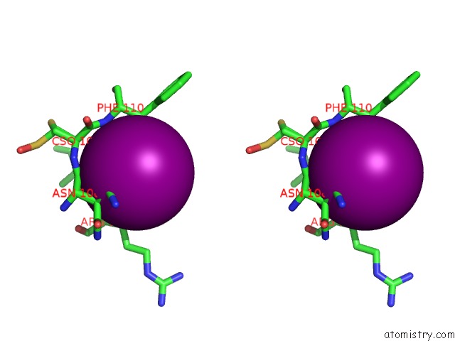
 Stereo pair view
|
A full contact list of Iodine with other atoms in the I binding
site number 10 of Crystal Structure of A Hypothetical Protein (BACUNI_04292) From Bacteroides Uniformis Atcc 8492 at 1.27 A Resolution within 5.0Å range:
probe
|
atom
|
residue
|
distance (Å)
|
B
|
Occ
|
B:I203
b:15.6
occ:0.50
|
O
|
A:HOH321
|
3.1
|
11.7
|
1.0
|
NE2
|
B:GLN122
|
3.2
|
22.3
|
1.0
|
OD1
|
B:ASN108
|
3.3
|
20.5
|
0.5
|
CG
|
B:GLN122
|
3.6
|
14.9
|
1.0
|
O
|
B:ASN108
|
3.7
|
17.0
|
1.0
|
CG
|
B:ASN108
|
3.7
|
20.1
|
0.5
|
CB
|
B:ASN108
|
3.8
|
12.8
|
0.5
|
CD
|
B:GLN122
|
3.8
|
20.5
|
1.0
|
CB
|
B:ASN108
|
3.8
|
16.2
|
0.5
|
CB
|
B:PHE110
|
3.9
|
12.1
|
1.0
|
CB
|
B:GLN122
|
4.0
|
12.4
|
1.0
|
C
|
B:ASN108
|
4.0
|
11.3
|
1.0
|
O
|
B:GLN122
|
4.0
|
10.8
|
1.0
|
CB
|
B:ARG124
|
4.1
|
13.8
|
1.0
|
N
|
B:PHE110
|
4.1
|
10.6
|
1.0
|
N
|
B:ARG124
|
4.1
|
10.0
|
1.0
|
C
|
B:GLN122
|
4.2
|
9.2
|
1.0
|
N
|
B:CSO109
|
4.5
|
10.9
|
1.0
|
C
|
B:CSO109
|
4.5
|
10.2
|
1.0
|
C
|
B:ILE123
|
4.6
|
9.8
|
1.0
|
N
|
B:ILE123
|
4.6
|
9.5
|
1.0
|
CA
|
B:PHE110
|
4.6
|
10.6
|
1.0
|
CA
|
B:ASN108
|
4.6
|
12.4
|
0.5
|
CA
|
B:ASN108
|
4.6
|
11.4
|
0.5
|
CA
|
B:ILE123
|
4.7
|
10.2
|
1.0
|
CA
|
B:CSO109
|
4.7
|
10.6
|
1.0
|
ND2
|
B:ASN108
|
4.7
|
23.6
|
0.5
|
CA
|
B:ARG124
|
4.7
|
11.8
|
1.0
|
CA
|
B:GLN122
|
4.8
|
9.5
|
1.0
|
CG
|
B:ASN108
|
4.9
|
13.3
|
0.5
|
OE1
|
B:GLN122
|
4.9
|
24.6
|
1.0
|
|
Reference:
Joint Center For Structural Genomics (Jcsg),
Joint Center For Structural Genomics (Jcsg).
N/A N/A.
Page generated: Sun Aug 11 19:40:52 2024
|