Iodine »
PDB 4p9t-4ttc »
4rcf »
Iodine in PDB 4rcf: Crystal Structure of BACE1 in Complex with 2-Aminooxazoline 4- Fluoroxanthene Inhibitor 49
Enzymatic activity of Crystal Structure of BACE1 in Complex with 2-Aminooxazoline 4- Fluoroxanthene Inhibitor 49
All present enzymatic activity of Crystal Structure of BACE1 in Complex with 2-Aminooxazoline 4- Fluoroxanthene Inhibitor 49:
3.4.23.46;
3.4.23.46;
Protein crystallography data
The structure of Crystal Structure of BACE1 in Complex with 2-Aminooxazoline 4- Fluoroxanthene Inhibitor 49, PDB code: 4rcf
was solved by
D.A.Whittington,
A.M.Long,
with X-Ray Crystallography technique. A brief refinement statistics is given in the table below:
| Resolution Low / High (Å) | 50.00 / 1.78 |
| Space group | P 61 2 2 |
| Cell size a, b, c (Å), α, β, γ (°) | 102.262, 102.262, 170.306, 90.00, 90.00, 120.00 |
| R / Rfree (%) | 20.3 / 22.9 |
Other elements in 4rcf:
The structure of Crystal Structure of BACE1 in Complex with 2-Aminooxazoline 4- Fluoroxanthene Inhibitor 49 also contains other interesting chemical elements:
| Fluorine | (F) | 2 atoms |
Iodine Binding Sites:
The binding sites of Iodine atom in the Crystal Structure of BACE1 in Complex with 2-Aminooxazoline 4- Fluoroxanthene Inhibitor 49
(pdb code 4rcf). This binding sites where shown within
5.0 Angstroms radius around Iodine atom.
In total 3 binding sites of Iodine where determined in the Crystal Structure of BACE1 in Complex with 2-Aminooxazoline 4- Fluoroxanthene Inhibitor 49, PDB code: 4rcf:
Jump to Iodine binding site number: 1; 2; 3;
In total 3 binding sites of Iodine where determined in the Crystal Structure of BACE1 in Complex with 2-Aminooxazoline 4- Fluoroxanthene Inhibitor 49, PDB code: 4rcf:
Jump to Iodine binding site number: 1; 2; 3;
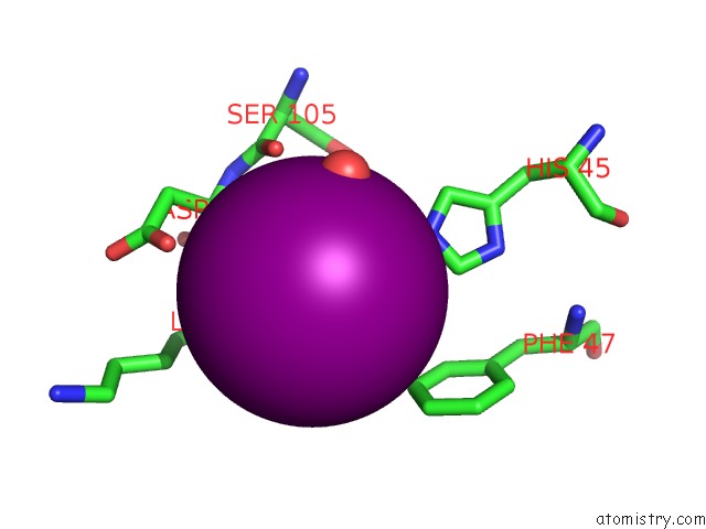
Iodine binding site 1 out of 3 in 4rcf
Go back to
Iodine binding site 1 out
of 3 in the Crystal Structure of BACE1 in Complex with 2-Aminooxazoline 4- Fluoroxanthene Inhibitor 49

Mono view

Stereo pair view
Mono view
Stereo pair view
A full contact list of Iodine with other atoms in the I binding
site number 1 of Crystal Structure of BACE1 in Complex with 2-Aminooxazoline 4- Fluoroxanthene Inhibitor 49 within 5.0Å range:
|
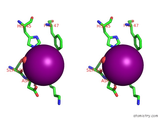
Iodine binding site 2 out of 3 in 4rcf
Go back to
Iodine binding site 2 out
of 3 in the Crystal Structure of BACE1 in Complex with 2-Aminooxazoline 4- Fluoroxanthene Inhibitor 49
Mono view
Stereo pair view
Mono view
Stereo pair view
A full contact list of Iodine with other atoms in the I binding
site number 2 of Crystal Structure of BACE1 in Complex with 2-Aminooxazoline 4- Fluoroxanthene Inhibitor 49 within 5.0Å range:
|
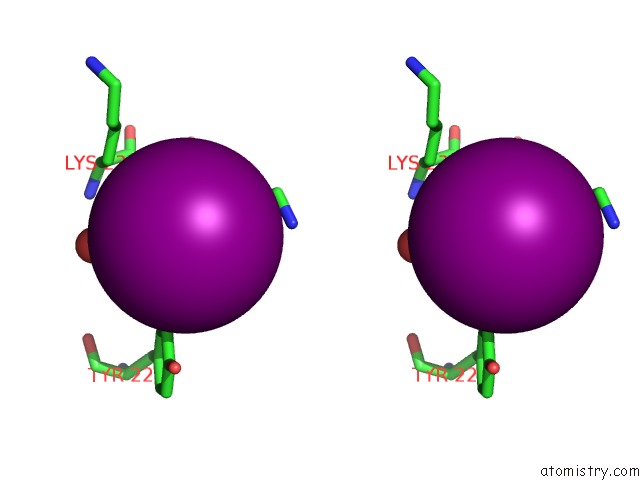
Iodine binding site 3 out of 3 in 4rcf
Go back to
Iodine binding site 3 out
of 3 in the Crystal Structure of BACE1 in Complex with 2-Aminooxazoline 4- Fluoroxanthene Inhibitor 49

Mono view
Stereo pair view
Mono view
Stereo pair view
A full contact list of Iodine with other atoms in the I binding
site number 3 of Crystal Structure of BACE1 in Complex with 2-Aminooxazoline 4- Fluoroxanthene Inhibitor 49 within 5.0Å range:
|
Reference:
O.Epstein,
M.C.Bryan,
A.C.Cheng,
K.Derakhchan,
T.A.Dineen,
D.Hickman,
Z.Hua,
J.B.Human,
C.Kreiman,
I.E.Marx,
M.M.Weiss,
R.C.Wahl,
P.H.Wen,
D.A.Whittington,
S.Wood,
X.M.Zheng,
R.T.Fremeau,
R.D.White,
V.F.Patel.
Lead Optimization and Modulation of Herg Activity in A Series of Aminooxazoline Xanthene Beta-Site Amyloid Precursor Protein Cleaving Enzyme (BACE1) Inhibitors. J.Med.Chem. 2014.
ISSN: ISSN 0022-2623
PubMed: 25389560
DOI: 10.1021/JM501266W
Page generated: Fri Aug 8 18:42:44 2025
ISSN: ISSN 0022-2623
PubMed: 25389560
DOI: 10.1021/JM501266W
Last articles
K in 4EVYK in 4EOU
K in 4ETM
K in 4ESK
K in 4ES8
K in 4ERT
K in 4ERD
K in 4ENC
K in 4EK1
K in 4ENB