Iodine »
PDB 4p9t-4ttc »
4s22 »
Iodine in PDB 4s22: Crystal Structure of K29 Linked Di-Ubiquitin
Protein crystallography data
The structure of Crystal Structure of K29 Linked Di-Ubiquitin, PDB code: 4s22
was solved by
Y.A.Kristariyanto,
S.A.Abdul Rehman,
D.G.Campbell,
N.A.Morrice,
C.Johnson,
R.Toth,
Y.Kulathu,
with X-Ray Crystallography technique. A brief refinement statistics is given in the table below:
| Resolution Low / High (Å) | 60.05 / 2.30 |
| Space group | P 1 21 1 |
| Cell size a, b, c (Å), α, β, γ (°) | 33.450, 69.245, 60.055, 90.00, 90.22, 90.00 |
| R / Rfree (%) | 19.8 / 24.6 |
Iodine Binding Sites:
The binding sites of Iodine atom in the Crystal Structure of K29 Linked Di-Ubiquitin
(pdb code 4s22). This binding sites where shown within
5.0 Angstroms radius around Iodine atom.
In total 6 binding sites of Iodine where determined in the Crystal Structure of K29 Linked Di-Ubiquitin, PDB code: 4s22:
Jump to Iodine binding site number: 1; 2; 3; 4; 5; 6;
In total 6 binding sites of Iodine where determined in the Crystal Structure of K29 Linked Di-Ubiquitin, PDB code: 4s22:
Jump to Iodine binding site number: 1; 2; 3; 4; 5; 6;
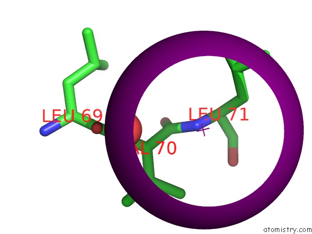
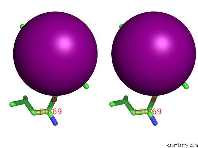
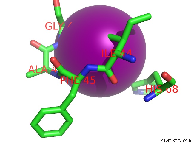
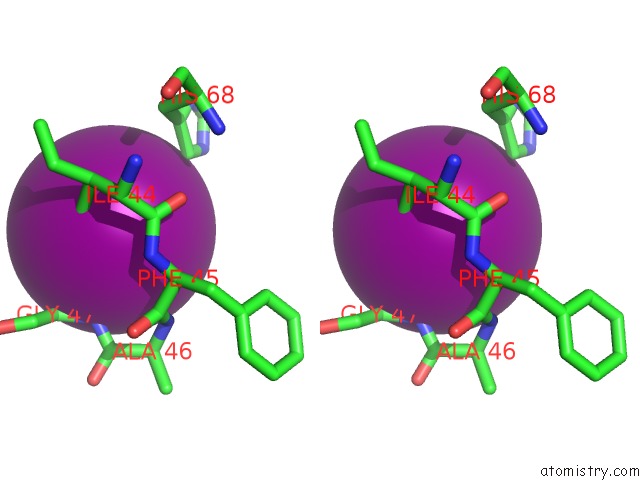
Iodine binding site 1 out of 6 in 4s22
Go back to
Iodine binding site 1 out
of 6 in the Crystal Structure of K29 Linked Di-Ubiquitin
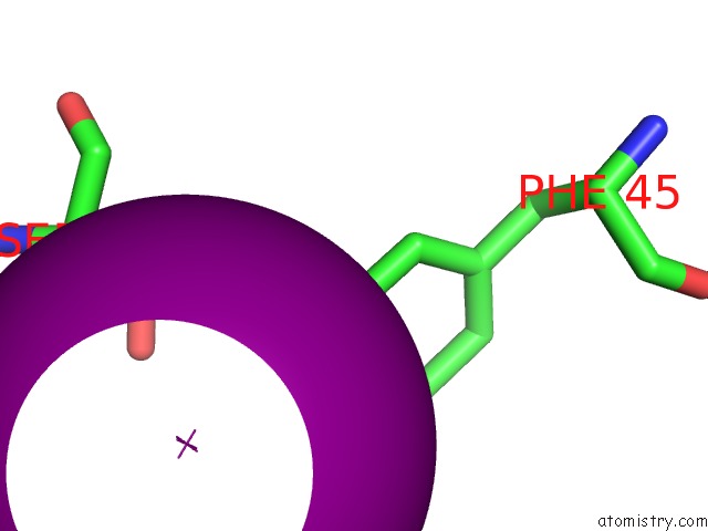
Mono view
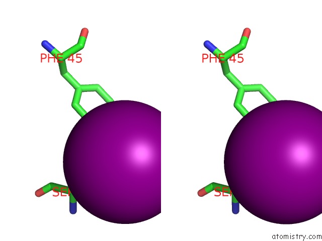
Stereo pair view
Mono view
Stereo pair view
A full contact list of Iodine with other atoms in the I binding
site number 1 of Crystal Structure of K29 Linked Di-Ubiquitin within 5.0Å range:
|
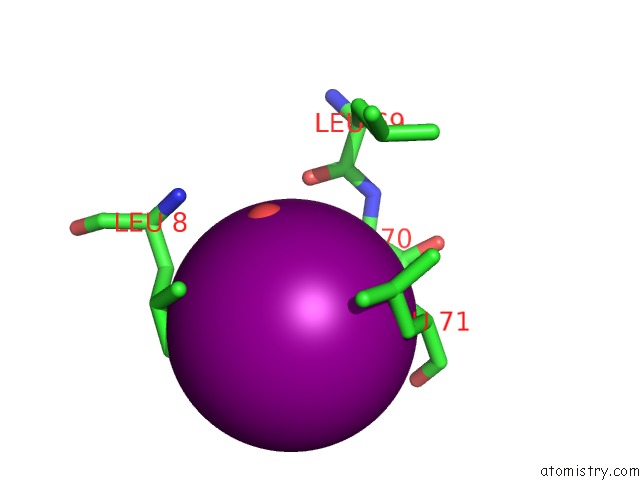
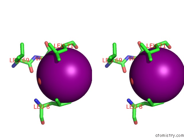
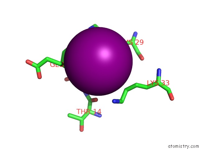
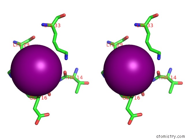
Iodine binding site 2 out of 6 in 4s22
Go back to
Iodine binding site 2 out
of 6 in the Crystal Structure of K29 Linked Di-Ubiquitin
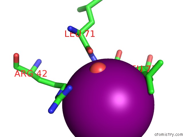
Mono view
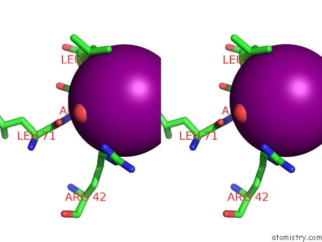
Stereo pair view
Mono view
Stereo pair view
A full contact list of Iodine with other atoms in the I binding
site number 2 of Crystal Structure of K29 Linked Di-Ubiquitin within 5.0Å range:
|
Iodine binding site 3 out of 6 in 4s22
Go back to
Iodine binding site 3 out
of 6 in the Crystal Structure of K29 Linked Di-Ubiquitin
Mono view
Stereo pair view
Mono view
Stereo pair view
A full contact list of Iodine with other atoms in the I binding
site number 3 of Crystal Structure of K29 Linked Di-Ubiquitin within 5.0Å range:
|
Iodine binding site 4 out of 6 in 4s22
Go back to
Iodine binding site 4 out
of 6 in the Crystal Structure of K29 Linked Di-Ubiquitin
Mono view
Stereo pair view
Mono view
Stereo pair view
A full contact list of Iodine with other atoms in the I binding
site number 4 of Crystal Structure of K29 Linked Di-Ubiquitin within 5.0Å range:
|
Iodine binding site 5 out of 6 in 4s22
Go back to
Iodine binding site 5 out
of 6 in the Crystal Structure of K29 Linked Di-Ubiquitin
Mono view
Stereo pair view
Mono view
Stereo pair view
A full contact list of Iodine with other atoms in the I binding
site number 5 of Crystal Structure of K29 Linked Di-Ubiquitin within 5.0Å range:
|
Iodine binding site 6 out of 6 in 4s22
Go back to
Iodine binding site 6 out
of 6 in the Crystal Structure of K29 Linked Di-Ubiquitin
Mono view
Stereo pair view
Mono view
Stereo pair view
A full contact list of Iodine with other atoms in the I binding
site number 6 of Crystal Structure of K29 Linked Di-Ubiquitin within 5.0Å range:
|
Reference:
Y.A.Kristariyanto,
S.A.Abdul Rehman,
D.G.Campbell,
N.A.Morrice,
C.Johnson,
R.Toth,
Y.Kulathu.
K29-Selective Ubiquitin Binding Domain Reveals Structural Basis of Specificity and Heterotypic Nature of K29 Polyubiquitin. Mol.Cell 2015.
ISSN: ISSN 1097-2765
PubMed: 25752573
DOI: 10.1016/J.MOLCEL.2015.01.041
Page generated: Sun Aug 11 19:48:00 2024
ISSN: ISSN 1097-2765
PubMed: 25752573
DOI: 10.1016/J.MOLCEL.2015.01.041
Last articles
Zn in 9J0NZn in 9J0O
Zn in 9J0P
Zn in 9FJX
Zn in 9EKB
Zn in 9C0F
Zn in 9CAH
Zn in 9CH0
Zn in 9CH3
Zn in 9CH1