Iodine »
PDB 4ttu-4xkz »
4wcn »
Iodine in PDB 4wcn: Crystal Structure of Tripeptide Bound Cell Shape Determinant CSD4 Protein From Helicobacter Pylori
Protein crystallography data
The structure of Crystal Structure of Tripeptide Bound Cell Shape Determinant CSD4 Protein From Helicobacter Pylori, PDB code: 4wcn
was solved by
A.C.Chan,
M.E.Murphy,
with X-Ray Crystallography technique. A brief refinement statistics is given in the table below:
| Resolution Low / High (Å) | 49.15 / 1.75 |
| Space group | P 21 21 21 |
| Cell size a, b, c (Å), α, β, γ (°) | 53.230, 66.830, 145.050, 90.00, 90.00, 90.00 |
| R / Rfree (%) | 17.7 / 20.8 |
Other elements in 4wcn:
The structure of Crystal Structure of Tripeptide Bound Cell Shape Determinant CSD4 Protein From Helicobacter Pylori also contains other interesting chemical elements:
| Zinc | (Zn) | 1 atom |
| Sodium | (Na) | 2 atoms |
Iodine Binding Sites:
Pages:
>>> Page 1 <<< Page 2, Binding sites: 11 - 20; Page 3, Binding sites: 21 - 30; Page 4, Binding sites: 31 - 40; Page 5, Binding sites: 41 - 44;Binding sites:
The binding sites of Iodine atom in the Crystal Structure of Tripeptide Bound Cell Shape Determinant CSD4 Protein From Helicobacter Pylori (pdb code 4wcn). This binding sites where shown within 5.0 Angstroms radius around Iodine atom.In total 44 binding sites of Iodine where determined in the Crystal Structure of Tripeptide Bound Cell Shape Determinant CSD4 Protein From Helicobacter Pylori, PDB code: 4wcn:
Jump to Iodine binding site number: 1; 2; 3; 4; 5; 6; 7; 8; 9; 10;
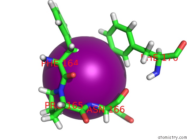
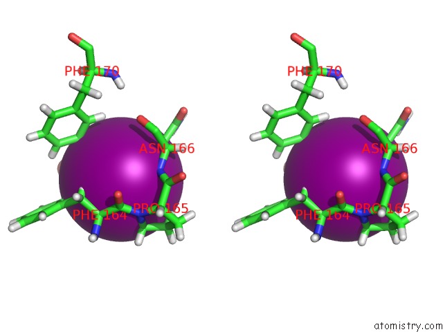
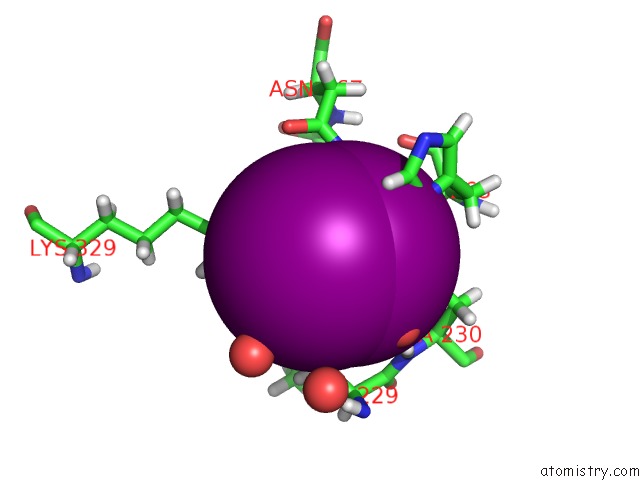
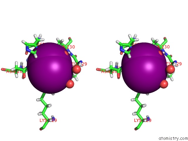
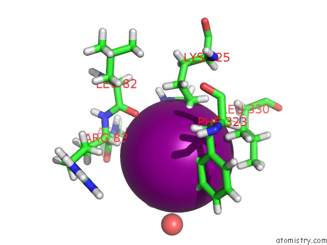
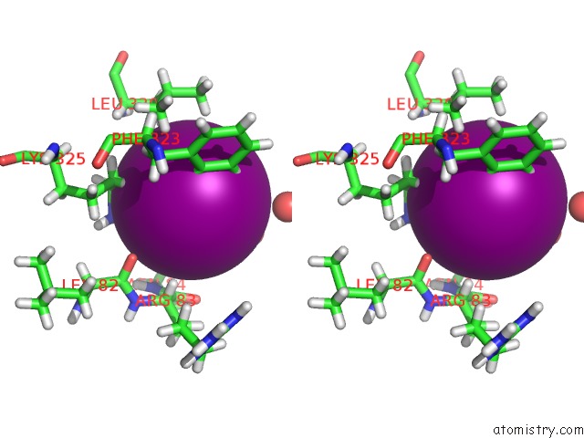
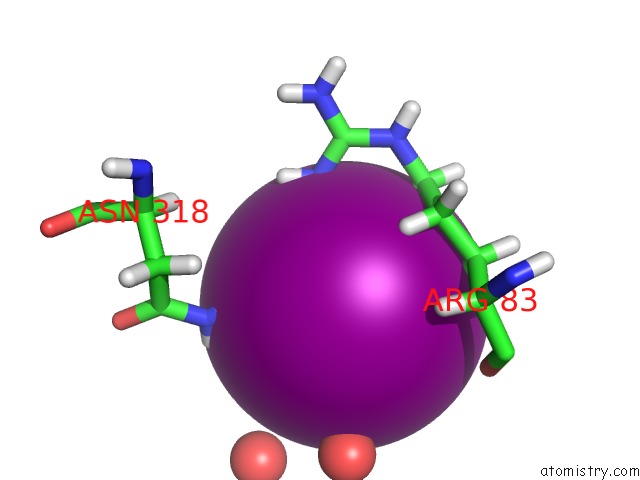
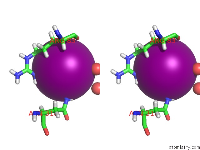
Iodine binding site 1 out of 44 in 4wcn
Go back to
Iodine binding site 1 out
of 44 in the Crystal Structure of Tripeptide Bound Cell Shape Determinant CSD4 Protein From Helicobacter Pylori
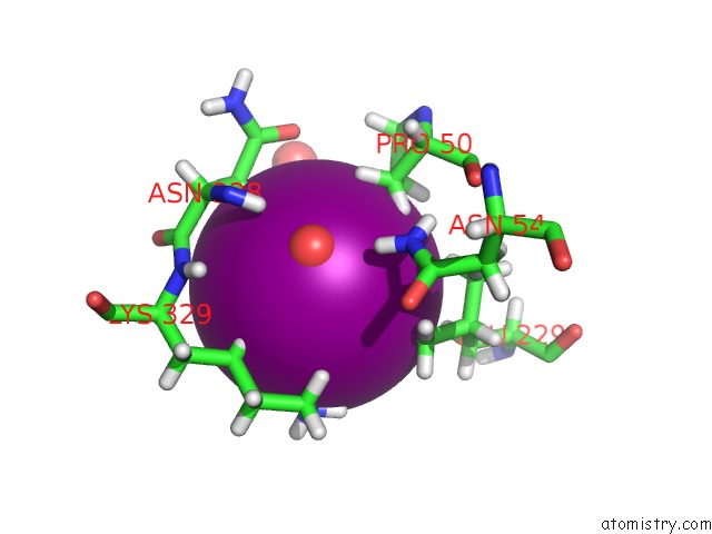
Mono view
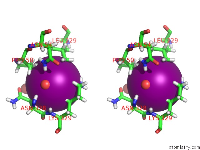
Stereo pair view
Mono view
Stereo pair view
A full contact list of Iodine with other atoms in the I binding
site number 1 of Crystal Structure of Tripeptide Bound Cell Shape Determinant CSD4 Protein From Helicobacter Pylori within 5.0Å range:
|
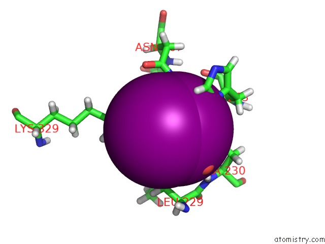
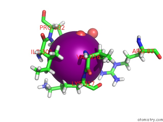
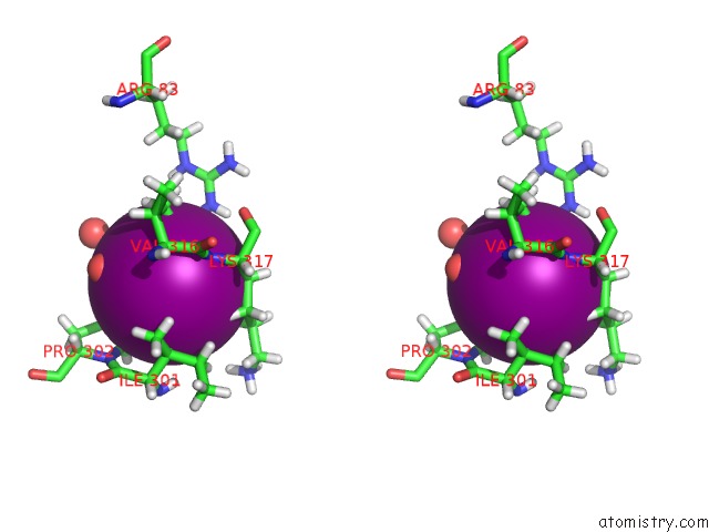
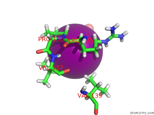
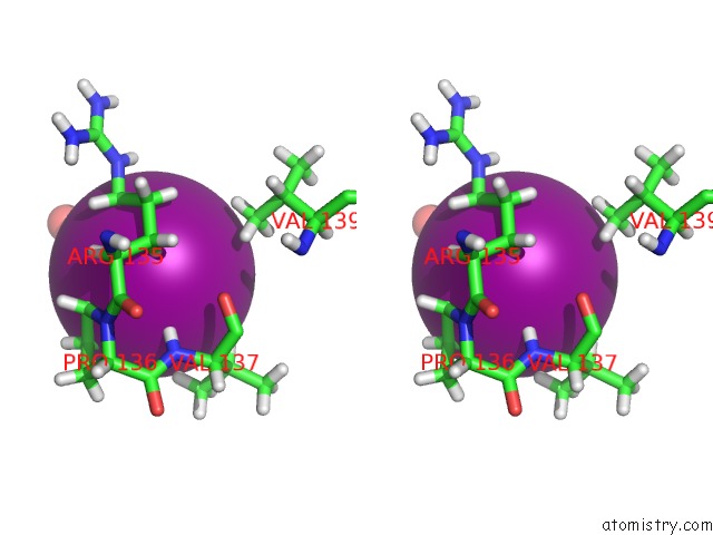
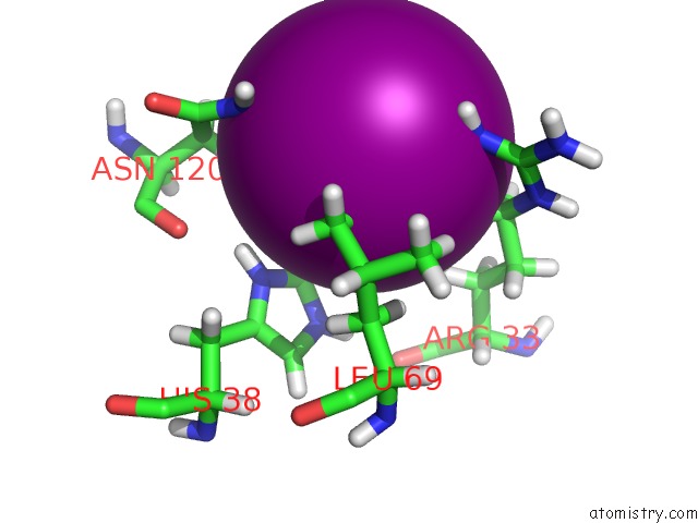
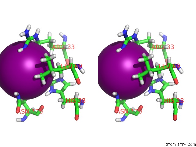
Iodine binding site 2 out of 44 in 4wcn
Go back to
Iodine binding site 2 out
of 44 in the Crystal Structure of Tripeptide Bound Cell Shape Determinant CSD4 Protein From Helicobacter Pylori
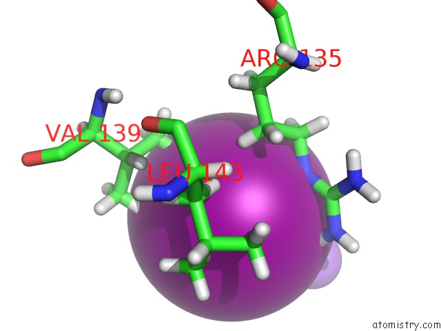
Mono view
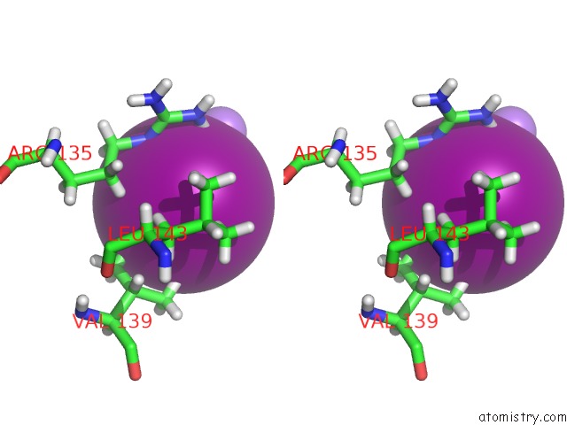
Stereo pair view
Mono view
Stereo pair view
A full contact list of Iodine with other atoms in the I binding
site number 2 of Crystal Structure of Tripeptide Bound Cell Shape Determinant CSD4 Protein From Helicobacter Pylori within 5.0Å range:
|
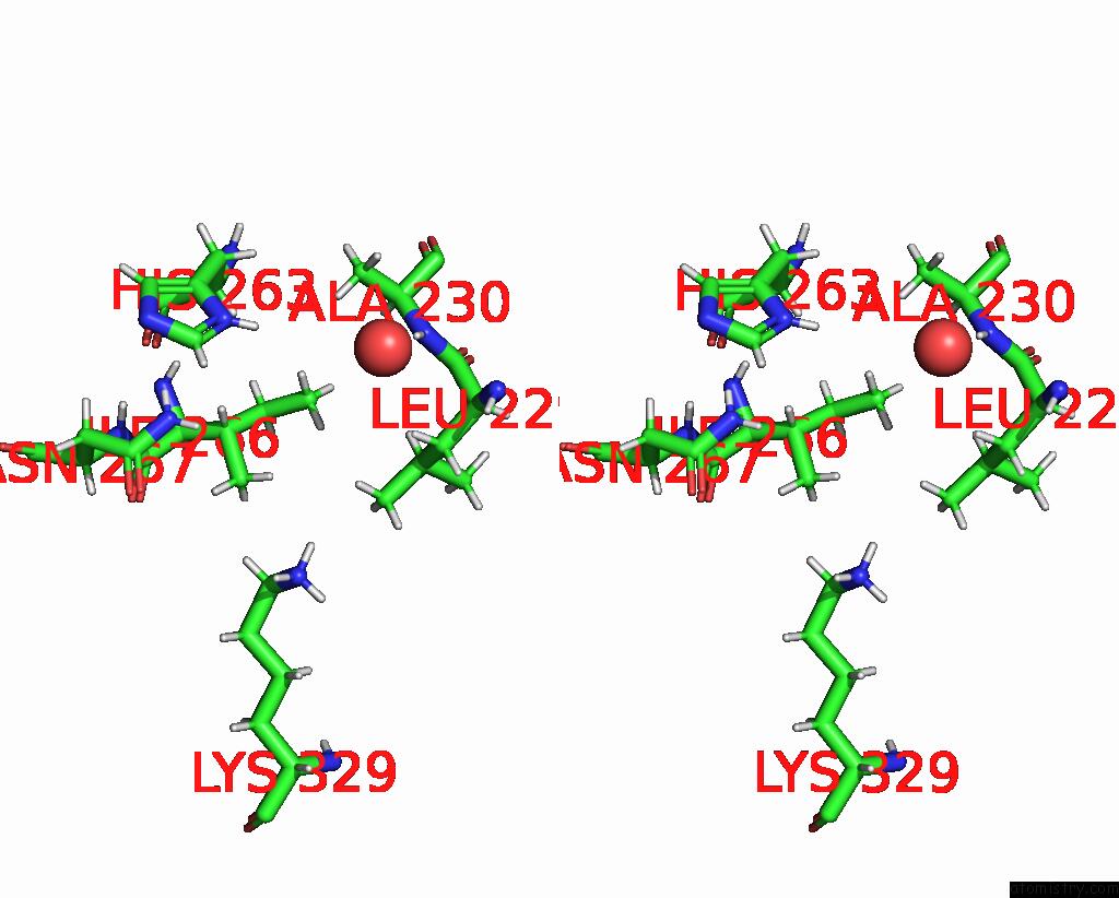
Iodine binding site 3 out of 44 in 4wcn
Go back to
Iodine binding site 3 out
of 44 in the Crystal Structure of Tripeptide Bound Cell Shape Determinant CSD4 Protein From Helicobacter Pylori
Mono view
Stereo pair view
Mono view
Stereo pair view
A full contact list of Iodine with other atoms in the I binding
site number 3 of Crystal Structure of Tripeptide Bound Cell Shape Determinant CSD4 Protein From Helicobacter Pylori within 5.0Å range:
|
Iodine binding site 4 out of 44 in 4wcn
Go back to
Iodine binding site 4 out
of 44 in the Crystal Structure of Tripeptide Bound Cell Shape Determinant CSD4 Protein From Helicobacter Pylori
Mono view
Stereo pair view
Mono view
Stereo pair view
A full contact list of Iodine with other atoms in the I binding
site number 4 of Crystal Structure of Tripeptide Bound Cell Shape Determinant CSD4 Protein From Helicobacter Pylori within 5.0Å range:
|
Iodine binding site 5 out of 44 in 4wcn
Go back to
Iodine binding site 5 out
of 44 in the Crystal Structure of Tripeptide Bound Cell Shape Determinant CSD4 Protein From Helicobacter Pylori
Mono view
Stereo pair view
Mono view
Stereo pair view
A full contact list of Iodine with other atoms in the I binding
site number 5 of Crystal Structure of Tripeptide Bound Cell Shape Determinant CSD4 Protein From Helicobacter Pylori within 5.0Å range:
|
Iodine binding site 6 out of 44 in 4wcn
Go back to
Iodine binding site 6 out
of 44 in the Crystal Structure of Tripeptide Bound Cell Shape Determinant CSD4 Protein From Helicobacter Pylori
Mono view
Stereo pair view
Mono view
Stereo pair view
A full contact list of Iodine with other atoms in the I binding
site number 6 of Crystal Structure of Tripeptide Bound Cell Shape Determinant CSD4 Protein From Helicobacter Pylori within 5.0Å range:
|
Iodine binding site 7 out of 44 in 4wcn
Go back to
Iodine binding site 7 out
of 44 in the Crystal Structure of Tripeptide Bound Cell Shape Determinant CSD4 Protein From Helicobacter Pylori
Mono view
Stereo pair view
Mono view
Stereo pair view
A full contact list of Iodine with other atoms in the I binding
site number 7 of Crystal Structure of Tripeptide Bound Cell Shape Determinant CSD4 Protein From Helicobacter Pylori within 5.0Å range:
|
Iodine binding site 8 out of 44 in 4wcn
Go back to
Iodine binding site 8 out
of 44 in the Crystal Structure of Tripeptide Bound Cell Shape Determinant CSD4 Protein From Helicobacter Pylori
Mono view
Stereo pair view
Mono view
Stereo pair view
A full contact list of Iodine with other atoms in the I binding
site number 8 of Crystal Structure of Tripeptide Bound Cell Shape Determinant CSD4 Protein From Helicobacter Pylori within 5.0Å range:
|
Iodine binding site 9 out of 44 in 4wcn
Go back to
Iodine binding site 9 out
of 44 in the Crystal Structure of Tripeptide Bound Cell Shape Determinant CSD4 Protein From Helicobacter Pylori
Mono view
Stereo pair view
Mono view
Stereo pair view
A full contact list of Iodine with other atoms in the I binding
site number 9 of Crystal Structure of Tripeptide Bound Cell Shape Determinant CSD4 Protein From Helicobacter Pylori within 5.0Å range:
|
Iodine binding site 10 out of 44 in 4wcn
Go back to
Iodine binding site 10 out
of 44 in the Crystal Structure of Tripeptide Bound Cell Shape Determinant CSD4 Protein From Helicobacter Pylori
Mono view
Stereo pair view
Mono view
Stereo pair view
A full contact list of Iodine with other atoms in the I binding
site number 10 of Crystal Structure of Tripeptide Bound Cell Shape Determinant CSD4 Protein From Helicobacter Pylori within 5.0Å range:
|
Reference:
A.C.Chan,
K.M.Blair,
Y.Liu,
E.Frirdich,
E.C.Gaynor,
M.E.Tanner,
N.R.Salama,
M.E.Murphy.
Helical Shape of Helicobacter Pylori Requires An Atypical Glutamine As A Zinc Ligand in the Carboxypeptidase CSD4 J.Biol.Chem. 2014.
ISSN: ESSN 1083-351X
DOI: 10.1074/JBC.M114.624734
Page generated: Fri Aug 8 18:56:46 2025
ISSN: ESSN 1083-351X
DOI: 10.1074/JBC.M114.624734
Last articles
I in 7B94I in 7BEN
I in 6Z52
I in 7B76
I in 6ZGQ
I in 7B10
I in 7B0T
I in 6Z79
I in 6Z6E
I in 6YVG