Iodine »
PDB 4ttu-4xkz »
4xfx »
Iodine in PDB 4xfx: Structure of the Native Full-Length Hiv-1 Capsid Protein
Protein crystallography data
The structure of Structure of the Native Full-Length Hiv-1 Capsid Protein, PDB code: 4xfx
was solved by
A.T.Gres,
K.A.Kirby,
S.G.Sarafianos,
with X-Ray Crystallography technique. A brief refinement statistics is given in the table below:
| Resolution Low / High (Å) | 19.00 / 2.43 |
| Space group | P 6 |
| Cell size a, b, c (Å), α, β, γ (°) | 92.328, 92.328, 57.340, 90.00, 90.00, 120.00 |
| R / Rfree (%) | 22.2 / 24.9 |
Other elements in 4xfx:
The structure of Structure of the Native Full-Length Hiv-1 Capsid Protein also contains other interesting chemical elements:
| Chlorine | (Cl) | 2 atoms |
Iodine Binding Sites:
The binding sites of Iodine atom in the Structure of the Native Full-Length Hiv-1 Capsid Protein
(pdb code 4xfx). This binding sites where shown within
5.0 Angstroms radius around Iodine atom.
In total 7 binding sites of Iodine where determined in the Structure of the Native Full-Length Hiv-1 Capsid Protein, PDB code: 4xfx:
Jump to Iodine binding site number: 1; 2; 3; 4; 5; 6; 7;
In total 7 binding sites of Iodine where determined in the Structure of the Native Full-Length Hiv-1 Capsid Protein, PDB code: 4xfx:
Jump to Iodine binding site number: 1; 2; 3; 4; 5; 6; 7;
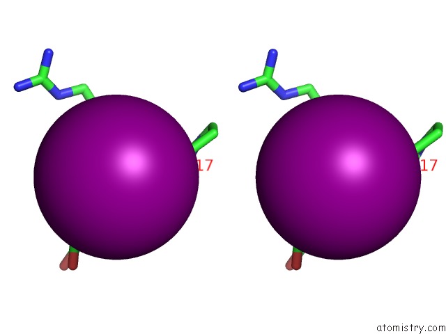
Iodine binding site 1 out of 7 in 4xfx
Go back to
Iodine binding site 1 out
of 7 in the Structure of the Native Full-Length Hiv-1 Capsid Protein

Mono view

Stereo pair view
Mono view
Stereo pair view
A full contact list of Iodine with other atoms in the I binding
site number 1 of Structure of the Native Full-Length Hiv-1 Capsid Protein within 5.0Å range:
|
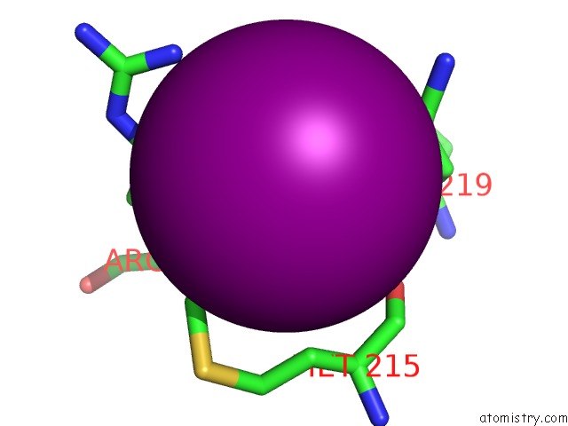
Iodine binding site 2 out of 7 in 4xfx
Go back to
Iodine binding site 2 out
of 7 in the Structure of the Native Full-Length Hiv-1 Capsid Protein

Mono view

Stereo pair view
Mono view
Stereo pair view
A full contact list of Iodine with other atoms in the I binding
site number 2 of Structure of the Native Full-Length Hiv-1 Capsid Protein within 5.0Å range:
|
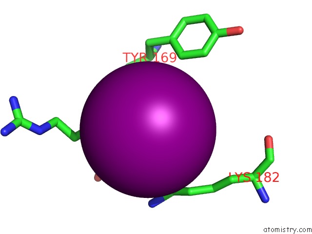
Iodine binding site 3 out of 7 in 4xfx
Go back to
Iodine binding site 3 out
of 7 in the Structure of the Native Full-Length Hiv-1 Capsid Protein

Mono view

Stereo pair view
Mono view
Stereo pair view
A full contact list of Iodine with other atoms in the I binding
site number 3 of Structure of the Native Full-Length Hiv-1 Capsid Protein within 5.0Å range:
|
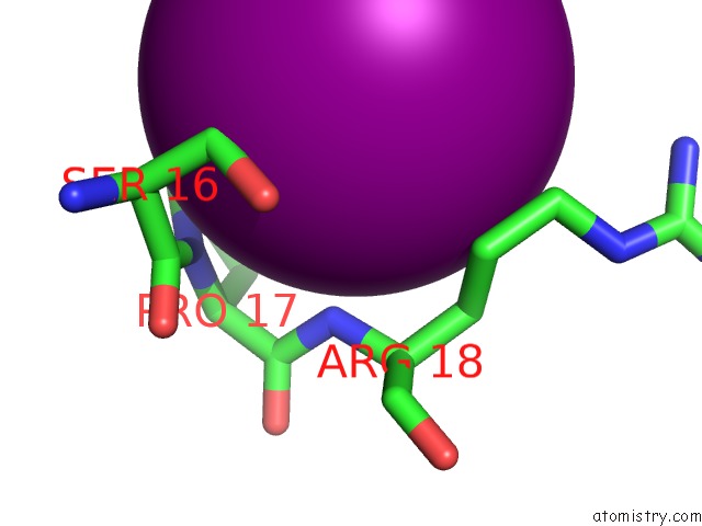
Iodine binding site 4 out of 7 in 4xfx
Go back to
Iodine binding site 4 out
of 7 in the Structure of the Native Full-Length Hiv-1 Capsid Protein

Mono view
Stereo pair view
Mono view
Stereo pair view
A full contact list of Iodine with other atoms in the I binding
site number 4 of Structure of the Native Full-Length Hiv-1 Capsid Protein within 5.0Å range:
|
Iodine binding site 5 out of 7 in 4xfx
Go back to
Iodine binding site 5 out
of 7 in the Structure of the Native Full-Length Hiv-1 Capsid Protein
Mono view

Stereo pair view
Mono view
Stereo pair view
A full contact list of Iodine with other atoms in the I binding
site number 5 of Structure of the Native Full-Length Hiv-1 Capsid Protein within 5.0Å range:
|
Iodine binding site 6 out of 7 in 4xfx
Go back to
Iodine binding site 6 out
of 7 in the Structure of the Native Full-Length Hiv-1 Capsid Protein
Mono view

Stereo pair view
Mono view
Stereo pair view
A full contact list of Iodine with other atoms in the I binding
site number 6 of Structure of the Native Full-Length Hiv-1 Capsid Protein within 5.0Å range:
|
Iodine binding site 7 out of 7 in 4xfx
Go back to
Iodine binding site 7 out
of 7 in the Structure of the Native Full-Length Hiv-1 Capsid Protein
Mono view

Stereo pair view
Mono view
Stereo pair view
A full contact list of Iodine with other atoms in the I binding
site number 7 of Structure of the Native Full-Length Hiv-1 Capsid Protein within 5.0Å range:
|
Reference:
A.T.Gres,
K.A.Kirby,
V.N.Kewalramani,
J.J.Tanner,
O.Pornillos,
S.G.Sarafianos.
Structural Virology. X-Ray Crystal Structures of Native Hiv-1 Capsid Protein Reveal Conformational Variability. Science V. 349 99 2015.
ISSN: ESSN 1095-9203
PubMed: 26044298
DOI: 10.1126/SCIENCE.AAA5936
Page generated: Fri Aug 8 19:07:27 2025
ISSN: ESSN 1095-9203
PubMed: 26044298
DOI: 10.1126/SCIENCE.AAA5936
Last articles
I in 6XNFI in 6XLO
I in 6WXM
I in 6X42
I in 6X2D
I in 6WYQ
I in 6WOK
I in 6WNY
I in 6W9D
I in 6WC8