Iodine in PDB 4xpv: Neutron and X-Ray Structure Analysis of Xylanase: N44D at PH6
Enzymatic activity of Neutron and X-Ray Structure Analysis of Xylanase: N44D at PH6
All present enzymatic activity of Neutron and X-Ray Structure Analysis of Xylanase: N44D at PH6:
3.2.1.8;
Protein crystallography data
The structure of Neutron and X-Ray Structure Analysis of Xylanase: N44D at PH6, PDB code: 4xpv
was solved by
Q.Wan,
J.M.Park,
D.M.Riccardi,
L.B.Hanson,
Z.Fisher,
J.C.Smith,
A.Ostermann,
T.Schrader,
D.E.Graham,
L.Coates,
P.Langan,
A.Y.Kovalevsky,
with X-Ray Crystallography technique. A brief refinement statistics is given in the table below:
Resolution Low / High (Å)
|
N/A /
1.70
|
Space group
|
P 21 21 21
|
Cell size a, b, c (Å), α, β, γ (°)
|
49.194,
60.287,
70.520,
90.00,
90.00,
90.00
|
R / Rfree (%)
|
26.4 /
30.4
|
Iodine Binding Sites:
The binding sites of Iodine atom in the Neutron and X-Ray Structure Analysis of Xylanase: N44D at PH6
(pdb code 4xpv). This binding sites where shown within
5.0 Angstroms radius around Iodine atom.
In total 3 binding sites of Iodine where determined in the
Neutron and X-Ray Structure Analysis of Xylanase: N44D at PH6, PDB code: 4xpv:
Jump to Iodine binding site number:
1;
2;
3;
Iodine binding site 1 out
of 3 in 4xpv
Go back to
Iodine Binding Sites List in 4xpv
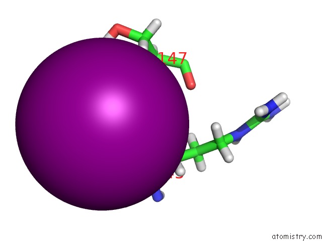
Iodine binding site 1 out
of 3 in the Neutron and X-Ray Structure Analysis of Xylanase: N44D at PH6
 Mono view
 Stereo pair view
|
A full contact list of Iodine with other atoms in the I binding
site number 1 of Neutron and X-Ray Structure Analysis of Xylanase: N44D at PH6 within 5.0Å range:
probe
|
atom
|
residue
|
distance (Å)
|
B
|
Occ
|
A:I201
b:14.7
occ:1.00
|
D
|
A:SER146
|
2.8
|
12.9
|
0.8
|
H
|
A:SER146
|
2.8
|
12.9
|
0.2
|
D1
|
A:DOD321
|
3.0
|
13.9
|
1.0
|
D1
|
A:DOD400
|
3.1
|
19.6
|
1.0
|
D2
|
A:DOD343
|
3.4
|
19.2
|
1.0
|
O
|
A:DOD321
|
3.5
|
13.2
|
1.0
|
HB2
|
A:ARG145
|
3.5
|
13.1
|
1.0
|
HA
|
A:ARG145
|
3.6
|
14.7
|
1.0
|
N
|
A:SER146
|
3.6
|
11.4
|
1.0
|
O
|
A:DOD400
|
3.7
|
24.4
|
1.0
|
H
|
A:SER147
|
3.7
|
12.9
|
0.5
|
D
|
A:SER147
|
3.7
|
12.9
|
0.5
|
D2
|
A:DOD400
|
3.7
|
21.0
|
1.0
|
HB2
|
A:SER146
|
3.8
|
10.9
|
1.0
|
HB2
|
A:SER147
|
4.2
|
13.8
|
1.0
|
O
|
A:DOD343
|
4.2
|
34.4
|
1.0
|
CA
|
A:ARG145
|
4.2
|
10.0
|
1.0
|
CB
|
A:ARG145
|
4.2
|
9.6
|
1.0
|
N
|
A:SER147
|
4.3
|
11.1
|
1.0
|
D2
|
A:DOD321
|
4.3
|
16.6
|
1.0
|
D1
|
A:DOD343
|
4.4
|
21.2
|
1.0
|
C
|
A:ARG145
|
4.4
|
10.5
|
1.0
|
CA
|
A:SER146
|
4.5
|
8.1
|
1.0
|
HB3
|
A:ARG145
|
4.5
|
11.3
|
1.0
|
CB
|
A:SER146
|
4.5
|
16.5
|
1.0
|
HG
|
A:SER147
|
4.7
|
14.5
|
0.0
|
DG
|
A:SER147
|
4.7
|
14.5
|
1.0
|
C
|
A:SER146
|
4.7
|
11.6
|
1.0
|
CB
|
A:SER147
|
4.9
|
13.6
|
1.0
|
OG
|
A:SER146
|
5.0
|
23.1
|
1.0
|
|
Iodine binding site 2 out
of 3 in 4xpv
Go back to
Iodine Binding Sites List in 4xpv
Iodine binding site 2 out
of 3 in the Neutron and X-Ray Structure Analysis of Xylanase: N44D at PH6
 Mono view
 Stereo pair view
|
A full contact list of Iodine with other atoms in the I binding
site number 2 of Neutron and X-Ray Structure Analysis of Xylanase: N44D at PH6 within 5.0Å range:
probe
|
atom
|
residue
|
distance (Å)
|
B
|
Occ
|
A:I202
b:35.7
occ:0.39
|
D
|
A:MET169
|
2.7
|
17.6
|
0.8
|
H
|
A:MET169
|
2.7
|
17.6
|
0.2
|
HG2
|
A:MET169
|
3.1
|
18.8
|
1.0
|
HE3
|
A:LYS56
|
3.2
|
20.0
|
1.0
|
HA
|
A:THR168
|
3.3
|
17.6
|
1.0
|
D1
|
A:DOD386
|
3.4
|
22.1
|
1.0
|
O
|
A:DOD352
|
3.4
|
30.1
|
1.0
|
D1
|
A:DOD352
|
3.5
|
21.6
|
1.0
|
N
|
A:MET169
|
3.5
|
13.2
|
1.0
|
HG23
|
A:THR168
|
3.6
|
18.2
|
1.0
|
HB2
|
A:MET169
|
3.8
|
15.4
|
1.0
|
O
|
A:DOD386
|
3.9
|
27.1
|
1.0
|
CG
|
A:MET169
|
4.0
|
17.1
|
1.0
|
CE
|
A:LYS56
|
4.1
|
21.3
|
1.0
|
CA
|
A:THR168
|
4.1
|
11.1
|
1.0
|
HG3
|
A:LYS56
|
4.1
|
19.0
|
1.0
|
D2
|
A:DOD352
|
4.2
|
23.0
|
1.0
|
CB
|
A:MET169
|
4.2
|
13.9
|
1.0
|
HG3
|
A:MET169
|
4.3
|
17.9
|
1.0
|
C
|
A:THR168
|
4.3
|
14.3
|
1.0
|
DZ1
|
A:LYS56
|
4.4
|
22.2
|
0.7
|
HZ1
|
A:LYS56
|
4.4
|
22.2
|
0.3
|
D2
|
A:DOD386
|
4.4
|
19.8
|
1.0
|
CG2
|
A:THR168
|
4.4
|
26.7
|
1.0
|
HG22
|
A:THR168
|
4.5
|
21.7
|
1.0
|
O
|
A:GLY167
|
4.5
|
19.6
|
1.0
|
CA
|
A:MET169
|
4.5
|
11.7
|
1.0
|
DZ3
|
A:LYS56
|
4.5
|
21.6
|
0.9
|
HZ3
|
A:LYS56
|
4.5
|
21.6
|
0.1
|
HD2
|
A:LYS56
|
4.6
|
20.5
|
1.0
|
NZ
|
A:LYS56
|
4.6
|
24.1
|
1.0
|
HE2
|
A:LYS56
|
4.7
|
19.4
|
1.0
|
CD
|
A:LYS56
|
4.8
|
19.6
|
1.0
|
CB
|
A:THR168
|
4.9
|
18.1
|
1.0
|
HA
|
A:THR55
|
4.9
|
18.4
|
1.0
|
CG
|
A:LYS56
|
4.9
|
17.2
|
1.0
|
|
Iodine binding site 3 out
of 3 in 4xpv
Go back to
Iodine Binding Sites List in 4xpv
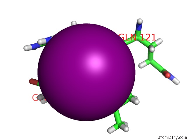
Iodine binding site 3 out
of 3 in the Neutron and X-Ray Structure Analysis of Xylanase: N44D at PH6
 Mono view
 Stereo pair view
|
A full contact list of Iodine with other atoms in the I binding
site number 3 of Neutron and X-Ray Structure Analysis of Xylanase: N44D at PH6 within 5.0Å range:
probe
|
atom
|
residue
|
distance (Å)
|
B
|
Occ
|
A:I203
b:45.3
occ:0.25
|
H
|
A:VAL123
|
2.4
|
21.8
|
0.1
|
D
|
A:VAL123
|
2.4
|
21.8
|
0.9
|
HA
|
A:ARG122
|
2.9
|
20.6
|
1.0
|
N
|
A:VAL123
|
3.2
|
19.7
|
1.0
|
DE21
|
A:GLN125
|
3.5
|
26.4
|
0.7
|
HE21
|
A:GLN125
|
3.5
|
26.4
|
0.3
|
HB
|
A:VAL123
|
3.5
|
21.9
|
1.0
|
HD2
|
A:ARG122
|
3.7
|
27.0
|
1.0
|
CA
|
A:ARG122
|
3.8
|
16.1
|
1.0
|
HG2
|
A:GLN125
|
3.8
|
23.6
|
1.0
|
HG23
|
A:VAL123
|
3.9
|
21.7
|
1.0
|
DH11
|
A:ARG122
|
3.9
|
34.1
|
1.0
|
HH11
|
A:ARG122
|
3.9
|
34.1
|
0.0
|
HG3
|
A:GLN125
|
3.9
|
25.4
|
1.0
|
C
|
A:ARG122
|
4.0
|
19.7
|
1.0
|
O
|
A:VAL123
|
4.0
|
19.2
|
1.0
|
O
|
A:GLN121
|
4.0
|
20.3
|
1.0
|
NE2
|
A:GLN125
|
4.0
|
29.2
|
1.0
|
CA
|
A:VAL123
|
4.1
|
15.9
|
1.0
|
CB
|
A:VAL123
|
4.2
|
21.2
|
1.0
|
CG
|
A:GLN125
|
4.3
|
25.1
|
1.0
|
HB3
|
A:ARG122
|
4.3
|
20.9
|
1.0
|
NH1
|
A:ARG122
|
4.4
|
40.1
|
1.0
|
CG2
|
A:VAL123
|
4.5
|
21.1
|
1.0
|
DE22
|
A:GLN125
|
4.5
|
28.6
|
0.7
|
HE22
|
A:GLN125
|
4.5
|
28.6
|
0.3
|
CD
|
A:GLN125
|
4.5
|
25.5
|
1.0
|
C
|
A:VAL123
|
4.5
|
18.6
|
1.0
|
CB
|
A:ARG122
|
4.6
|
20.7
|
1.0
|
CD
|
A:ARG122
|
4.6
|
27.1
|
1.0
|
DH12
|
A:ARG122
|
4.6
|
35.4
|
0.7
|
HH12
|
A:ARG122
|
4.6
|
35.4
|
0.3
|
N
|
A:ARG122
|
4.7
|
14.7
|
1.0
|
C
|
A:GLN121
|
4.8
|
20.1
|
1.0
|
HG21
|
A:VAL123
|
4.9
|
23.3
|
1.0
|
HA
|
A:VAL123
|
5.0
|
20.0
|
1.0
|
|
Reference:
Q.Wan,
J.M.Parks,
B.L.Hanson,
S.Z.Fisher,
A.Ostermann,
T.E.Schrader,
D.E.Graham,
L.Coates,
P.Langan,
A.Kovalevsky.
Direct Determination of Protonation States and Visualization of Hydrogen Bonding in A Glycoside Hydrolase with Neutron Crystallography. Proc.Natl.Acad.Sci.Usa V. 112 12384 2015.
ISSN: ESSN 1091-6490
PubMed: 26392527
DOI: 10.1073/PNAS.1504986112
Page generated: Sun Aug 11 20:21:40 2024
|