Iodine »
PDB 4xnw-5ax3 »
4xq4 »
Iodine in PDB 4xq4: X-Ray Structure Analysis of Xylanase - N44D
Enzymatic activity of X-Ray Structure Analysis of Xylanase - N44D
All present enzymatic activity of X-Ray Structure Analysis of Xylanase - N44D:
3.2.1.8;
3.2.1.8;
Protein crystallography data
The structure of X-Ray Structure Analysis of Xylanase - N44D, PDB code: 4xq4
was solved by
Q.Wan,
J.M.Park,
D.M.Riccardi,
L.B.Hanson,
Z.Fisher,
J.C.Smith,
A.Ostermann,
T.Schrader,
D.E.Graham,
L.Coates,
P.Langan,
A.Y.Kovalevsky,
with X-Ray Crystallography technique. A brief refinement statistics is given in the table below:
| Resolution Low / High (Å) | 27.20 / 1.25 |
| Space group | C 1 2 1 |
| Cell size a, b, c (Å), α, β, γ (°) | 80.940, 69.750, 78.281, 90.00, 117.45, 90.00 |
| R / Rfree (%) | 14.1 / 16.8 |
Iodine Binding Sites:
The binding sites of Iodine atom in the X-Ray Structure Analysis of Xylanase - N44D
(pdb code 4xq4). This binding sites where shown within
5.0 Angstroms radius around Iodine atom.
In total 5 binding sites of Iodine where determined in the X-Ray Structure Analysis of Xylanase - N44D, PDB code: 4xq4:
Jump to Iodine binding site number: 1; 2; 3; 4; 5;
In total 5 binding sites of Iodine where determined in the X-Ray Structure Analysis of Xylanase - N44D, PDB code: 4xq4:
Jump to Iodine binding site number: 1; 2; 3; 4; 5;
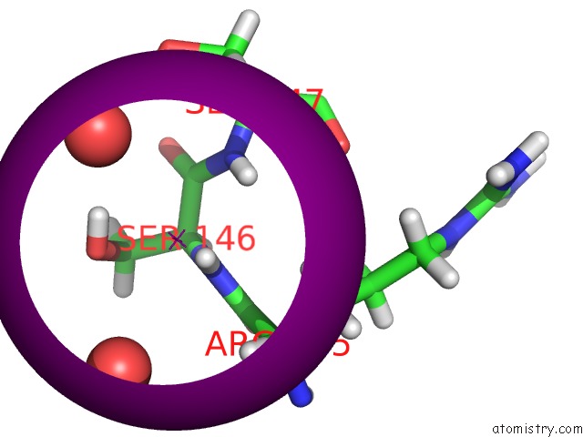
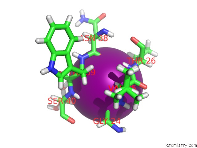
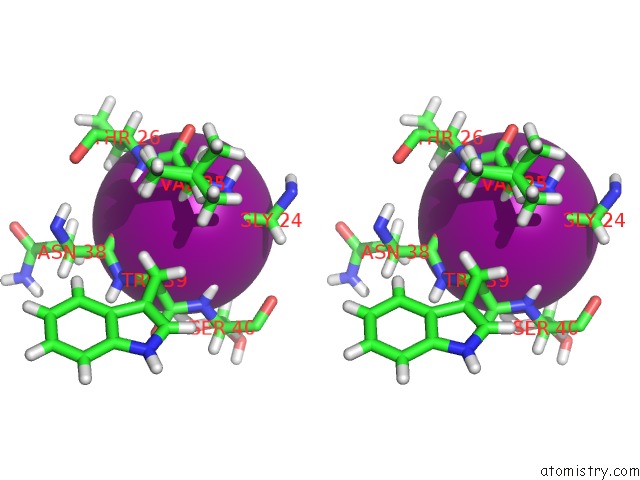
Iodine binding site 1 out of 5 in 4xq4
Go back to
Iodine binding site 1 out
of 5 in the X-Ray Structure Analysis of Xylanase - N44D
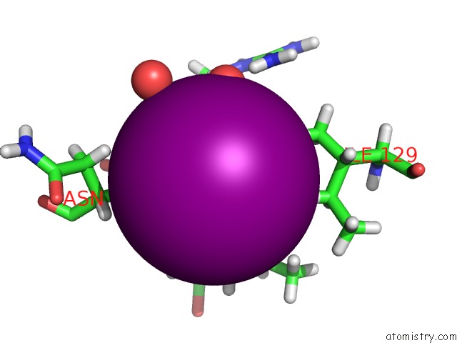
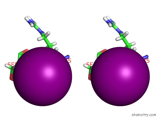
Mono view
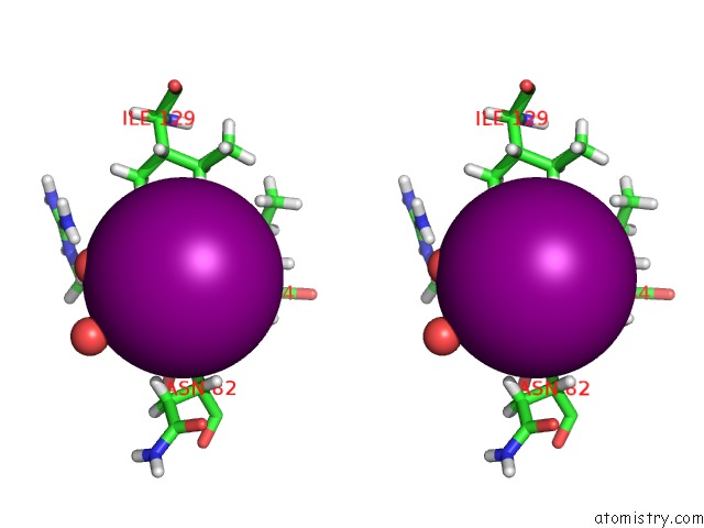
Stereo pair view
Mono view
Stereo pair view
A full contact list of Iodine with other atoms in the I binding
site number 1 of X-Ray Structure Analysis of Xylanase - N44D within 5.0Å range:
|
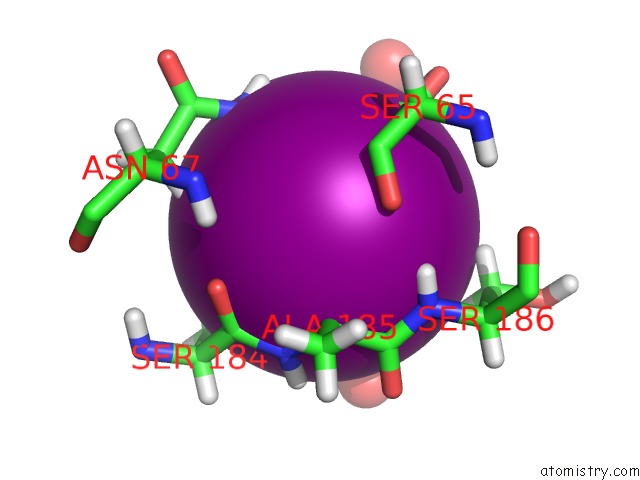
Iodine binding site 2 out of 5 in 4xq4
Go back to
Iodine binding site 2 out
of 5 in the X-Ray Structure Analysis of Xylanase - N44D
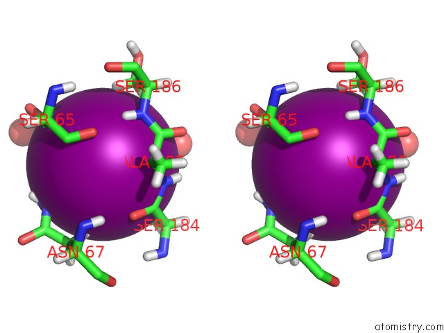
Mono view
Stereo pair view
Mono view
Stereo pair view
A full contact list of Iodine with other atoms in the I binding
site number 2 of X-Ray Structure Analysis of Xylanase - N44D within 5.0Å range:
|
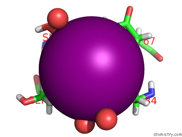
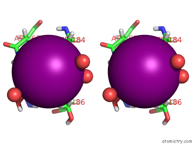
Iodine binding site 3 out of 5 in 4xq4
Go back to
Iodine binding site 3 out
of 5 in the X-Ray Structure Analysis of Xylanase - N44D
Mono view
Stereo pair view
Mono view
Stereo pair view
A full contact list of Iodine with other atoms in the I binding
site number 3 of X-Ray Structure Analysis of Xylanase - N44D within 5.0Å range:
|
Iodine binding site 4 out of 5 in 4xq4
Go back to
Iodine binding site 4 out
of 5 in the X-Ray Structure Analysis of Xylanase - N44D
Mono view
Stereo pair view
Mono view
Stereo pair view
A full contact list of Iodine with other atoms in the I binding
site number 4 of X-Ray Structure Analysis of Xylanase - N44D within 5.0Å range:
|
Iodine binding site 5 out of 5 in 4xq4
Go back to
Iodine binding site 5 out
of 5 in the X-Ray Structure Analysis of Xylanase - N44D
Mono view
Stereo pair view
Mono view
Stereo pair view
A full contact list of Iodine with other atoms in the I binding
site number 5 of X-Ray Structure Analysis of Xylanase - N44D within 5.0Å range:
|
Reference:
Q.Wan,
J.M.Parks,
B.L.Hanson,
S.Z.Fisher,
A.Ostermann,
T.E.Schrader,
D.E.Graham,
L.Coates,
P.Langan,
A.Kovalevsky.
Direct Determination of Protonation States and Visualization of Hydrogen Bonding in A Glycoside Hydrolase with Neutron Crystallography. Proc.Natl.Acad.Sci.Usa V. 112 12384 2015.
ISSN: ESSN 1091-6490
PubMed: 26392527
DOI: 10.1073/PNAS.1504986112
Page generated: Sun Aug 11 20:21:40 2024
ISSN: ESSN 1091-6490
PubMed: 26392527
DOI: 10.1073/PNAS.1504986112
Last articles
Zn in 9J0NZn in 9J0O
Zn in 9J0P
Zn in 9FJX
Zn in 9EKB
Zn in 9C0F
Zn in 9CAH
Zn in 9CH0
Zn in 9CH3
Zn in 9CH1