Iodine »
PDB 4xnw-5ax3 »
4zij »
Iodine in PDB 4zij: Crystal Structure of E.Coli Dsba in Complex with 2-(4- Iodophenylsulfonamido) Benzoic Acid
Protein crystallography data
The structure of Crystal Structure of E.Coli Dsba in Complex with 2-(4- Iodophenylsulfonamido) Benzoic Acid, PDB code: 4zij
was solved by
M.Vazirani,
O.V.Ilyichova,
M.J.Scanlon,
with X-Ray Crystallography technique. A brief refinement statistics is given in the table below:
| Resolution Low / High (Å) | 41.50 / 1.78 |
| Space group | C 1 2 1 |
| Cell size a, b, c (Å), α, β, γ (°) | 123.965, 46.982, 62.288, 90.00, 96.20, 90.00 |
| R / Rfree (%) | 17.4 / 21.7 |
Iodine Binding Sites:
The binding sites of Iodine atom in the Crystal Structure of E.Coli Dsba in Complex with 2-(4- Iodophenylsulfonamido) Benzoic Acid
(pdb code 4zij). This binding sites where shown within
5.0 Angstroms radius around Iodine atom.
In total only one binding site of Iodine was determined in the Crystal Structure of E.Coli Dsba in Complex with 2-(4- Iodophenylsulfonamido) Benzoic Acid, PDB code: 4zij:
In total only one binding site of Iodine was determined in the Crystal Structure of E.Coli Dsba in Complex with 2-(4- Iodophenylsulfonamido) Benzoic Acid, PDB code: 4zij:
Iodine binding site 1 out of 1 in 4zij
Go back to
Iodine binding site 1 out
of 1 in the Crystal Structure of E.Coli Dsba in Complex with 2-(4- Iodophenylsulfonamido) Benzoic Acid
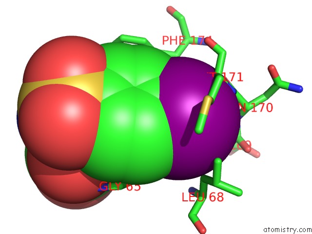
Mono view
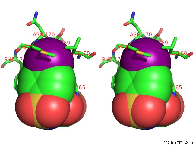
Stereo pair view
Mono view
Stereo pair view
A full contact list of Iodine with other atoms in the I binding
site number 1 of Crystal Structure of E.Coli Dsba in Complex with 2-(4- Iodophenylsulfonamido) Benzoic Acid within 5.0Å range:
|
Reference:
B.Mohanty,
M.L.Williams,
B.C.Doak,
M.Vazirani,
O.Ilyichova,
G.Wang,
W.Bermel,
J.S.Simpson,
D.K.Chalmers,
G.F.King,
M.Mobli,
M.J.Scanlon.
Determination of Ligand Binding Modes in Weak Protein-Ligand Complexes Using Sparse uc(Nmr) Data. J.Biomol.uc(Nmr) V. 66 195 2016.
ISSN: ISSN 0925-2738
PubMed: 27778134
DOI: 10.1007/S10858-016-0067-4
Page generated: Fri Aug 8 19:16:14 2025
ISSN: ISSN 0925-2738
PubMed: 27778134
DOI: 10.1007/S10858-016-0067-4
Last articles
I in 6XNFI in 6XLO
I in 6WXM
I in 6X42
I in 6X2D
I in 6WYQ
I in 6WOK
I in 6WNY
I in 6W9D
I in 6WC8