Iodine »
PDB 4xnw-5ax3 »
5aoj »
Iodine in PDB 5aoj: Structure of the P53 Cancer Mutant Y220C in Complex with 2-Hydroxy-3, 5-Diiodo-4-(1H-Pyrrol-1-Yl)Benzoic Acid
Protein crystallography data
The structure of Structure of the P53 Cancer Mutant Y220C in Complex with 2-Hydroxy-3, 5-Diiodo-4-(1H-Pyrrol-1-Yl)Benzoic Acid, PDB code: 5aoj
was solved by
A.C.Joerger,
M.G.Baud,
M.R.Bauer,
A.R.Fersht,
with X-Ray Crystallography technique. A brief refinement statistics is given in the table below:
| Resolution Low / High (Å) | 29.47 / 1.47 |
| Space group | P 21 21 21 |
| Cell size a, b, c (Å), α, β, γ (°) | 65.216, 71.108, 105.353, 90.00, 90.00, 90.00 |
| R / Rfree (%) | 14.9 / 17.8 |
Other elements in 5aoj:
The structure of Structure of the P53 Cancer Mutant Y220C in Complex with 2-Hydroxy-3, 5-Diiodo-4-(1H-Pyrrol-1-Yl)Benzoic Acid also contains other interesting chemical elements:
| Zinc | (Zn) | 2 atoms |
Iodine Binding Sites:
The binding sites of Iodine atom in the Structure of the P53 Cancer Mutant Y220C in Complex with 2-Hydroxy-3, 5-Diiodo-4-(1H-Pyrrol-1-Yl)Benzoic Acid
(pdb code 5aoj). This binding sites where shown within
5.0 Angstroms radius around Iodine atom.
In total 4 binding sites of Iodine where determined in the Structure of the P53 Cancer Mutant Y220C in Complex with 2-Hydroxy-3, 5-Diiodo-4-(1H-Pyrrol-1-Yl)Benzoic Acid, PDB code: 5aoj:
Jump to Iodine binding site number: 1; 2; 3; 4;
In total 4 binding sites of Iodine where determined in the Structure of the P53 Cancer Mutant Y220C in Complex with 2-Hydroxy-3, 5-Diiodo-4-(1H-Pyrrol-1-Yl)Benzoic Acid, PDB code: 5aoj:
Jump to Iodine binding site number: 1; 2; 3; 4;
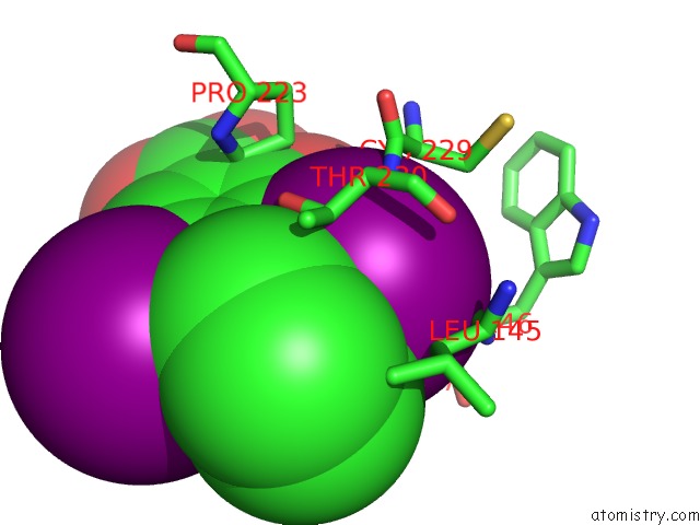
Iodine binding site 1 out of 4 in 5aoj
Go back to
Iodine binding site 1 out
of 4 in the Structure of the P53 Cancer Mutant Y220C in Complex with 2-Hydroxy-3, 5-Diiodo-4-(1H-Pyrrol-1-Yl)Benzoic Acid
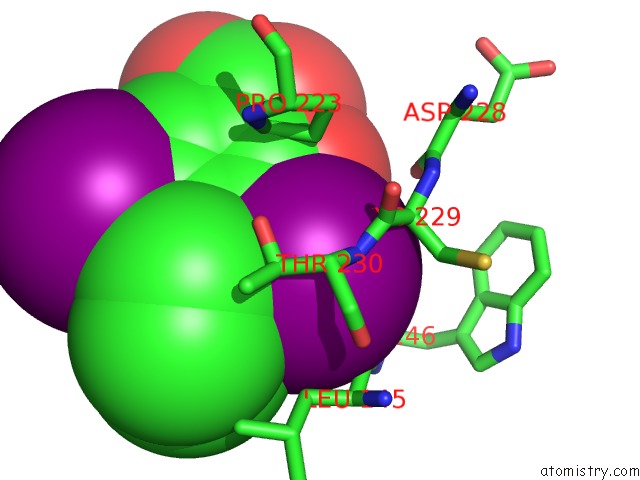
Mono view
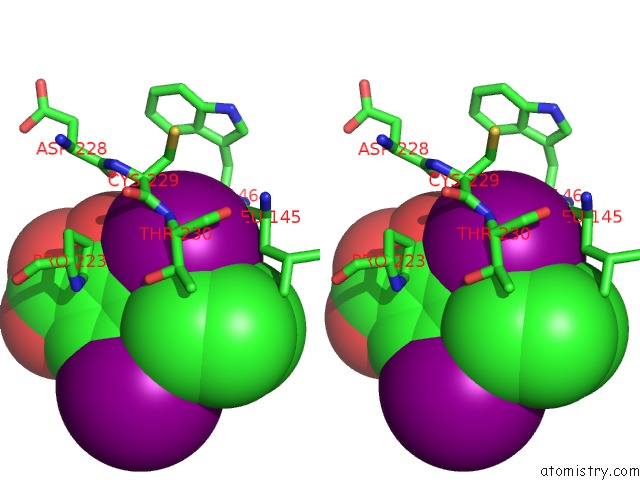
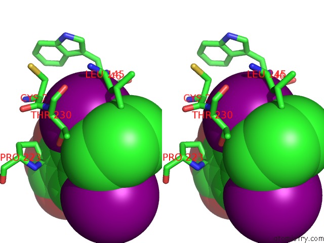
Stereo pair view
Mono view
Stereo pair view
A full contact list of Iodine with other atoms in the I binding
site number 1 of Structure of the P53 Cancer Mutant Y220C in Complex with 2-Hydroxy-3, 5-Diiodo-4-(1H-Pyrrol-1-Yl)Benzoic Acid within 5.0Å range:
|
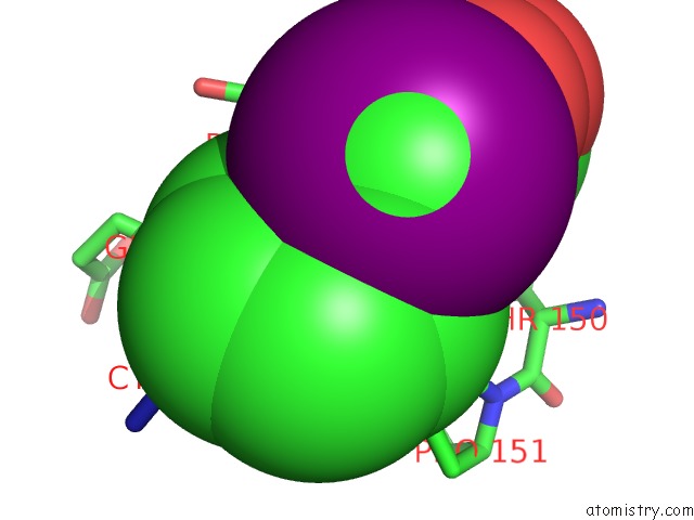
Iodine binding site 2 out of 4 in 5aoj
Go back to
Iodine binding site 2 out
of 4 in the Structure of the P53 Cancer Mutant Y220C in Complex with 2-Hydroxy-3, 5-Diiodo-4-(1H-Pyrrol-1-Yl)Benzoic Acid
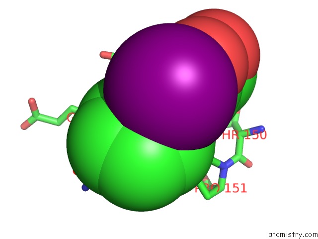
Mono view
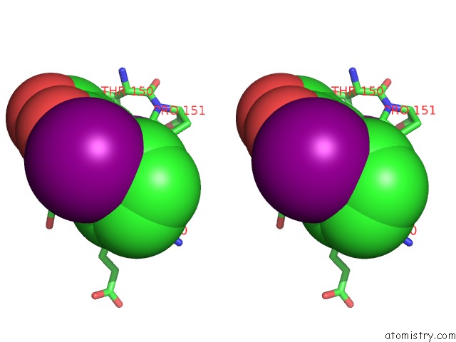
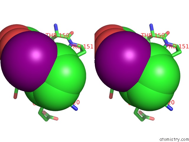
Stereo pair view
Mono view
Stereo pair view
A full contact list of Iodine with other atoms in the I binding
site number 2 of Structure of the P53 Cancer Mutant Y220C in Complex with 2-Hydroxy-3, 5-Diiodo-4-(1H-Pyrrol-1-Yl)Benzoic Acid within 5.0Å range:
|
Iodine binding site 3 out of 4 in 5aoj
Go back to
Iodine binding site 3 out
of 4 in the Structure of the P53 Cancer Mutant Y220C in Complex with 2-Hydroxy-3, 5-Diiodo-4-(1H-Pyrrol-1-Yl)Benzoic Acid
Mono view
Stereo pair view
Mono view
Stereo pair view
A full contact list of Iodine with other atoms in the I binding
site number 3 of Structure of the P53 Cancer Mutant Y220C in Complex with 2-Hydroxy-3, 5-Diiodo-4-(1H-Pyrrol-1-Yl)Benzoic Acid within 5.0Å range:
|
Iodine binding site 4 out of 4 in 5aoj
Go back to
Iodine binding site 4 out
of 4 in the Structure of the P53 Cancer Mutant Y220C in Complex with 2-Hydroxy-3, 5-Diiodo-4-(1H-Pyrrol-1-Yl)Benzoic Acid
Mono view
Stereo pair view
Mono view
Stereo pair view
A full contact list of Iodine with other atoms in the I binding
site number 4 of Structure of the P53 Cancer Mutant Y220C in Complex with 2-Hydroxy-3, 5-Diiodo-4-(1H-Pyrrol-1-Yl)Benzoic Acid within 5.0Å range:
|
Reference:
A.C.Joerger,
M.R.Bauer,
R.Wilcken,
M.G.J.Baud,
H.Harbrecht,
T.E.Exner,
F.M.Boeckler,
J.Spencer,
A.R.Fersht.
Exploiting Transient Protein States For the Design of Small-Molecule Stabilizers of Mutant P53. Structure V. 23 2246 2015.
ISSN: ISSN 0969-2126
PubMed: 26636255
DOI: 10.1016/J.STR.2015.10.016
Page generated: Sun Aug 11 20:33:16 2024
ISSN: ISSN 0969-2126
PubMed: 26636255
DOI: 10.1016/J.STR.2015.10.016
Last articles
F in 7M0MF in 7M0V
F in 7M0W
F in 7M0U
F in 7M0N
F in 7M0T
F in 7M00
F in 7M01
F in 7M03
F in 7M02