Iodine »
PDB 5b34-5ems »
5cqf »
Iodine in PDB 5cqf: Crystal Structure of L-Lysine 6-Monooxygenase From Pseudomonas Syringae
Protein crystallography data
The structure of Crystal Structure of L-Lysine 6-Monooxygenase From Pseudomonas Syringae, PDB code: 5cqf
was solved by
K.Michalska,
L.Bigelow,
R.Jedrzejczak,
R.S.Weerth,
H.Cao,
R.Yennamalli,
G.N.Phillips Jr.,
M.G.Thomas,
A.Joachimiak,
Midwest Center Forstructural Genomics (Mcsg),
Enzyme Discovery For Natural Productbiosynthesis (Natpro),
with X-Ray Crystallography technique. A brief refinement statistics is given in the table below:
| Resolution Low / High (Å) | 29.10 / 2.28 |
| Space group | I 2 2 2 |
| Cell size a, b, c (Å), α, β, γ (°) | 77.218, 85.495, 139.016, 90.00, 90.00, 90.00 |
| R / Rfree (%) | 18.4 / 23.3 |
Iodine Binding Sites:
Pages:
>>> Page 1 <<< Page 2, Binding sites: 11 - 12;Binding sites:
The binding sites of Iodine atom in the Crystal Structure of L-Lysine 6-Monooxygenase From Pseudomonas Syringae (pdb code 5cqf). This binding sites where shown within 5.0 Angstroms radius around Iodine atom.In total 12 binding sites of Iodine where determined in the Crystal Structure of L-Lysine 6-Monooxygenase From Pseudomonas Syringae, PDB code: 5cqf:
Jump to Iodine binding site number: 1; 2; 3; 4; 5; 6; 7; 8; 9; 10;
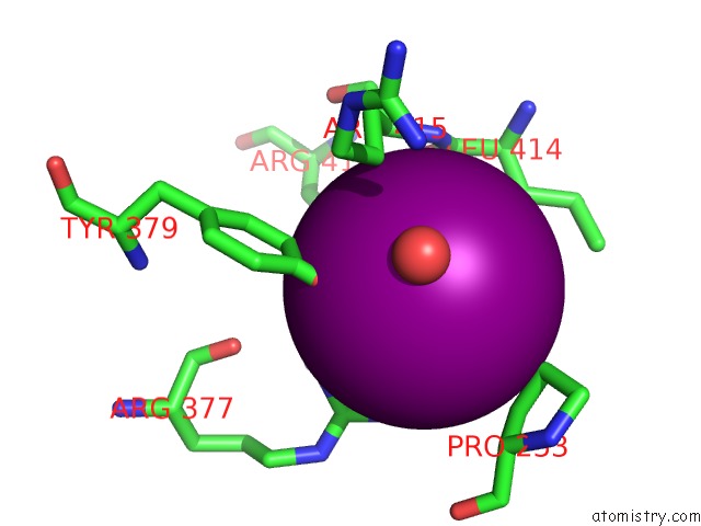
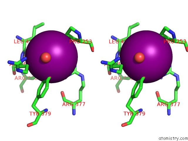
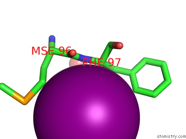
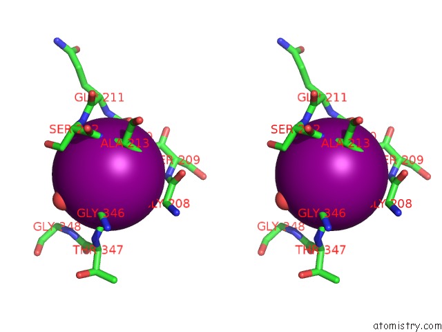
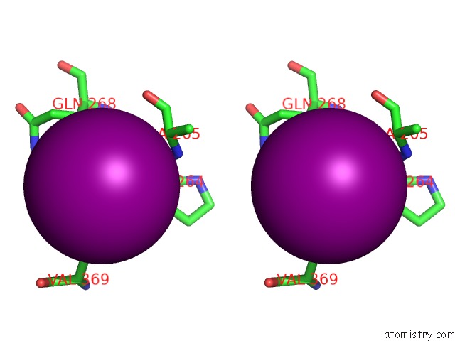
Iodine binding site 1 out of 12 in 5cqf
Go back to
Iodine binding site 1 out
of 12 in the Crystal Structure of L-Lysine 6-Monooxygenase From Pseudomonas Syringae
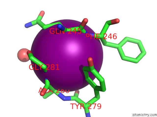
Mono view
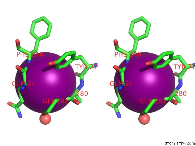
Stereo pair view
Mono view
Stereo pair view
A full contact list of Iodine with other atoms in the I binding
site number 1 of Crystal Structure of L-Lysine 6-Monooxygenase From Pseudomonas Syringae within 5.0Å range:
|
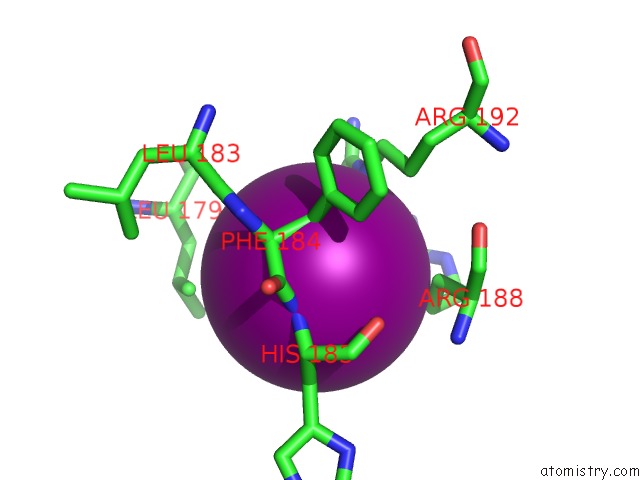
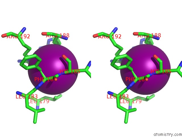
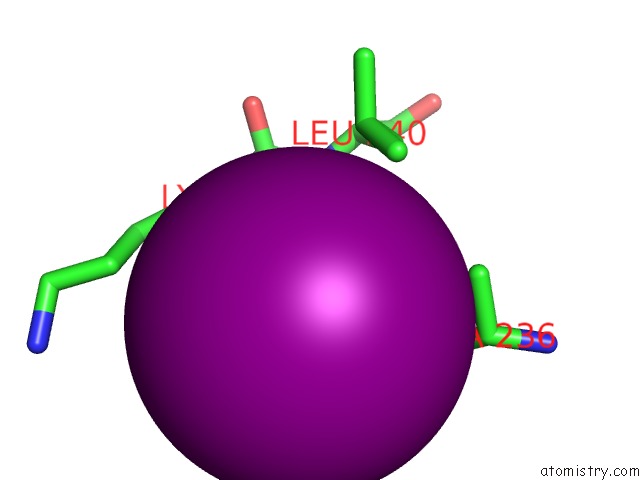
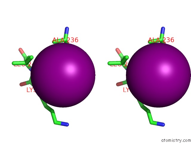
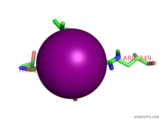
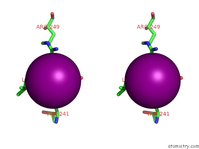
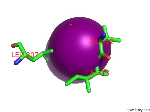
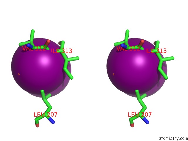
Iodine binding site 2 out of 12 in 5cqf
Go back to
Iodine binding site 2 out
of 12 in the Crystal Structure of L-Lysine 6-Monooxygenase From Pseudomonas Syringae
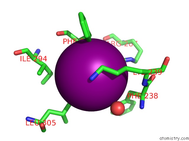
Mono view
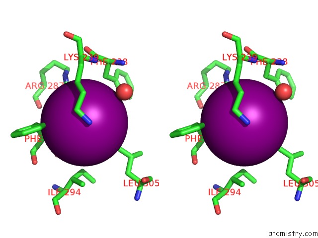
Stereo pair view
Mono view
Stereo pair view
A full contact list of Iodine with other atoms in the I binding
site number 2 of Crystal Structure of L-Lysine 6-Monooxygenase From Pseudomonas Syringae within 5.0Å range:
|
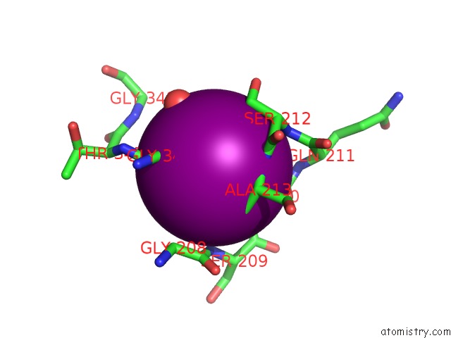
Iodine binding site 3 out of 12 in 5cqf
Go back to
Iodine binding site 3 out
of 12 in the Crystal Structure of L-Lysine 6-Monooxygenase From Pseudomonas Syringae
Mono view
Stereo pair view
Mono view
Stereo pair view
A full contact list of Iodine with other atoms in the I binding
site number 3 of Crystal Structure of L-Lysine 6-Monooxygenase From Pseudomonas Syringae within 5.0Å range:
|
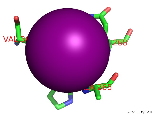
Iodine binding site 4 out of 12 in 5cqf
Go back to
Iodine binding site 4 out
of 12 in the Crystal Structure of L-Lysine 6-Monooxygenase From Pseudomonas Syringae
Mono view
Stereo pair view
Mono view
Stereo pair view
A full contact list of Iodine with other atoms in the I binding
site number 4 of Crystal Structure of L-Lysine 6-Monooxygenase From Pseudomonas Syringae within 5.0Å range:
|
Iodine binding site 5 out of 12 in 5cqf
Go back to
Iodine binding site 5 out
of 12 in the Crystal Structure of L-Lysine 6-Monooxygenase From Pseudomonas Syringae
Mono view

Stereo pair view
Mono view
Stereo pair view
A full contact list of Iodine with other atoms in the I binding
site number 5 of Crystal Structure of L-Lysine 6-Monooxygenase From Pseudomonas Syringae within 5.0Å range:
|
Iodine binding site 6 out of 12 in 5cqf
Go back to
Iodine binding site 6 out
of 12 in the Crystal Structure of L-Lysine 6-Monooxygenase From Pseudomonas Syringae
Mono view
Stereo pair view
Mono view
Stereo pair view
A full contact list of Iodine with other atoms in the I binding
site number 6 of Crystal Structure of L-Lysine 6-Monooxygenase From Pseudomonas Syringae within 5.0Å range:
|
Iodine binding site 7 out of 12 in 5cqf
Go back to
Iodine binding site 7 out
of 12 in the Crystal Structure of L-Lysine 6-Monooxygenase From Pseudomonas Syringae
Mono view
Stereo pair view
Mono view
Stereo pair view
A full contact list of Iodine with other atoms in the I binding
site number 7 of Crystal Structure of L-Lysine 6-Monooxygenase From Pseudomonas Syringae within 5.0Å range:
|
Iodine binding site 8 out of 12 in 5cqf
Go back to
Iodine binding site 8 out
of 12 in the Crystal Structure of L-Lysine 6-Monooxygenase From Pseudomonas Syringae
Mono view
Stereo pair view
Mono view
Stereo pair view
A full contact list of Iodine with other atoms in the I binding
site number 8 of Crystal Structure of L-Lysine 6-Monooxygenase From Pseudomonas Syringae within 5.0Å range:
|
Iodine binding site 9 out of 12 in 5cqf
Go back to
Iodine binding site 9 out
of 12 in the Crystal Structure of L-Lysine 6-Monooxygenase From Pseudomonas Syringae
Mono view
Stereo pair view
Mono view
Stereo pair view
A full contact list of Iodine with other atoms in the I binding
site number 9 of Crystal Structure of L-Lysine 6-Monooxygenase From Pseudomonas Syringae within 5.0Å range:
|
Iodine binding site 10 out of 12 in 5cqf
Go back to
Iodine binding site 10 out
of 12 in the Crystal Structure of L-Lysine 6-Monooxygenase From Pseudomonas Syringae
Mono view
Stereo pair view
Mono view
Stereo pair view
A full contact list of Iodine with other atoms in the I binding
site number 10 of Crystal Structure of L-Lysine 6-Monooxygenase From Pseudomonas Syringae within 5.0Å range:
|
Reference:
K.Michalska,
L.Bigelow,
R.Jedrzejczak,
R.S.Weerth,
H.Cao,
R.Yennamalli,
G.N.Phillips Jr.,
M.G.Thomas,
A.Joachimiak,
Midwest Center For Structural Genomics (Mcsg),
Enzyme Discovery For Natural Product Biosynthesis (Natpro).
Crystal Structure of L-Lysine 6-Monooxygenase From Pseudomonas Syringae To Be Published.
Page generated: Sun Aug 11 20:39:14 2024
Last articles
Zn in 9J0NZn in 9J0O
Zn in 9J0P
Zn in 9FJX
Zn in 9EKB
Zn in 9C0F
Zn in 9CAH
Zn in 9CH0
Zn in 9CH3
Zn in 9CH1