Iodine »
PDB 5b34-5ems »
5e5t »
Iodine in PDB 5e5t: Quasi-Racemic Snakin-1 in P1 After Radiation Damage
Protein crystallography data
The structure of Quasi-Racemic Snakin-1 in P1 After Radiation Damage, PDB code: 5e5t
was solved by
H.Yeung,
C.J.Squire,
Y.Yosaatmadja,
S.Panjikar,
E.N.Baker,
P.W.R.Harris,
M.A.Brimble,
with X-Ray Crystallography technique. A brief refinement statistics is given in the table below:
| Resolution Low / High (Å) | 36.49 / 1.57 |
| Space group | P 1 |
| Cell size a, b, c (Å), α, β, γ (°) | 31.256, 37.442, 50.391, 92.96, 90.55, 102.58 |
| R / Rfree (%) | 20.5 / 24.3 |
Iodine Binding Sites:
The binding sites of Iodine atom in the Quasi-Racemic Snakin-1 in P1 After Radiation Damage
(pdb code 5e5t). This binding sites where shown within
5.0 Angstroms radius around Iodine atom.
In total 2 binding sites of Iodine where determined in the Quasi-Racemic Snakin-1 in P1 After Radiation Damage, PDB code: 5e5t:
Jump to Iodine binding site number: 1; 2;
In total 2 binding sites of Iodine where determined in the Quasi-Racemic Snakin-1 in P1 After Radiation Damage, PDB code: 5e5t:
Jump to Iodine binding site number: 1; 2;
Iodine binding site 1 out of 2 in 5e5t
Go back to
Iodine binding site 1 out
of 2 in the Quasi-Racemic Snakin-1 in P1 After Radiation Damage
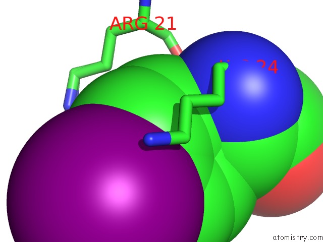
Mono view
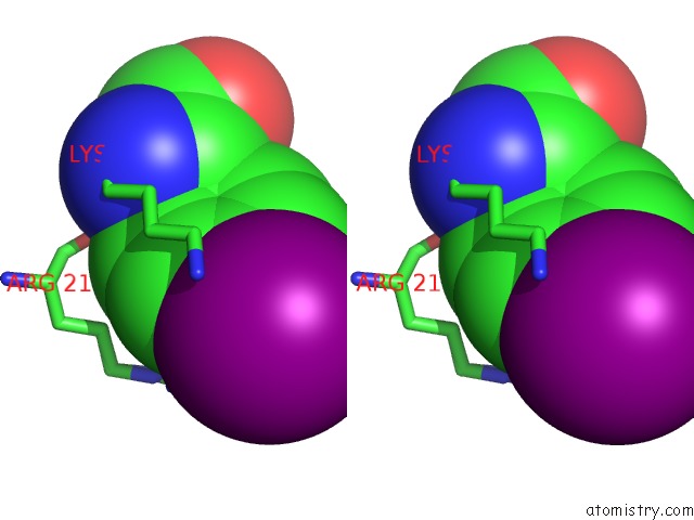
Stereo pair view
Mono view
Stereo pair view
A full contact list of Iodine with other atoms in the I binding
site number 1 of Quasi-Racemic Snakin-1 in P1 After Radiation Damage within 5.0Å range:
|
Iodine binding site 2 out of 2 in 5e5t
Go back to
Iodine binding site 2 out
of 2 in the Quasi-Racemic Snakin-1 in P1 After Radiation Damage
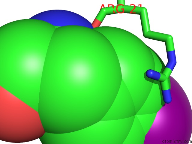
Mono view
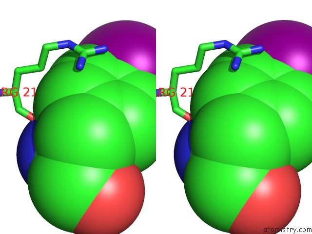
Stereo pair view
Mono view
Stereo pair view
A full contact list of Iodine with other atoms in the I binding
site number 2 of Quasi-Racemic Snakin-1 in P1 After Radiation Damage within 5.0Å range:
|
Reference:
H.Yeung,
C.J.Squire,
Y.Yosaatmadja,
S.Panjikar,
G.Lopez,
A.Molina,
E.N.Baker,
P.W.Harris,
M.A.Brimble.
Radiation Damage and Racemic Protein Crystallography Reveal the Unique Structure of the Gasa/Snakin Protein Superfamily. Angew.Chem.Int.Ed.Engl. V. 55 7930 2016.
ISSN: ESSN 1521-3773
PubMed: 27145301
DOI: 10.1002/ANIE.201602719
Page generated: Sun Aug 11 20:50:59 2024
ISSN: ESSN 1521-3773
PubMed: 27145301
DOI: 10.1002/ANIE.201602719
Last articles
Zn in 9J0NZn in 9J0O
Zn in 9J0P
Zn in 9FJX
Zn in 9EKB
Zn in 9C0F
Zn in 9CAH
Zn in 9CH0
Zn in 9CH3
Zn in 9CH1