Iodine »
PDB 5b34-5ems »
5ei1 »
Iodine in PDB 5ei1: Crystal Structure of the Er-Alpha Ligand-Binding Domain (Y537S) in Complex with the Imidazopyridine Derivative 2-(4-Hydroxyphenyl)-3- Iodanyl-Imidazo[1,2-A]Pyridin-6-Ol
Protein crystallography data
The structure of Crystal Structure of the Er-Alpha Ligand-Binding Domain (Y537S) in Complex with the Imidazopyridine Derivative 2-(4-Hydroxyphenyl)-3- Iodanyl-Imidazo[1,2-A]Pyridin-6-Ol, PDB code: 5ei1
was solved by
J.C.Nwachukwu,
S.Srinivasan,
Y.Zheng,
S.Wang,
J.Min,
C.Dong,
Z.Liao,
V.Cavett,
J.Nowak,
R.Houtman,
K.E.Carlson,
J.S.Josan,
O.Elemento,
J.A.Katzenellenbogen,
H.B.Zhou,
K.W.Nettles,
with X-Ray Crystallography technique. A brief refinement statistics is given in the table below:
| Resolution Low / High (Å) | 46.43 / 2.40 |
| Space group | P 1 21 1 |
| Cell size a, b, c (Å), α, β, γ (°) | 54.737, 82.484, 58.308, 90.00, 110.80, 90.00 |
| R / Rfree (%) | 17.8 / 22.8 |
Iodine Binding Sites:
The binding sites of Iodine atom in the Crystal Structure of the Er-Alpha Ligand-Binding Domain (Y537S) in Complex with the Imidazopyridine Derivative 2-(4-Hydroxyphenyl)-3- Iodanyl-Imidazo[1,2-A]Pyridin-6-Ol
(pdb code 5ei1). This binding sites where shown within
5.0 Angstroms radius around Iodine atom.
In total 2 binding sites of Iodine where determined in the Crystal Structure of the Er-Alpha Ligand-Binding Domain (Y537S) in Complex with the Imidazopyridine Derivative 2-(4-Hydroxyphenyl)-3- Iodanyl-Imidazo[1,2-A]Pyridin-6-Ol, PDB code: 5ei1:
Jump to Iodine binding site number: 1; 2;
In total 2 binding sites of Iodine where determined in the Crystal Structure of the Er-Alpha Ligand-Binding Domain (Y537S) in Complex with the Imidazopyridine Derivative 2-(4-Hydroxyphenyl)-3- Iodanyl-Imidazo[1,2-A]Pyridin-6-Ol, PDB code: 5ei1:
Jump to Iodine binding site number: 1; 2;
Iodine binding site 1 out of 2 in 5ei1
Go back to
Iodine binding site 1 out
of 2 in the Crystal Structure of the Er-Alpha Ligand-Binding Domain (Y537S) in Complex with the Imidazopyridine Derivative 2-(4-Hydroxyphenyl)-3- Iodanyl-Imidazo[1,2-A]Pyridin-6-Ol
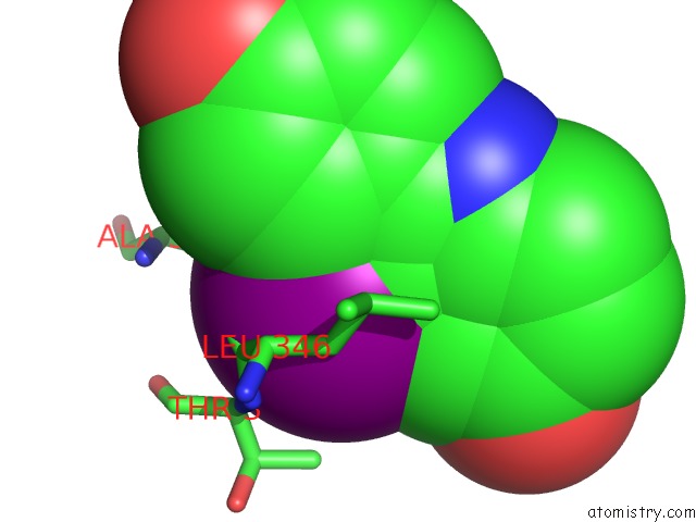
Mono view

Stereo pair view
Mono view
Stereo pair view
A full contact list of Iodine with other atoms in the I binding
site number 1 of Crystal Structure of the Er-Alpha Ligand-Binding Domain (Y537S) in Complex with the Imidazopyridine Derivative 2-(4-Hydroxyphenyl)-3- Iodanyl-Imidazo[1,2-A]Pyridin-6-Ol within 5.0Å range:
|
Iodine binding site 2 out of 2 in 5ei1
Go back to
Iodine binding site 2 out
of 2 in the Crystal Structure of the Er-Alpha Ligand-Binding Domain (Y537S) in Complex with the Imidazopyridine Derivative 2-(4-Hydroxyphenyl)-3- Iodanyl-Imidazo[1,2-A]Pyridin-6-Ol
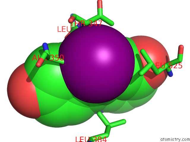
Mono view
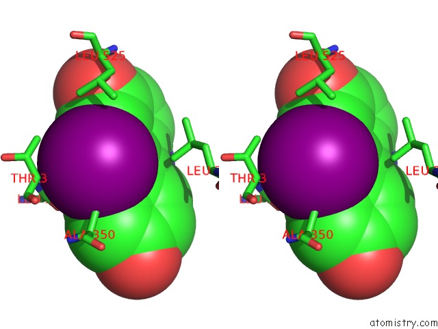
Stereo pair view
Mono view
Stereo pair view
A full contact list of Iodine with other atoms in the I binding
site number 2 of Crystal Structure of the Er-Alpha Ligand-Binding Domain (Y537S) in Complex with the Imidazopyridine Derivative 2-(4-Hydroxyphenyl)-3- Iodanyl-Imidazo[1,2-A]Pyridin-6-Ol within 5.0Å range:
|
Reference:
J.C.Nwachukwu,
S.Srinivasan,
Y.Zheng,
S.Wang,
J.Min,
C.Dong,
Z.Liao,
J.Nowak,
N.J.Wright,
R.Houtman,
K.E.Carlson,
J.S.Josan,
O.Elemento,
J.A.Katzenellenbogen,
H.B.Zhou,
K.W.Nettles.
Predictive Features of Ligand-Specific Signaling Through the Estrogen Receptor. Mol.Syst.Biol. V. 12 864 2016.
ISSN: ESSN 1744-4292
PubMed: 27107013
DOI: 10.15252/MSB.20156701
Page generated: Sun Aug 11 20:53:43 2024
ISSN: ESSN 1744-4292
PubMed: 27107013
DOI: 10.15252/MSB.20156701
Last articles
Zn in 9MJ5Zn in 9HNW
Zn in 9G0L
Zn in 9FNE
Zn in 9DZN
Zn in 9E0I
Zn in 9D32
Zn in 9DAK
Zn in 8ZXC
Zn in 8ZUF