Iodine in PDB 5g5g: Escherichia Coli Periplasmic Aldehyde Oxidase
Protein crystallography data
The structure of Escherichia Coli Periplasmic Aldehyde Oxidase, PDB code: 5g5g
was solved by
M.A.S.Correia,
A.R.Otrelo-Cardoso,
M.J.Romao,
T.Santos-Silva,
with X-Ray Crystallography technique. A brief refinement statistics is given in the table below:
Resolution Low / High (Å)
|
48.32 /
1.70
|
Space group
|
C 1 2 1
|
Cell size a, b, c (Å), α, β, γ (°)
|
109.681,
78.342,
151.909,
90.00,
99.69,
90.00
|
R / Rfree (%)
|
13.8 /
16.7
|
Other elements in 5g5g:
The structure of Escherichia Coli Periplasmic Aldehyde Oxidase also contains other interesting chemical elements:
Iodine Binding Sites:
The binding sites of Iodine atom in the Escherichia Coli Periplasmic Aldehyde Oxidase
(pdb code 5g5g). This binding sites where shown within
5.0 Angstroms radius around Iodine atom.
In total 7 binding sites of Iodine where determined in the
Escherichia Coli Periplasmic Aldehyde Oxidase, PDB code: 5g5g:
Jump to Iodine binding site number:
1;
2;
3;
4;
5;
6;
7;
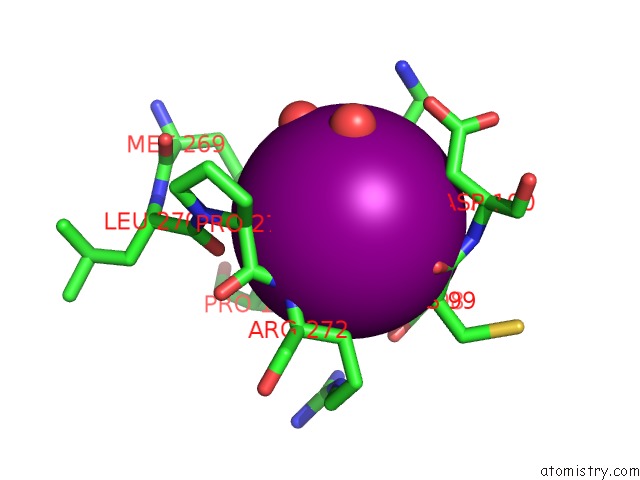
Iodine binding site 1 out
of 7 in 5g5g
Go back to
Iodine Binding Sites List in 5g5g
Iodine binding site 1 out
of 7 in the Escherichia Coli Periplasmic Aldehyde Oxidase
Mono view
Stereo pair view
|
A full contact list of Iodine with other atoms in the I binding
site number 1 of Escherichia Coli Periplasmic Aldehyde Oxidase within 5.0Å range:
probe
|
atom
|
residue
|
distance (Å)
|
B
|
Occ
|
A:I1227
b:13.1
occ:0.68
|
O
|
A:HOH2067
|
3.1
|
31.3
|
1.0
|
N
|
C:ARG272
|
3.5
|
12.7
|
1.0
|
O
|
A:HOH2048
|
3.7
|
22.7
|
1.0
|
C
|
A:CYS99
|
3.7
|
11.8
|
1.0
|
O
|
A:HOH2072
|
3.8
|
23.9
|
1.0
|
O
|
A:CYS99
|
3.8
|
12.7
|
1.0
|
CG
|
C:PRO192
|
3.9
|
11.0
|
1.0
|
CD
|
A:LYS97
|
3.9
|
14.2
|
1.0
|
CG
|
A:LYS97
|
3.9
|
13.1
|
1.0
|
CA
|
A:CYS99
|
4.0
|
11.3
|
1.0
|
N
|
A:CYS99
|
4.1
|
11.1
|
1.0
|
CA
|
C:PRO271
|
4.1
|
14.4
|
1.0
|
CB
|
A:ASP100
|
4.1
|
11.9
|
1.0
|
SD
|
C:MET269
|
4.1
|
23.4
|
1.0
|
CB
|
C:ARG272
|
4.2
|
12.2
|
1.0
|
N
|
A:ASP100
|
4.2
|
11.2
|
1.0
|
C
|
C:PRO271
|
4.3
|
13.1
|
1.0
|
CA
|
C:ARG272
|
4.4
|
12.0
|
1.0
|
O
|
C:LEU270
|
4.5
|
15.6
|
1.0
|
C
|
A:GLY98
|
4.5
|
11.2
|
1.0
|
CB
|
C:PRO192
|
4.5
|
11.2
|
1.0
|
O
|
A:GLY98
|
4.7
|
12.1
|
1.0
|
O
|
A:LYS97
|
4.8
|
11.7
|
1.0
|
CA
|
A:ASP100
|
4.8
|
11.4
|
1.0
|
CG
|
A:ASP100
|
4.9
|
12.5
|
1.0
|
OD2
|
A:ASP100
|
4.9
|
13.0
|
1.0
|
CB
|
C:PRO271
|
5.0
|
15.9
|
1.0
|
CG
|
C:MET269
|
5.0
|
18.9
|
1.0
|
|
Iodine binding site 2 out
of 7 in 5g5g
Go back to
Iodine Binding Sites List in 5g5g
Iodine binding site 2 out
of 7 in the Escherichia Coli Periplasmic Aldehyde Oxidase
Mono view
Stereo pair view
|
A full contact list of Iodine with other atoms in the I binding
site number 2 of Escherichia Coli Periplasmic Aldehyde Oxidase within 5.0Å range:
probe
|
atom
|
residue
|
distance (Å)
|
B
|
Occ
|
A:I3000
b:30.5
occ:0.70
|
O
|
A:HOH2173
|
3.7
|
40.0
|
1.0
|
CE1
|
A:HIS144
|
3.8
|
18.9
|
1.0
|
CE
|
A:MET146
|
4.2
|
16.0
|
1.0
|
CB
|
A:ALA223
|
4.3
|
23.4
|
1.0
|
CD1
|
A:LEU173
|
4.5
|
20.1
|
1.0
|
ND1
|
A:HIS144
|
4.6
|
18.3
|
1.0
|
CB
|
A:MET146
|
4.6
|
15.3
|
1.0
|
N
|
A:MET146
|
4.7
|
16.0
|
1.0
|
O
|
A:ALA223
|
4.7
|
26.7
|
1.0
|
CB
|
A:PRO145
|
4.7
|
19.1
|
1.0
|
NE2
|
A:HIS144
|
4.8
|
19.2
|
1.0
|
O
|
A:HOH2130
|
4.9
|
29.1
|
1.0
|
CG2
|
A:VAL170
|
4.9
|
19.3
|
1.0
|
C
|
A:ALA223
|
5.0
|
24.6
|
1.0
|
|
Iodine binding site 3 out
of 7 in 5g5g
Go back to
Iodine Binding Sites List in 5g5g
Iodine binding site 3 out
of 7 in the Escherichia Coli Periplasmic Aldehyde Oxidase
Mono view
Stereo pair view
|
A full contact list of Iodine with other atoms in the I binding
site number 3 of Escherichia Coli Periplasmic Aldehyde Oxidase within 5.0Å range:
probe
|
atom
|
residue
|
distance (Å)
|
B
|
Occ
|
B:I1317
b:28.8
occ:0.77
|
NE
|
B:ARG255
|
3.7
|
23.0
|
1.0
|
O
|
B:HOH2211
|
3.8
|
39.2
|
1.0
|
NE2
|
B:GLN249
|
3.8
|
28.1
|
1.0
|
CA
|
B:GLY254
|
4.0
|
25.5
|
1.0
|
O
|
B:HOH2213
|
4.0
|
41.4
|
1.0
|
CG
|
B:ARG255
|
4.1
|
21.5
|
1.0
|
CG
|
B:GLN249
|
4.2
|
28.7
|
1.0
|
C
|
B:GLY254
|
4.2
|
24.7
|
1.0
|
CD
|
B:ARG255
|
4.4
|
21.5
|
1.0
|
CD1
|
B:LEU222
|
4.4
|
23.3
|
1.0
|
CG2
|
B:ILE247
|
4.5
|
22.2
|
1.0
|
NH2
|
B:ARG255
|
4.5
|
24.8
|
1.0
|
CZ
|
B:ARG255
|
4.5
|
23.3
|
1.0
|
N
|
B:ARG255
|
4.5
|
22.8
|
1.0
|
CD
|
B:GLN249
|
4.5
|
28.8
|
1.0
|
CD2
|
B:LEU222
|
4.6
|
24.7
|
1.0
|
O
|
B:GLY254
|
4.7
|
25.6
|
1.0
|
O
|
B:ILE247
|
4.7
|
22.1
|
1.0
|
N
|
B:GLY254
|
4.8
|
27.1
|
1.0
|
CB
|
B:LEU222
|
4.8
|
23.0
|
1.0
|
CG
|
B:LEU222
|
4.8
|
23.2
|
1.0
|
CB
|
B:ARG255
|
4.9
|
21.4
|
1.0
|
|
Iodine binding site 4 out
of 7 in 5g5g
Go back to
Iodine Binding Sites List in 5g5g
Iodine binding site 4 out
of 7 in the Escherichia Coli Periplasmic Aldehyde Oxidase
Mono view
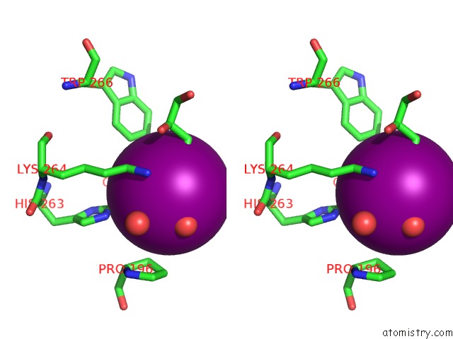
Stereo pair view
|
A full contact list of Iodine with other atoms in the I binding
site number 4 of Escherichia Coli Periplasmic Aldehyde Oxidase within 5.0Å range:
probe
|
atom
|
residue
|
distance (Å)
|
B
|
Occ
|
B:I3000
b:30.9
occ:0.76
|
O3
|
B:GOL1322
|
3.2
|
42.3
|
1.0
|
O
|
B:HOH2101
|
3.6
|
39.1
|
1.0
|
CE1
|
B:HIS263
|
3.9
|
21.0
|
1.0
|
C3
|
B:GOL1322
|
4.0
|
41.8
|
1.0
|
CA
|
B:GLY152
|
4.3
|
23.2
|
1.0
|
CZ3
|
B:TRP266
|
4.3
|
20.0
|
1.0
|
CH2
|
B:TRP266
|
4.3
|
20.8
|
1.0
|
O
|
B:HOH2171
|
4.4
|
20.7
|
1.0
|
NZ
|
B:LYS264
|
4.5
|
22.2
|
1.0
|
CE
|
B:LYS264
|
4.5
|
21.6
|
1.0
|
NE2
|
B:HIS263
|
4.5
|
21.1
|
1.0
|
CB
|
B:PRO196
|
4.6
|
27.1
|
1.0
|
O
|
B:GLY152
|
4.7
|
25.1
|
1.0
|
C2
|
B:GOL1322
|
4.9
|
41.1
|
1.0
|
C
|
B:GLY152
|
5.0
|
24.1
|
1.0
|
|
Iodine binding site 5 out
of 7 in 5g5g
Go back to
Iodine Binding Sites List in 5g5g
Iodine binding site 5 out
of 7 in the Escherichia Coli Periplasmic Aldehyde Oxidase
Mono view
Stereo pair view
|
A full contact list of Iodine with other atoms in the I binding
site number 5 of Escherichia Coli Periplasmic Aldehyde Oxidase within 5.0Å range:
probe
|
atom
|
residue
|
distance (Å)
|
B
|
Occ
|
C:I1732
b:23.6
occ:0.60
|
N
|
C:ASN47
|
3.4
|
20.0
|
1.0
|
O
|
C:HOH2059
|
3.6
|
42.0
|
1.0
|
CB
|
C:ASN47
|
3.7
|
26.3
|
1.0
|
CG
|
C:PRO265
|
3.7
|
17.8
|
1.0
|
CD
|
C:PRO265
|
3.8
|
18.5
|
1.0
|
CA
|
C:ASN47
|
3.8
|
22.7
|
1.0
|
C
|
C:ASN47
|
3.9
|
19.8
|
1.0
|
N
|
C:PRO46
|
3.9
|
18.3
|
1.0
|
C
|
C:ALA45
|
3.9
|
17.0
|
1.0
|
CA
|
C:ALA45
|
4.0
|
16.7
|
1.0
|
CD
|
C:PRO46
|
4.1
|
19.0
|
1.0
|
N
|
C:ALA48
|
4.2
|
16.6
|
1.0
|
O
|
C:HOH2060
|
4.3
|
38.1
|
1.0
|
O
|
C:ASN47
|
4.3
|
19.2
|
1.0
|
OD1
|
C:ASN47
|
4.3
|
44.4
|
1.0
|
CB
|
C:ALA45
|
4.3
|
16.6
|
1.0
|
CG
|
C:ASN47
|
4.4
|
34.4
|
1.0
|
O
|
C:ALA45
|
4.5
|
16.4
|
1.0
|
C
|
C:PRO46
|
4.5
|
19.4
|
1.0
|
CA
|
C:PRO46
|
4.8
|
19.4
|
1.0
|
CB
|
C:PRO265
|
4.8
|
16.9
|
1.0
|
CA
|
C:ALA48
|
4.9
|
15.8
|
1.0
|
|
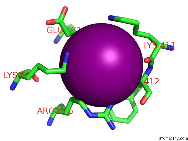
Iodine binding site 6 out
of 7 in 5g5g
Go back to
Iodine Binding Sites List in 5g5g
Iodine binding site 6 out
of 7 in the Escherichia Coli Periplasmic Aldehyde Oxidase
Mono view
Stereo pair view
|
A full contact list of Iodine with other atoms in the I binding
site number 6 of Escherichia Coli Periplasmic Aldehyde Oxidase within 5.0Å range:
probe
|
atom
|
residue
|
distance (Å)
|
B
|
Occ
|
C:I1733
b:25.8
occ:0.87
|
NH1
|
C:ARG606
|
3.7
|
21.1
|
1.0
|
NZ
|
C:LYS669
|
3.9
|
42.2
|
1.0
|
CE
|
C:LYS669
|
4.0
|
37.8
|
1.0
|
CD
|
C:ARG606
|
4.1
|
22.1
|
1.0
|
CB
|
C:LEU608
|
4.2
|
14.3
|
1.0
|
CD1
|
C:PHE412
|
4.2
|
17.8
|
1.0
|
CG
|
C:ARG606
|
4.2
|
21.2
|
1.0
|
CA
|
C:PHE412
|
4.3
|
18.0
|
1.0
|
CG
|
C:GLU671
|
4.4
|
24.3
|
1.0
|
O
|
C:LYS411
|
4.4
|
18.7
|
1.0
|
CG
|
C:LYS411
|
4.5
|
25.4
|
1.0
|
CD2
|
C:LEU608
|
4.5
|
15.3
|
1.0
|
NZ
|
C:LYS411
|
4.5
|
34.9
|
1.0
|
CB
|
C:GLU671
|
4.6
|
19.2
|
1.0
|
CG
|
C:LEU608
|
4.7
|
15.4
|
1.0
|
CD1
|
C:LEU608
|
4.7
|
15.8
|
1.0
|
CZ
|
C:ARG606
|
4.7
|
21.5
|
1.0
|
C
|
C:LYS411
|
4.8
|
19.7
|
1.0
|
N
|
C:PHE412
|
4.8
|
18.1
|
1.0
|
CE
|
C:LYS411
|
4.8
|
33.7
|
1.0
|
CB
|
C:PHE412
|
4.9
|
17.8
|
1.0
|
NE
|
C:ARG606
|
4.9
|
22.1
|
1.0
|
CG
|
C:PHE412
|
4.9
|
17.0
|
1.0
|
|
Iodine binding site 7 out
of 7 in 5g5g
Go back to
Iodine Binding Sites List in 5g5g
Iodine binding site 7 out
of 7 in the Escherichia Coli Periplasmic Aldehyde Oxidase
Mono view
Stereo pair view
|
A full contact list of Iodine with other atoms in the I binding
site number 7 of Escherichia Coli Periplasmic Aldehyde Oxidase within 5.0Å range:
probe
|
atom
|
residue
|
distance (Å)
|
B
|
Occ
|
C:I1734
b:27.7
occ:0.67
|
O
|
C:HOH2480
|
3.4
|
28.9
|
1.0
|
O
|
C:HOH2006
|
3.7
|
29.3
|
1.0
|
ND2
|
C:ASN10
|
3.7
|
18.2
|
1.0
|
CB
|
C:ALA7
|
3.8
|
20.3
|
1.0
|
CB
|
C:PRO684
|
3.9
|
15.4
|
1.0
|
CA
|
C:ALA7
|
4.0
|
20.5
|
1.0
|
OH
|
C:TYR520
|
4.1
|
20.2
|
1.0
|
CA
|
C:PRO684
|
4.2
|
15.0
|
1.0
|
CG
|
C:PRO684
|
4.2
|
15.7
|
1.0
|
N
|
C:GLY8
|
4.4
|
22.2
|
1.0
|
CE1
|
C:PHE3
|
4.4
|
18.1
|
1.0
|
O
|
C:HOH2007
|
4.6
|
41.5
|
1.0
|
CZ
|
C:TYR520
|
4.7
|
18.4
|
1.0
|
C
|
C:ALA7
|
4.7
|
20.8
|
1.0
|
CG
|
C:ASN10
|
4.8
|
18.0
|
1.0
|
CB
|
C:ASN10
|
4.8
|
18.9
|
1.0
|
CE1
|
C:TYR520
|
4.8
|
18.0
|
1.0
|
CD1
|
C:PHE3
|
4.8
|
18.7
|
1.0
|
|
Reference:
M.A.Correia,
A.R.Otrelo-Cardoso,
V.Schwuchow,
K.G.Sigfridsson Clauss,
M.Haumann,
M.J.Romao,
S.Leimkuhler,
T.Santos-Silva.
The Escherichia Coli Periplasmic Aldehyde Oxidoreductase Is An Exceptional Member of the Xanthine Oxidase Family of Molybdoenzymes. Acs Chem.Biol. V. 11 2923 2016.
ISSN: ISSN 1554-8929
PubMed: 27622978
DOI: 10.1021/ACSCHEMBIO.6B00572
Page generated: Sun Aug 11 20:59:17 2024
|