Iodine »
PDB 5enk-5kio »
5ijw »
Iodine in PDB 5ijw: Glutamate Racemase (Muri) From Mycobacterium Smegmatis with Bound D- Glutamate, 1.8 Angstrom Resolution, X-Ray Diffraction
Enzymatic activity of Glutamate Racemase (Muri) From Mycobacterium Smegmatis with Bound D- Glutamate, 1.8 Angstrom Resolution, X-Ray Diffraction
All present enzymatic activity of Glutamate Racemase (Muri) From Mycobacterium Smegmatis with Bound D- Glutamate, 1.8 Angstrom Resolution, X-Ray Diffraction:
5.1.1.3;
5.1.1.3;
Protein crystallography data
The structure of Glutamate Racemase (Muri) From Mycobacterium Smegmatis with Bound D- Glutamate, 1.8 Angstrom Resolution, X-Ray Diffraction, PDB code: 5ijw
was solved by
S.Poen,
Y.Nakatani,
K.Krause,
with X-Ray Crystallography technique. A brief refinement statistics is given in the table below:
| Resolution Low / High (Å) | 52.40 / 1.76 |
| Space group | P 21 21 21 |
| Cell size a, b, c (Å), α, β, γ (°) | 61.100, 90.100, 101.700, 90.00, 90.00, 90.00 |
| R / Rfree (%) | 17 / 20.5 |
Iodine Binding Sites:
The binding sites of Iodine atom in the Glutamate Racemase (Muri) From Mycobacterium Smegmatis with Bound D- Glutamate, 1.8 Angstrom Resolution, X-Ray Diffraction
(pdb code 5ijw). This binding sites where shown within
5.0 Angstroms radius around Iodine atom.
In total 3 binding sites of Iodine where determined in the Glutamate Racemase (Muri) From Mycobacterium Smegmatis with Bound D- Glutamate, 1.8 Angstrom Resolution, X-Ray Diffraction, PDB code: 5ijw:
Jump to Iodine binding site number: 1; 2; 3;
In total 3 binding sites of Iodine where determined in the Glutamate Racemase (Muri) From Mycobacterium Smegmatis with Bound D- Glutamate, 1.8 Angstrom Resolution, X-Ray Diffraction, PDB code: 5ijw:
Jump to Iodine binding site number: 1; 2; 3;
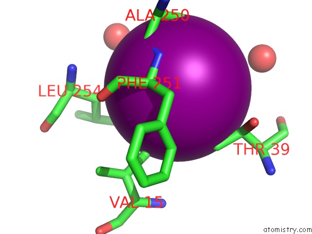
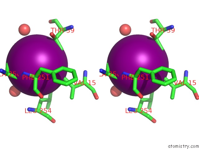
Iodine binding site 1 out of 3 in 5ijw
Go back to
Iodine binding site 1 out
of 3 in the Glutamate Racemase (Muri) From Mycobacterium Smegmatis with Bound D- Glutamate, 1.8 Angstrom Resolution, X-Ray Diffraction
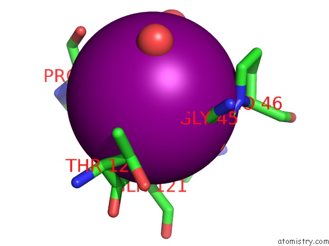
Mono view
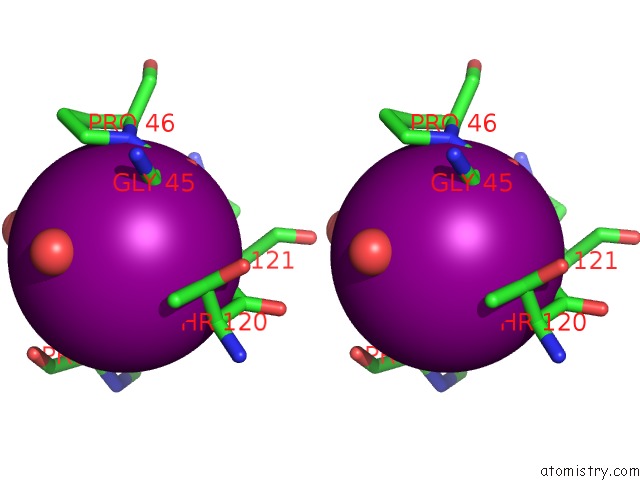
Stereo pair view
Mono view
Stereo pair view
A full contact list of Iodine with other atoms in the I binding
site number 1 of Glutamate Racemase (Muri) From Mycobacterium Smegmatis with Bound D- Glutamate, 1.8 Angstrom Resolution, X-Ray Diffraction within 5.0Å range:
|
Iodine binding site 2 out of 3 in 5ijw
Go back to
Iodine binding site 2 out
of 3 in the Glutamate Racemase (Muri) From Mycobacterium Smegmatis with Bound D- Glutamate, 1.8 Angstrom Resolution, X-Ray Diffraction
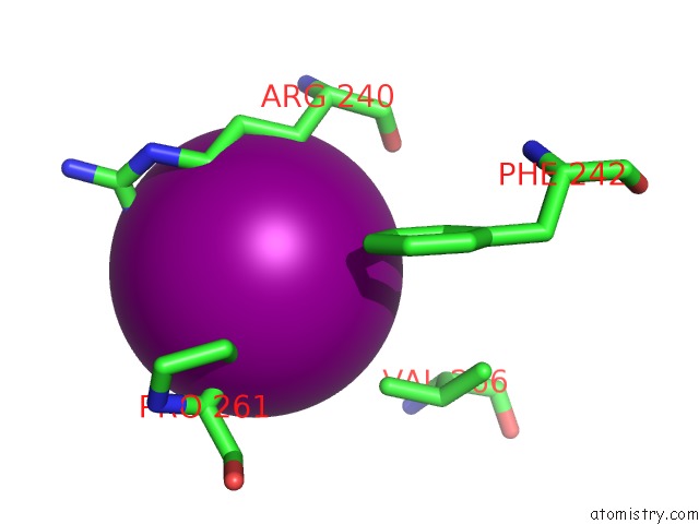
Mono view
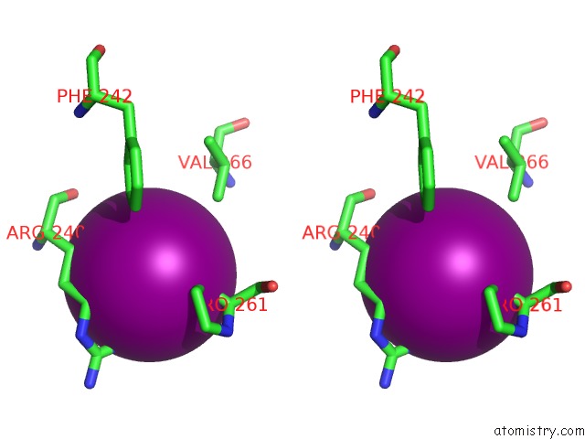
Stereo pair view
Mono view
Stereo pair view
A full contact list of Iodine with other atoms in the I binding
site number 2 of Glutamate Racemase (Muri) From Mycobacterium Smegmatis with Bound D- Glutamate, 1.8 Angstrom Resolution, X-Ray Diffraction within 5.0Å range:
|
Iodine binding site 3 out of 3 in 5ijw
Go back to
Iodine binding site 3 out
of 3 in the Glutamate Racemase (Muri) From Mycobacterium Smegmatis with Bound D- Glutamate, 1.8 Angstrom Resolution, X-Ray Diffraction
Mono view
Stereo pair view
Mono view
Stereo pair view
A full contact list of Iodine with other atoms in the I binding
site number 3 of Glutamate Racemase (Muri) From Mycobacterium Smegmatis with Bound D- Glutamate, 1.8 Angstrom Resolution, X-Ray Diffraction within 5.0Å range:
|
Reference:
S.Poen,
Y.Nakatani,
H.K.Opel-Reading,
M.Lasse,
R.C.Dobson,
K.L.Krause.
Exploring the Structure of Glutamate Racemase From Mycobacterium Tuberculosis As A Template For Anti-Mycobacterial Drug Discovery. Biochem. J. V. 473 1267 2016.
ISSN: ESSN 1470-8728
PubMed: 26964898
DOI: 10.1042/BCJ20160186
Page generated: Sun Aug 11 21:04:50 2024
ISSN: ESSN 1470-8728
PubMed: 26964898
DOI: 10.1042/BCJ20160186
Last articles
Zn in 9MJ5Zn in 9HNW
Zn in 9G0L
Zn in 9FNE
Zn in 9DZN
Zn in 9E0I
Zn in 9D32
Zn in 9DAK
Zn in 8ZXC
Zn in 8ZUF