Iodine »
PDB 5enk-5kio »
5jrv »
Iodine in PDB 5jrv: Crystal Structure of Fe(II) No-Bound H-Nox Protein From C. Subterraneus
Protein crystallography data
The structure of Crystal Structure of Fe(II) No-Bound H-Nox Protein From C. Subterraneus, PDB code: 5jrv
was solved by
J.Bruegger,
C.Hespen,
C.M.Phillips-Piro,
M.A.Marletta,
with X-Ray Crystallography technique. A brief refinement statistics is given in the table below:
| Resolution Low / High (Å) | 49.98 / 1.95 |
| Space group | P 21 21 2 |
| Cell size a, b, c (Å), α, β, γ (°) | 80.385, 127.626, 42.937, 90.00, 90.00, 90.00 |
| R / Rfree (%) | 20.2 / 23.1 |
Other elements in 5jrv:
The structure of Crystal Structure of Fe(II) No-Bound H-Nox Protein From C. Subterraneus also contains other interesting chemical elements:
| Iron | (Fe) | 2 atoms |
Iodine Binding Sites:
The binding sites of Iodine atom in the Crystal Structure of Fe(II) No-Bound H-Nox Protein From C. Subterraneus
(pdb code 5jrv). This binding sites where shown within
5.0 Angstroms radius around Iodine atom.
In total only one binding site of Iodine was determined in the Crystal Structure of Fe(II) No-Bound H-Nox Protein From C. Subterraneus, PDB code: 5jrv:
In total only one binding site of Iodine was determined in the Crystal Structure of Fe(II) No-Bound H-Nox Protein From C. Subterraneus, PDB code: 5jrv:
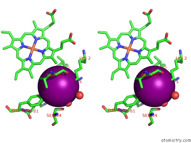
Iodine binding site 1 out of 1 in 5jrv
Go back to
Iodine binding site 1 out
of 1 in the Crystal Structure of Fe(II) No-Bound H-Nox Protein From C. Subterraneus

Mono view
Stereo pair view
Mono view
Stereo pair view
A full contact list of Iodine with other atoms in the I binding
site number 1 of Crystal Structure of Fe(II) No-Bound H-Nox Protein From C. Subterraneus within 5.0Å range:
|
Reference:
C.W.Hespen,
J.J.Bruegger,
C.M.Phillips-Piro,
M.A.Marletta.
Structural and Functional Evidence Indicates Selective Oxygen Signaling in Caldanaerobacter Subterraneus H-Nox. Acs Chem.Biol. V. 11 2337 2016.
ISSN: ESSN 1554-8937
PubMed: 27328180
DOI: 10.1021/ACSCHEMBIO.6B00431
Page generated: Sun Aug 11 21:09:11 2024
ISSN: ESSN 1554-8937
PubMed: 27328180
DOI: 10.1021/ACSCHEMBIO.6B00431
Last articles
Zn in 9J0NZn in 9J0O
Zn in 9J0P
Zn in 9FJX
Zn in 9EKB
Zn in 9C0F
Zn in 9CAH
Zn in 9CH0
Zn in 9CH3
Zn in 9CH1