Atomistry » Iodine » PDB 5enk-5kio » 5jsi
Atomistry »
  Iodine »
    PDB 5enk-5kio »
      5jsi »

Iodine in PDB 5jsi: Structure of Membrane Protein

Protein crystallography data

The structure of Structure of Membrane Protein, PDB code: 5jsi was solved by I.Melnikov, V.Polovinkin, K.Kovalev, V.Shevchenko, I.Gushchin, A.Popov, V.Gordeliy, with X-Ray Crystallography technique. A brief refinement statistics is given in the table below:

Resolution Low / High (Å) 50.80 / 2.00
Space group P 1
Cell size a, b, c (Å), α, β, γ (°) 40.793, 56.771, 57.286, 63.61, 78.48, 80.21
R / Rfree (%) 17.7 / 22

Iodine Binding Sites:

Pages:

>>> Page 1 <<< Page 2, Binding sites: 11 - 20; Page 3, Binding sites: 21 - 22;

Binding sites:

The binding sites of Iodine atom in the Structure of Membrane Protein (pdb code 5jsi). This binding sites where shown within 5.0 Angstroms radius around Iodine atom.
In total 22 binding sites of Iodine where determined in the Structure of Membrane Protein, PDB code: 5jsi:
Jump to Iodine binding site number: 1; 2; 3; 4; 5; 6; 7; 8; 9; 10;

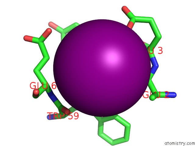
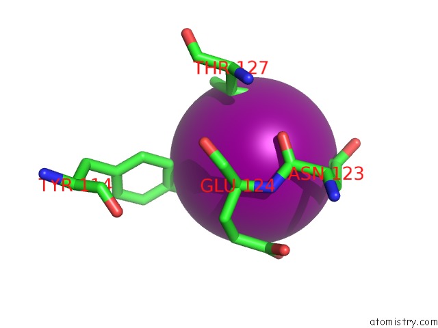
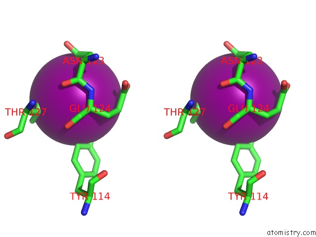
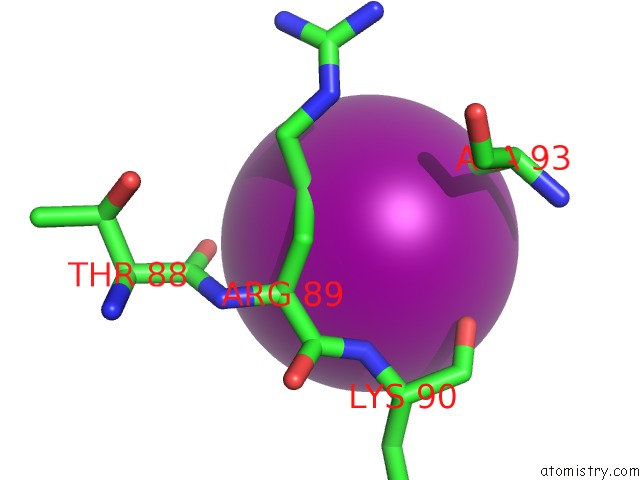
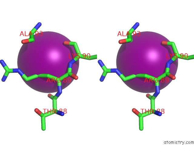
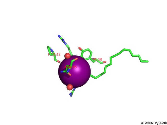
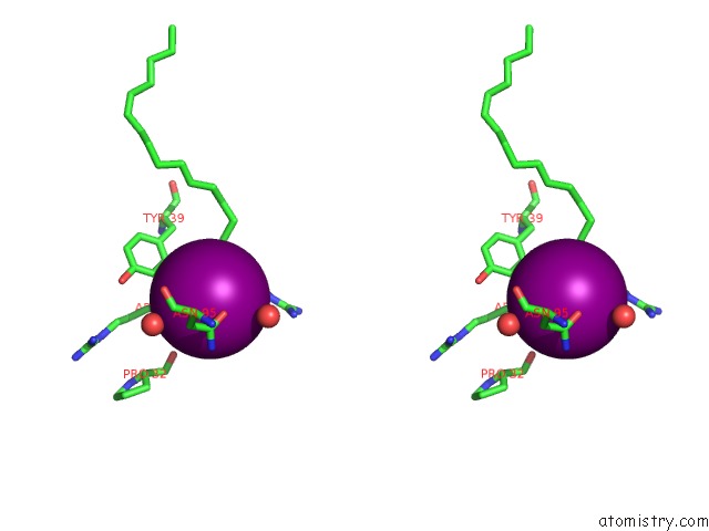
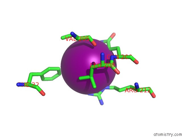
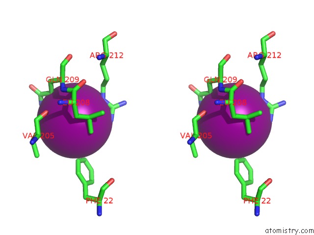
Iodine binding site 1 out of 22 in 5jsi

Go back to Iodine Binding Sites List in 5jsi
Iodine binding site 1 out of 22 in the Structure of Membrane Protein


Mono view


Stereo pair view

A full contact list of Iodine with other atoms in the I binding site number 1 of Structure of Membrane Protein within 5.0Å range:
probe atom residue distance (Å) B Occ
A:I313

b:63.6
occ:0.46
CD1 A:TRP59 3.7 47.8 1.0
OE1 A:GLU3 3.9 69.8 1.0
O A:GLU2 3.9 57.6 1.0
NE1 A:TRP59 3.9 49.7 1.0
CB A:TYR6 3.9 55.8 1.0
C A:GLU2 4.3 61.2 1.0
CG A:TYR6 4.4 57.0 1.0
CA A:GLU3 4.6 64.2 1.0
O A:TRP59 4.7 57.0 1.0
N A:GLU3 4.7 60.5 1.0
CB A:GLU2 4.7 62.5 1.0
CD A:GLU3 4.9 70.6 1.0
CA A:GLU60 4.9 69.4 1.0
CD2 A:TYR6 4.9 58.6 1.0
CG A:TRP59 4.9 47.0 1.0

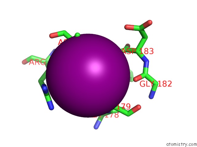
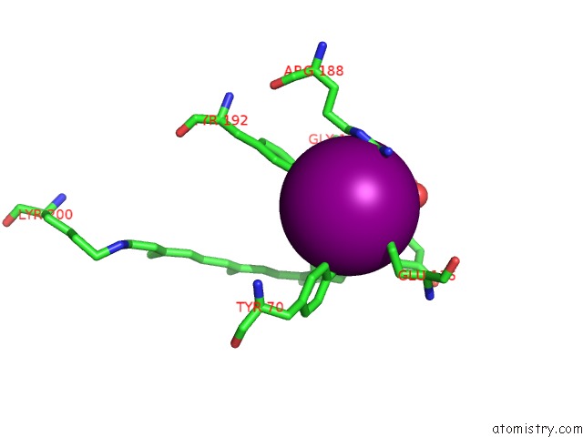
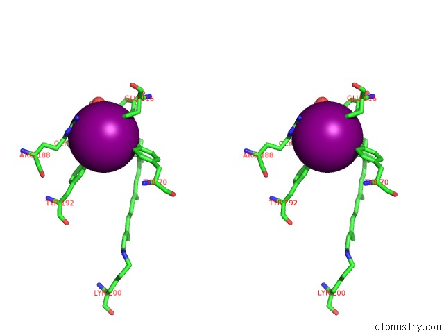
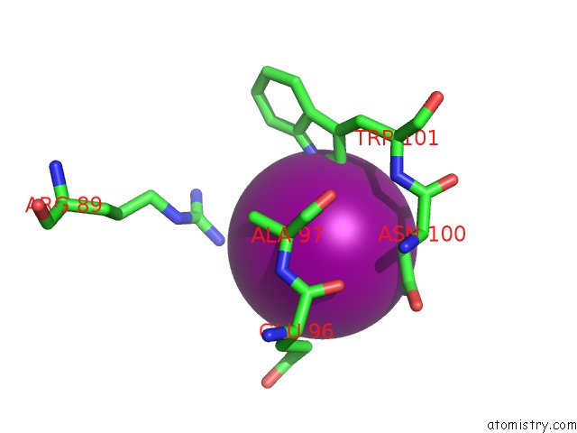
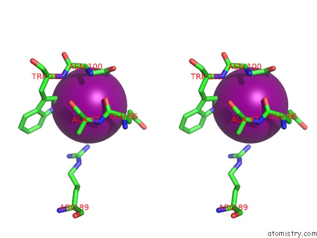
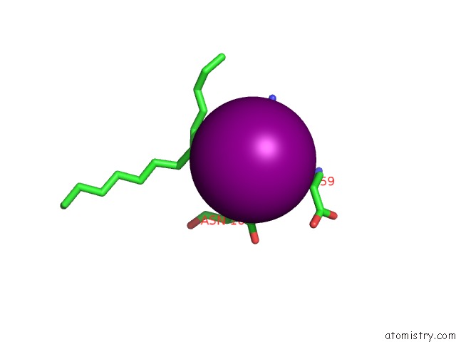
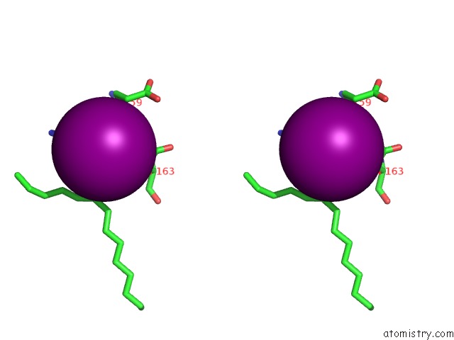
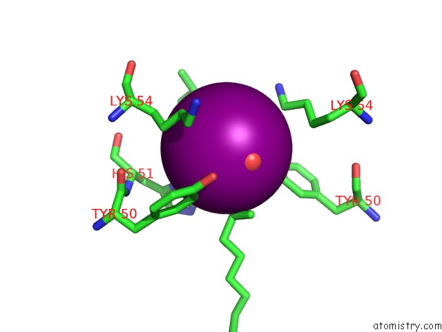
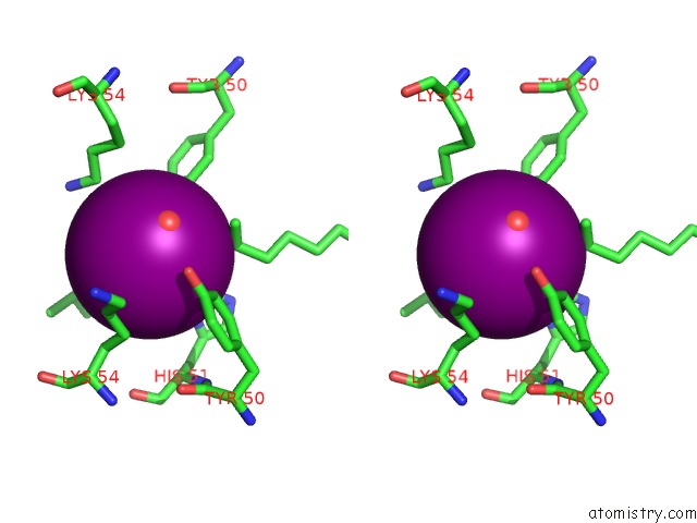
Iodine binding site 2 out of 22 in 5jsi

Go back to Iodine Binding Sites List in 5jsi
Iodine binding site 2 out of 22 in the Structure of Membrane Protein


Mono view


Stereo pair view

A full contact list of Iodine with other atoms in the I binding site number 2 of Structure of Membrane Protein within 5.0Å range:
probe atom residue distance (Å) B Occ
A:I314

b:0.3
occ:0.52
N A:ASP185 3.2 80.6 1.0
C A:PHE184 3.2 82.3 1.0
CA A:ASP185 3.5 72.5 1.0
CG A:PRO179 3.5 60.5 1.0
O A:PHE184 3.5 93.0 1.0
CD A:PRO179 3.6 63.1 1.0
CB A:PRO179 3.6 61.0 1.0
N A:PHE184 3.7 83.7 1.0
CA A:PHE184 3.8 81.4 1.0
CA A:PRO179 3.8 62.0 1.0
CB A:PHE184 3.9 78.7 1.0
N A:PRO179 4.0 60.7 1.0
CB A:ASP185 4.0 72.9 1.0
CZ A:ARG188 4.2 54.3 1.0
NE A:ARG188 4.4 53.5 1.0
NH2 A:ARG188 4.4 60.4 1.0
NH1 A:ARG188 4.6 57.0 1.0
O A:GLY182 4.6 76.8 1.0
C A:ASP185 4.8 69.3 1.0
CD A:ARG188 4.8 48.4 1.0
C A:ALA178 4.9 57.2 1.0
C A:ASP183 5.0 83.0 1.0
CB A:ARG188 5.0 41.8 1.0

Iodine binding site 3 out of 22 in 5jsi

Go back to Iodine Binding Sites List in 5jsi
Iodine binding site 3 out of 22 in the Structure of Membrane Protein


Mono view


Stereo pair view

A full contact list of Iodine with other atoms in the I binding site number 3 of Structure of Membrane Protein within 5.0Å range:
probe atom residue distance (Å) B Occ
A:I315

b:40.2
occ:0.48
ND2 A:ASN123 3.1 71.8 1.0
OH A:TYR114 3.2 39.8 1.0
OG1 A:THR127 3.5 38.9 1.0
CB A:THR127 4.0 29.9 1.0
CG2 A:THR127 4.2 29.1 1.0
CZ A:TYR114 4.2 40.4 1.0
CG A:ASN123 4.3 69.4 1.0
CA A:GLU124 4.3 39.6 1.0
CE2 A:TYR114 4.3 37.5 1.0
O A:ASN123 4.4 39.0 1.0
N A:GLU124 4.5 41.2 1.0
C A:ASN123 4.5 40.7 1.0
CG A:GLU124 4.6 59.9 1.0
OE2 A:GLU124 4.8 71.8 1.0
CB A:ASN123 4.9 56.7 1.0
CD A:GLU124 4.9 61.9 1.0
CB A:GLU124 5.0 50.4 1.0

Iodine binding site 4 out of 22 in 5jsi

Go back to Iodine Binding Sites List in 5jsi
Iodine binding site 4 out of 22 in the Structure of Membrane Protein


Mono view


Stereo pair view

A full contact list of Iodine with other atoms in the I binding site number 4 of Structure of Membrane Protein within 5.0Å range:
probe atom residue distance (Å) B Occ
A:I316

b:48.4
occ:0.28
OE2 A:GLU116 2.0 55.7 1.0
CD A:GLU116 2.9 59.1 1.0
NH1 A:ARG188 3.2 57.0 1.0
OE1 A:GLU116 3.2 60.8 1.0
OH A:TYR70 3.4 30.2 1.0
OH A:TYR192 3.6 35.5 1.0
CZ A:ARG188 3.7 54.3 1.0
CE2 A:TYR192 3.8 29.7 1.0
O A:HOH412 3.9 37.4 1.0
CD2 A:TYR176 4.0 23.0 1.0
CA A:TYR176 4.1 30.0 1.0
CZ A:TYR192 4.1 30.4 1.0
NE A:ARG188 4.2 53.5 1.0
CB A:TYR176 4.3 24.2 1.0
CG A:GLU116 4.3 52.0 1.0
CD A:ARG188 4.3 48.4 1.0
NH2 A:ARG188 4.4 60.4 1.0
CE1 A:TYR70 4.4 36.0 1.0
CZ A:TYR70 4.4 30.3 1.0
N A:TYR176 4.5 26.6 1.0
CG A:TYR176 4.6 24.5 1.0
C19 A:LYR200 4.7 24.1 1.0
O A:GLY175 4.8 28.8 1.0
C A:GLY175 4.8 29.3 1.0
CE2 A:TYR176 5.0 24.7 1.0
CD2 A:TYR192 5.0 30.1 1.0

Iodine binding site 5 out of 22 in 5jsi

Go back to Iodine Binding Sites List in 5jsi
Iodine binding site 5 out of 22 in the Structure of Membrane Protein


Mono view


Stereo pair view

A full contact list of Iodine with other atoms in the I binding site number 5 of Structure of Membrane Protein within 5.0Å range:
probe atom residue distance (Å) B Occ
A:I317

b:94.8
occ:0.62
CG A:ARG89 3.7 51.7 1.0
CB A:ALA93 3.9 51.1 1.0
CA A:ARG89 4.2 48.7 1.0
N A:LYS90 4.2 48.5 1.0
O A:LYS90 4.3 45.0 1.0
CB A:ARG89 4.4 51.5 1.0
O A:THR88 4.5 51.3 1.0
C A:ARG89 4.8 45.7 1.0

Iodine binding site 6 out of 22 in 5jsi

Go back to Iodine Binding Sites List in 5jsi
Iodine binding site 6 out of 22 in the Structure of Membrane Protein


Mono view


Stereo pair view

A full contact list of Iodine with other atoms in the I binding site number 6 of Structure of Membrane Protein within 5.0Å range:
probe atom residue distance (Å) B Occ
A:I318

b:68.0
occ:0.79
NE1 A:TRP101 3.6 44.7 1.0
CD1 A:TRP101 4.0 45.9 1.0
ND2 A:ASN100 4.0 45.3 1.0
CA A:ALA97 4.1 48.3 1.0
CB A:ASN100 4.2 38.5 1.0
NH1 A:ARG89 4.3 48.9 1.0
OE1 A:GLU96 4.5 74.5 1.0
CG A:ASN100 4.5 47.6 1.0
O A:GLU96 4.5 46.0 1.0
CB A:ALA97 4.6 52.0 1.0
N A:ALA97 4.6 51.5 1.0
CG A:GLU96 4.7 56.1 1.0
C A:GLU96 4.8 50.4 1.0
CE2 A:TRP101 4.9 42.7 1.0

Iodine binding site 7 out of 22 in 5jsi

Go back to Iodine Binding Sites List in 5jsi
Iodine binding site 7 out of 22 in the Structure of Membrane Protein


Mono view


Stereo pair view

A full contact list of Iodine with other atoms in the I binding site number 7 of Structure of Membrane Protein within 5.0Å range:
probe atom residue distance (Å) B Occ
A:I319

b:0.7
occ:0.84
CD2 A:TYR39 4.0 43.2 1.0
C15 A:LFA308 4.0 39.5 1.0
CE2 A:TYR39 4.1 42.5 1.0
CA A:ARG36 4.1 38.0 1.0
CB A:ARG36 4.1 42.0 1.0
N A:ARG36 4.4 34.8 1.0
O A:HOH428 4.4 45.0 1.0
CG A:ARG36 4.5 49.5 1.0
OD1 B:ASN95 4.7 59.7 1.0
NE A:ARG36 4.7 66.7 1.0
O B:HOH422 4.9 60.8 1.0
O A:PRO32 4.9 49.6 1.0
C A:ARG35 5.0 35.5 1.0

Iodine binding site 8 out of 22 in 5jsi

Go back to Iodine Binding Sites List in 5jsi
Iodine binding site 8 out of 22 in the Structure of Membrane Protein


Mono view


Stereo pair view

A full contact list of Iodine with other atoms in the I binding site number 8 of Structure of Membrane Protein within 5.0Å range:
probe atom residue distance (Å) B Occ
A:I320

b:33.2
occ:0.23
OD1 A:ASN160 3.0 26.8 1.0
ND2 A:ASN163 3.4 30.1 1.0
CB A:ASN163 4.1 25.5 1.0
CG A:ASN160 4.1 24.8 1.0
CA A:ASN160 4.1 22.9 1.0
O A:GLU159 4.2 23.6 1.0
C11 A:LFA307 4.2 44.0 1.0
CG A:ASN163 4.2 31.0 1.0
CG A:GLU159 4.4 31.0 1.0
N A:ASN160 4.4 20.8 1.0
C A:GLU159 4.4 22.8 1.0
CB A:ASN160 4.6 21.7 1.0
C13 A:LFA307 4.8 39.6 1.0

Iodine binding site 9 out of 22 in 5jsi

Go back to Iodine Binding Sites List in 5jsi
Iodine binding site 9 out of 22 in the Structure of Membrane Protein


Mono view


Stereo pair view

A full contact list of Iodine with other atoms in the I binding site number 9 of Structure of Membrane Protein within 5.0Å range:
probe atom residue distance (Å) B Occ
A:I321

b:52.1
occ:0.57
NE A:ARG212 3.8 44.6 1.0
NH1 A:ARG212 3.9 59.0 1.0
CG A:GLN209 3.9 38.4 1.0
NE2 A:GLN209 3.9 43.2 1.0
CD2 A:LEU208 4.2 33.0 1.0
CZ A:ARG212 4.3 50.6 1.0
CZ A:PHE22 4.3 34.6 1.0
CG1 A:VAL205 4.4 30.8 1.0
CD A:GLN209 4.5 43.4 1.0
CG A:LEU208 4.5 31.2 1.0
CE2 A:PHE22 4.6 36.3 1.0
CD A:ARG212 4.8 43.2 1.0
O A:VAL205 4.8 24.4 1.0

Iodine binding site 10 out of 22 in 5jsi

Go back to Iodine Binding Sites List in 5jsi
Iodine binding site 10 out of 22 in the Structure of Membrane Protein


Mono view


Stereo pair view

A full contact list of Iodine with other atoms in the I binding site number 10 of Structure of Membrane Protein within 5.0Å range:
probe atom residue distance (Å) B Occ
B:I311

b:53.9
occ:0.46
O B:HOH438 2.6 45.1 1.0
OH B:TYR50 3.1 41.2 1.0
O A:HOH405 3.4 41.4 1.0
CD2 A:LEU68 3.5 43.0 1.0
ND1 A:HIS51 3.9 45.5 1.0
CE1 A:HIS51 3.9 34.9 1.0
CE2 B:TYR50 4.0 36.2 1.0
CZ B:TYR50 4.1 39.0 1.0
C19 A:LFA301 4.1 48.4 1.0
CE2 A:TYR50 4.3 42.6 1.0
C20 A:LFA301 4.4 48.4 1.0
CE B:LYS54 4.4 53.7 1.0
CD A:LYS54 4.5 53.2 1.0
CG A:LYS54 4.5 47.4 1.0
OH A:TYR50 4.7 46.4 1.0
CB A:LYS54 4.9 39.4 1.0
CZ A:TYR50 4.9 43.1 1.0

Reference:

I.Melnikov, V.Polovinkin, K.Kovalev, I.Gushchin, M.Shevtsov, V.Shevchenko, A.Mishin, A.Alekseev, F.Rodriguez-Valera, V.Borshchevskiy, V.Cherezov, G.A.Leonard, V.Gordeliy, A.Popov. Fast Iodide-Sad Phasing For High-Throughput Membrane Protein Structure Determination. Sci Adv V. 3 02952 2017.
ISSN: ESSN 2375-2548
PubMed: 28508075
DOI: 10.1126/SCIADV.1602952
Page generated: Sun Dec 13 19:39:36 2020

Last articles

Zn in 8WB0
Zn in 8WAX
Zn in 8WAU
Zn in 8WAZ
Zn in 8WAY
Zn in 8WAV
Zn in 8WAW
Zn in 8WAT
Zn in 8W7M
Zn in 8WD3
© Copyright 2008-2020 by atomistry.com
Home   |    Site Map   |    Copyright   |    Contact us   |    Privacy