Iodine »
PDB 5enk-5kio »
5k7p »
Iodine in PDB 5k7p: Microed Structure of Xylanase at 2.3 A Resolution
Enzymatic activity of Microed Structure of Xylanase at 2.3 A Resolution
All present enzymatic activity of Microed Structure of Xylanase at 2.3 A Resolution:
3.2.1.8;
3.2.1.8;
Iodine Binding Sites:
The binding sites of Iodine atom in the Microed Structure of Xylanase at 2.3 A Resolution
(pdb code 5k7p). This binding sites where shown within
5.0 Angstroms radius around Iodine atom.
In total 2 binding sites of Iodine where determined in the Microed Structure of Xylanase at 2.3 A Resolution, PDB code: 5k7p:
Jump to Iodine binding site number: 1; 2;
In total 2 binding sites of Iodine where determined in the Microed Structure of Xylanase at 2.3 A Resolution, PDB code: 5k7p:
Jump to Iodine binding site number: 1; 2;
Iodine binding site 1 out of 2 in 5k7p
Go back to
Iodine binding site 1 out
of 2 in the Microed Structure of Xylanase at 2.3 A Resolution
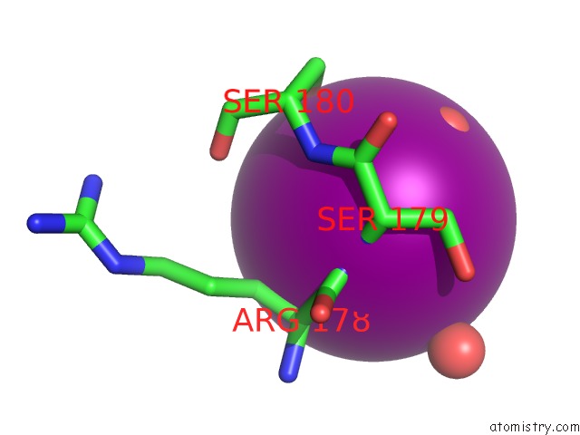
Mono view
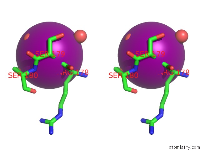
Stereo pair view
Mono view
Stereo pair view
A full contact list of Iodine with other atoms in the I binding
site number 1 of Microed Structure of Xylanase at 2.3 A Resolution within 5.0Å range:
|
Iodine binding site 2 out of 2 in 5k7p
Go back to
Iodine binding site 2 out
of 2 in the Microed Structure of Xylanase at 2.3 A Resolution
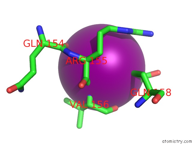
Mono view
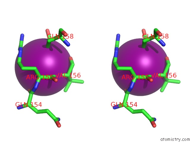
Stereo pair view
Mono view
Stereo pair view
A full contact list of Iodine with other atoms in the I binding
site number 2 of Microed Structure of Xylanase at 2.3 A Resolution within 5.0Å range:
|
Reference:
M.J.De La Cruz,
J.Hattne,
D.Shi,
P.Seidler,
J.Rodriguez,
F.E.Reyes,
M.R.Sawaya,
D.Cascio,
S.C.Weiss,
S.K.Kim,
C.S.Hinck,
A.P.Hinck,
G.Calero,
D.Eisenberg,
T.Gonen.
Atomic-Resolution Structures From Fragmented Protein Crystals with the Cryoem Method Microed. Nat. Methods V. 14 399 2017.
ISSN: ESSN 1548-7105
PubMed: 28192420
DOI: 10.1038/NMETH.4178
Page generated: Sun Aug 11 21:15:28 2024
ISSN: ESSN 1548-7105
PubMed: 28192420
DOI: 10.1038/NMETH.4178
Last articles
Zn in 9J0NZn in 9J0O
Zn in 9J0P
Zn in 9FJX
Zn in 9EKB
Zn in 9C0F
Zn in 9CAH
Zn in 9CH0
Zn in 9CH3
Zn in 9CH1