Iodine »
PDB 5nje-5say »
5od2 »
Iodine in PDB 5od2: Crystal Structure of Adp-Dependent Glucokinase From Methanocaldococcus Jannaschii
Enzymatic activity of Crystal Structure of Adp-Dependent Glucokinase From Methanocaldococcus Jannaschii
All present enzymatic activity of Crystal Structure of Adp-Dependent Glucokinase From Methanocaldococcus Jannaschii:
2.7.1.146; 2.7.1.147;
2.7.1.146; 2.7.1.147;
Protein crystallography data
The structure of Crystal Structure of Adp-Dependent Glucokinase From Methanocaldococcus Jannaschii, PDB code: 5od2
was solved by
M.Wisniewska,
P.Tokarz,
P.Grudnik,
with X-Ray Crystallography technique. A brief refinement statistics is given in the table below:
| Resolution Low / High (Å) | 47.00 / 1.98 |
| Space group | P 31 |
| Cell size a, b, c (Å), α, β, γ (°) | 154.565, 154.565, 50.483, 90.00, 90.00, 120.00 |
| R / Rfree (%) | 18.6 / 22.4 |
Other elements in 5od2:
The structure of Crystal Structure of Adp-Dependent Glucokinase From Methanocaldococcus Jannaschii also contains other interesting chemical elements:
| Magnesium | (Mg) | 3 atoms |
Iodine Binding Sites:
The binding sites of Iodine atom in the Crystal Structure of Adp-Dependent Glucokinase From Methanocaldococcus Jannaschii
(pdb code 5od2). This binding sites where shown within
5.0 Angstroms radius around Iodine atom.
In total 3 binding sites of Iodine where determined in the Crystal Structure of Adp-Dependent Glucokinase From Methanocaldococcus Jannaschii, PDB code: 5od2:
Jump to Iodine binding site number: 1; 2; 3;
In total 3 binding sites of Iodine where determined in the Crystal Structure of Adp-Dependent Glucokinase From Methanocaldococcus Jannaschii, PDB code: 5od2:
Jump to Iodine binding site number: 1; 2; 3;
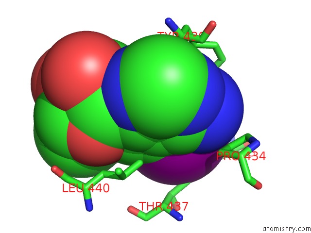
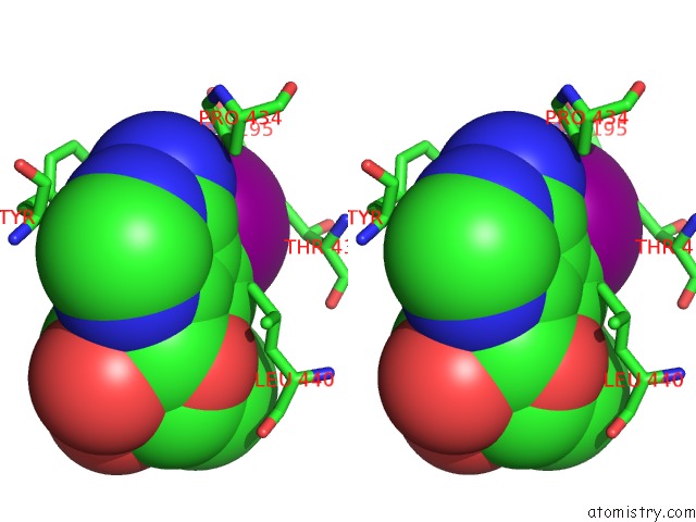
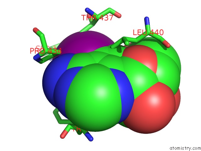
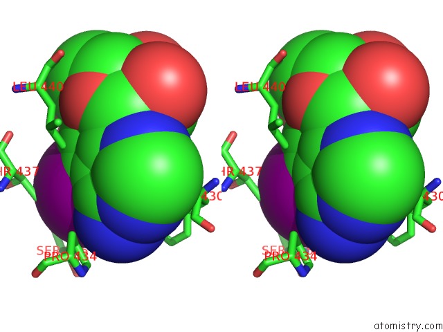
Iodine binding site 1 out of 3 in 5od2
Go back to
Iodine binding site 1 out
of 3 in the Crystal Structure of Adp-Dependent Glucokinase From Methanocaldococcus Jannaschii
Mono view
Stereo pair view
Mono view
Stereo pair view
A full contact list of Iodine with other atoms in the I binding
site number 1 of Crystal Structure of Adp-Dependent Glucokinase From Methanocaldococcus Jannaschii within 5.0Å range:
|
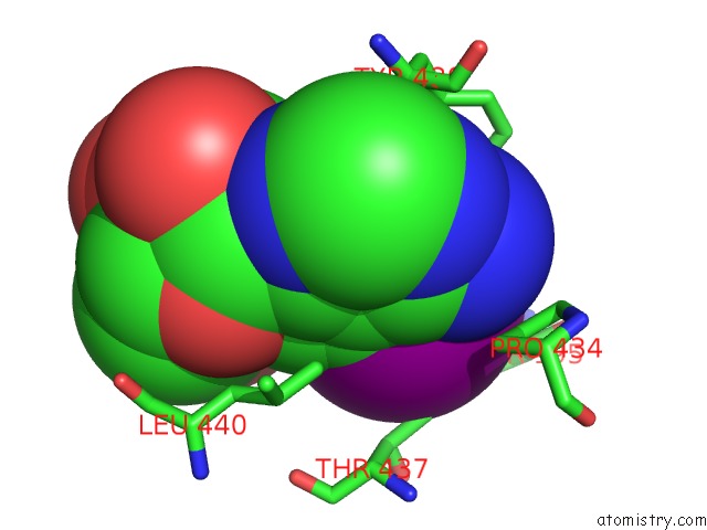
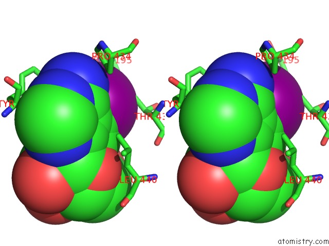
Iodine binding site 2 out of 3 in 5od2
Go back to
Iodine binding site 2 out
of 3 in the Crystal Structure of Adp-Dependent Glucokinase From Methanocaldococcus Jannaschii
Mono view
Stereo pair view
Mono view
Stereo pair view
A full contact list of Iodine with other atoms in the I binding
site number 2 of Crystal Structure of Adp-Dependent Glucokinase From Methanocaldococcus Jannaschii within 5.0Å range:
|
Iodine binding site 3 out of 3 in 5od2
Go back to
Iodine binding site 3 out
of 3 in the Crystal Structure of Adp-Dependent Glucokinase From Methanocaldococcus Jannaschii
Mono view
Stereo pair view
Mono view
Stereo pair view
A full contact list of Iodine with other atoms in the I binding
site number 3 of Crystal Structure of Adp-Dependent Glucokinase From Methanocaldococcus Jannaschii within 5.0Å range:
|
Reference:
P.Tokarz,
M.Wisniewska,
M.M.Kaminski,
G.Dubin,
P.Grudnik.
Crystal Structure of Adp-Dependent Glucokinase From Methanocaldococcus Jannaschii in Complex with 5-Iodotubercidin Reveals Phosphoryl Transfer Mechanism. Protein Sci. V. 27 790 2018.
ISSN: ESSN 1469-896X
PubMed: 29352744
DOI: 10.1002/PRO.3377
Page generated: Sun Aug 11 21:42:01 2024
ISSN: ESSN 1469-896X
PubMed: 29352744
DOI: 10.1002/PRO.3377
Last articles
Zn in 9J0NZn in 9J0O
Zn in 9J0P
Zn in 9FJX
Zn in 9EKB
Zn in 9C0F
Zn in 9CAH
Zn in 9CH0
Zn in 9CH3
Zn in 9CH1