Iodine »
PDB 5sb0-5w1i »
5ug6 »
Iodine in PDB 5ug6: Perforin C2 Domain - T431D
Protein crystallography data
The structure of Perforin C2 Domain - T431D, PDB code: 5ug6
was solved by
R.H.P.Law,
P.J.Conroy,
I.Voskoboinik,
J.C.Whisstock,
with X-Ray Crystallography technique. A brief refinement statistics is given in the table below:
| Resolution Low / High (Å) | 33.01 / 2.00 |
| Space group | P 21 21 21 |
| Cell size a, b, c (Å), α, β, γ (°) | 27.774, 66.027, 74.867, 90.00, 90.00, 90.00 |
| R / Rfree (%) | 19.1 / 22.2 |
Iodine Binding Sites:
The binding sites of Iodine atom in the Perforin C2 Domain - T431D
(pdb code 5ug6). This binding sites where shown within
5.0 Angstroms radius around Iodine atom.
In total only one binding site of Iodine was determined in the Perforin C2 Domain - T431D, PDB code: 5ug6:
In total only one binding site of Iodine was determined in the Perforin C2 Domain - T431D, PDB code: 5ug6:
Iodine binding site 1 out of 1 in 5ug6
Go back to
Iodine binding site 1 out
of 1 in the Perforin C2 Domain - T431D
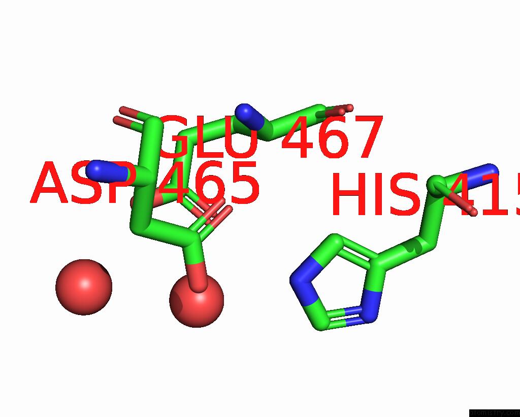
Mono view
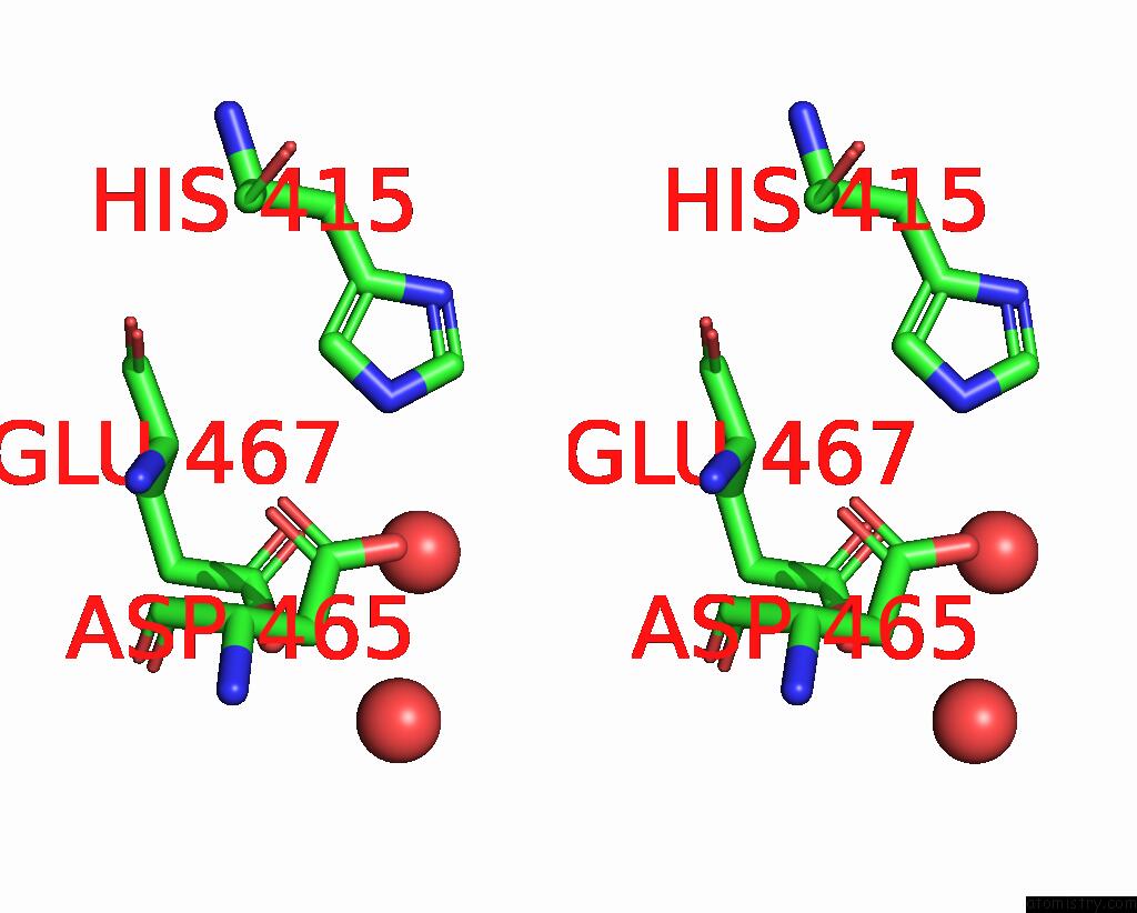
Stereo pair view
Mono view
Stereo pair view
A full contact list of Iodine with other atoms in the I binding
site number 1 of Perforin C2 Domain - T431D within 5.0Å range:
|
Reference:
A.J.Brennan,
R.H.P.Law,
P.J.Conroy,
T.Noori,
N.Lukoyanova,
H.Saibil,
H.Yagita,
A.Ciccone,
S.Verschoor,
J.C.Whisstock,
J.A.Trapani,
I.Voskoboinik.
Perforin Proteostasis Is Regulated Through Its C2 Domain: Supra-Physiological Cell Death Mediated By T431D-Perforin. Cell Death Differ. V. 25 1517 2018.
ISSN: ISSN 1476-5403
PubMed: 29416110
DOI: 10.1038/S41418-018-0057-Z
Page generated: Sun Aug 11 22:00:43 2024
ISSN: ISSN 1476-5403
PubMed: 29416110
DOI: 10.1038/S41418-018-0057-Z
Last articles
F in 4G5JF in 4G3G
F in 4G3F
F in 4G2I
F in 4G2H
F in 4G31
F in 4G1W
F in 4G16
F in 4FXY
F in 4FXQ