Iodine »
PDB 5w4i-6axx »
5zh9 »
Iodine in PDB 5zh9: Crystal Structures of Mutant Endo-Beta-1,4-Xylanase II (Y88F)
Enzymatic activity of Crystal Structures of Mutant Endo-Beta-1,4-Xylanase II (Y88F)
All present enzymatic activity of Crystal Structures of Mutant Endo-Beta-1,4-Xylanase II (Y88F):
3.2.1.8;
3.2.1.8;
Protein crystallography data
The structure of Crystal Structures of Mutant Endo-Beta-1,4-Xylanase II (Y88F), PDB code: 5zh9
was solved by
X.Zhang,
Q.Wan,
with X-Ray Crystallography technique. A brief refinement statistics is given in the table below:
| Resolution Low / High (Å) | 39.78 / 1.15 |
| Space group | P 21 21 21 |
| Cell size a, b, c (Å), α, β, γ (°) | 48.331, 59.327, 70.056, 90.00, 90.00, 90.00 |
| R / Rfree (%) | 13.7 / 14.8 |
Iodine Binding Sites:
The binding sites of Iodine atom in the Crystal Structures of Mutant Endo-Beta-1,4-Xylanase II (Y88F)
(pdb code 5zh9). This binding sites where shown within
5.0 Angstroms radius around Iodine atom.
In total 3 binding sites of Iodine where determined in the Crystal Structures of Mutant Endo-Beta-1,4-Xylanase II (Y88F), PDB code: 5zh9:
Jump to Iodine binding site number: 1; 2; 3;
In total 3 binding sites of Iodine where determined in the Crystal Structures of Mutant Endo-Beta-1,4-Xylanase II (Y88F), PDB code: 5zh9:
Jump to Iodine binding site number: 1; 2; 3;
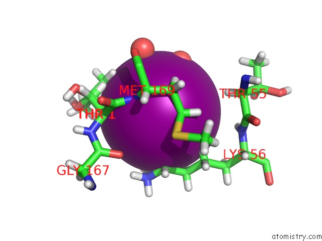
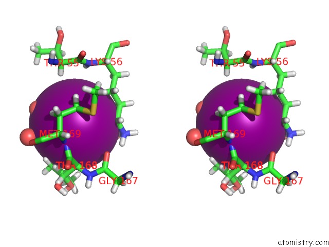
Iodine binding site 1 out of 3 in 5zh9
Go back to
Iodine binding site 1 out
of 3 in the Crystal Structures of Mutant Endo-Beta-1,4-Xylanase II (Y88F)
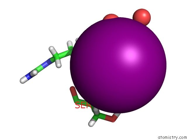
Mono view
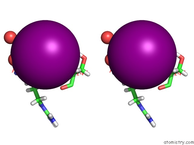
Stereo pair view
Mono view
Stereo pair view
A full contact list of Iodine with other atoms in the I binding
site number 1 of Crystal Structures of Mutant Endo-Beta-1,4-Xylanase II (Y88F) within 5.0Å range:
|
Iodine binding site 2 out of 3 in 5zh9
Go back to
Iodine binding site 2 out
of 3 in the Crystal Structures of Mutant Endo-Beta-1,4-Xylanase II (Y88F)
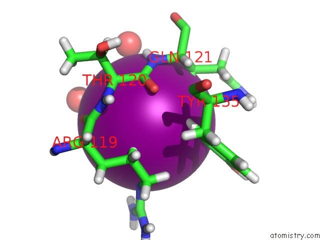
Mono view
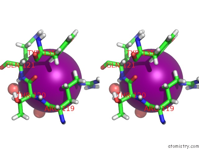
Stereo pair view
Mono view
Stereo pair view
A full contact list of Iodine with other atoms in the I binding
site number 2 of Crystal Structures of Mutant Endo-Beta-1,4-Xylanase II (Y88F) within 5.0Å range:
|
Iodine binding site 3 out of 3 in 5zh9
Go back to
Iodine binding site 3 out
of 3 in the Crystal Structures of Mutant Endo-Beta-1,4-Xylanase II (Y88F)
Mono view
Stereo pair view
Mono view
Stereo pair view
A full contact list of Iodine with other atoms in the I binding
site number 3 of Crystal Structures of Mutant Endo-Beta-1,4-Xylanase II (Y88F) within 5.0Å range:
|
Reference:
X.Zhang,
Q.Wan.
Crystal Structures of Mutant Endo-Beta-1,4-Xylanase II To Be Published.
Page generated: Sun Aug 11 22:25:23 2024
Last articles
Ca in 2PT3Ca in 2PTN
Ca in 2PSR
Ca in 2PTC
Ca in 2PRK
Ca in 2PR7
Ca in 2PR3
Ca in 2PPL
Ca in 2PQY
Ca in 2PQX