Iodine »
PDB 5w4i-6axx »
6an0 »
Iodine in PDB 6an0: Crystal Structure of Histidinol Dehydrogenase From Elizabethkingia Anophelis
Enzymatic activity of Crystal Structure of Histidinol Dehydrogenase From Elizabethkingia Anophelis
All present enzymatic activity of Crystal Structure of Histidinol Dehydrogenase From Elizabethkingia Anophelis:
1.1.1.23;
1.1.1.23;
Protein crystallography data
The structure of Crystal Structure of Histidinol Dehydrogenase From Elizabethkingia Anophelis, PDB code: 6an0
was solved by
Seattle Structural Genomics Center For Infectious Disease (Ssgcid),
with X-Ray Crystallography technique. A brief refinement statistics is given in the table below:
| Resolution Low / High (Å) | 31.45 / 1.85 |
| Space group | C 2 2 21 |
| Cell size a, b, c (Å), α, β, γ (°) | 82.520, 170.080, 65.900, 90.00, 90.00, 90.00 |
| R / Rfree (%) | 15.7 / 19.2 |
Other elements in 6an0:
The structure of Crystal Structure of Histidinol Dehydrogenase From Elizabethkingia Anophelis also contains other interesting chemical elements:
| Zinc | (Zn) | 1 atom |
Iodine Binding Sites:
Pages:
>>> Page 1 <<< Page 2, Binding sites: 11 - 13;Binding sites:
The binding sites of Iodine atom in the Crystal Structure of Histidinol Dehydrogenase From Elizabethkingia Anophelis (pdb code 6an0). This binding sites where shown within 5.0 Angstroms radius around Iodine atom.In total 13 binding sites of Iodine where determined in the Crystal Structure of Histidinol Dehydrogenase From Elizabethkingia Anophelis, PDB code: 6an0:
Jump to Iodine binding site number: 1; 2; 3; 4; 5; 6; 7; 8; 9; 10;
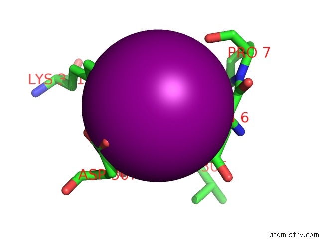
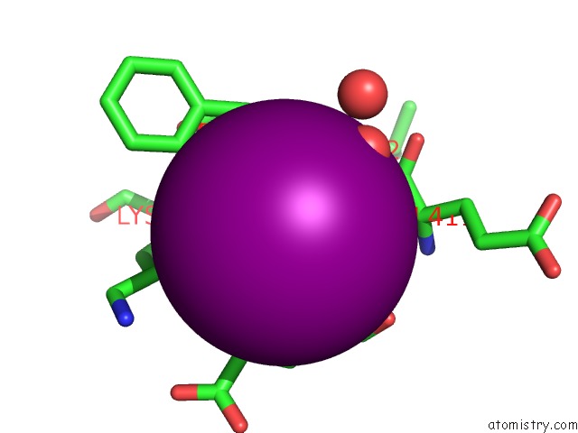
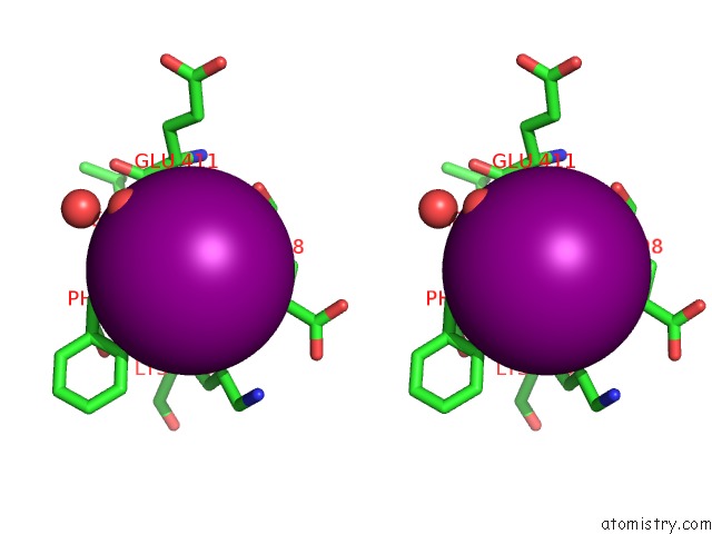
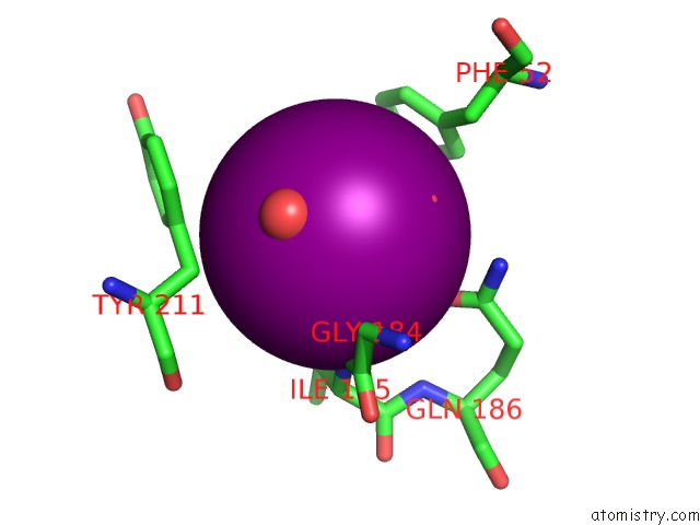
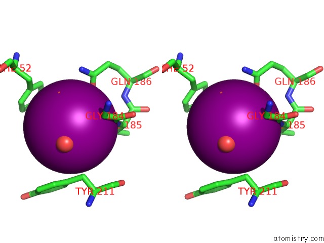
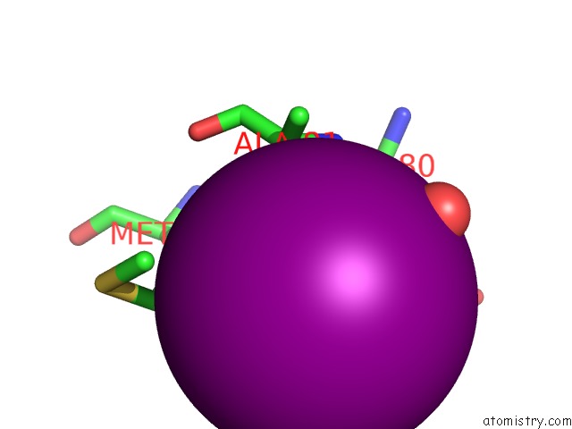
Iodine binding site 1 out of 13 in 6an0
Go back to
Iodine binding site 1 out
of 13 in the Crystal Structure of Histidinol Dehydrogenase From Elizabethkingia Anophelis
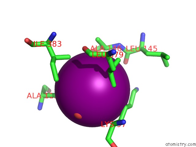
Mono view
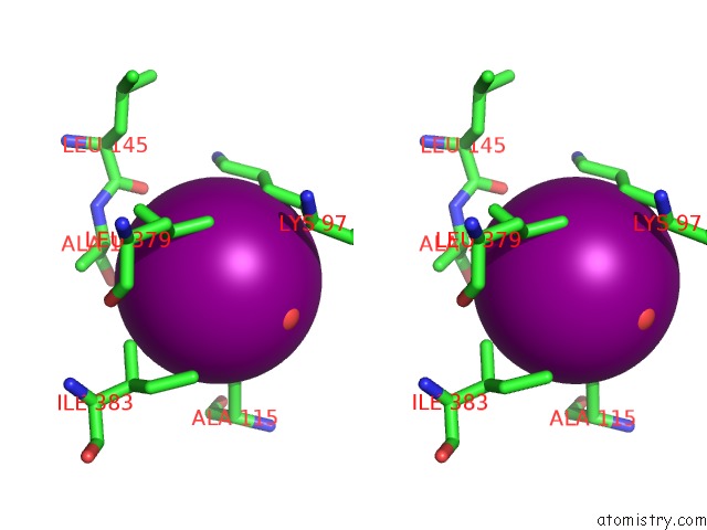
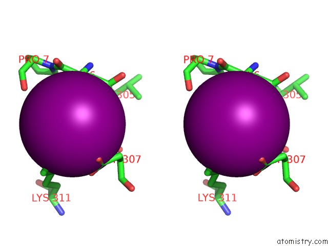
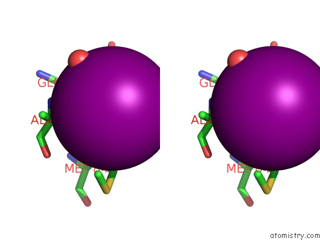
Stereo pair view
Mono view
Stereo pair view
A full contact list of Iodine with other atoms in the I binding
site number 1 of Crystal Structure of Histidinol Dehydrogenase From Elizabethkingia Anophelis within 5.0Å range:
|
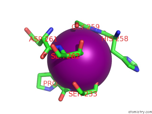
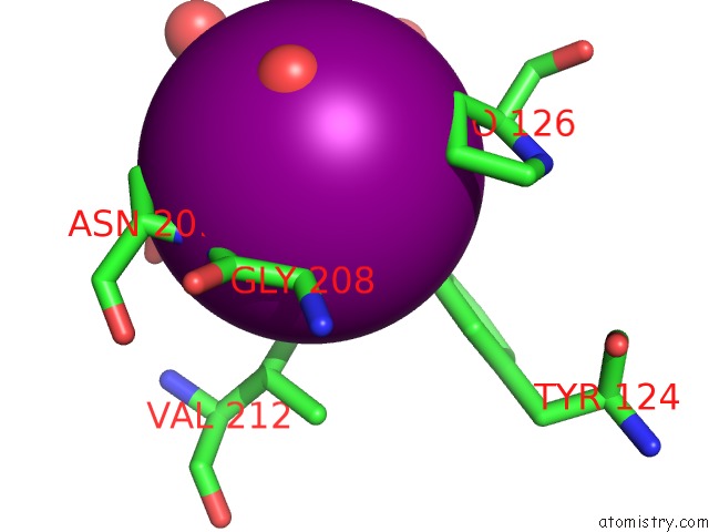
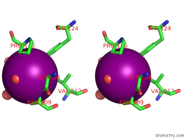
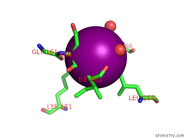
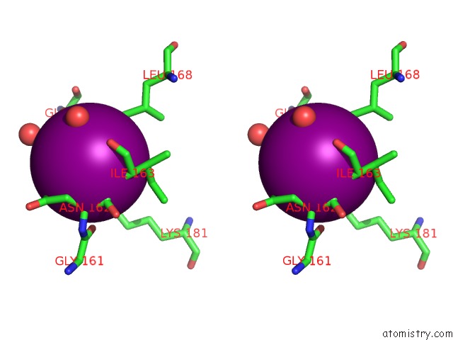
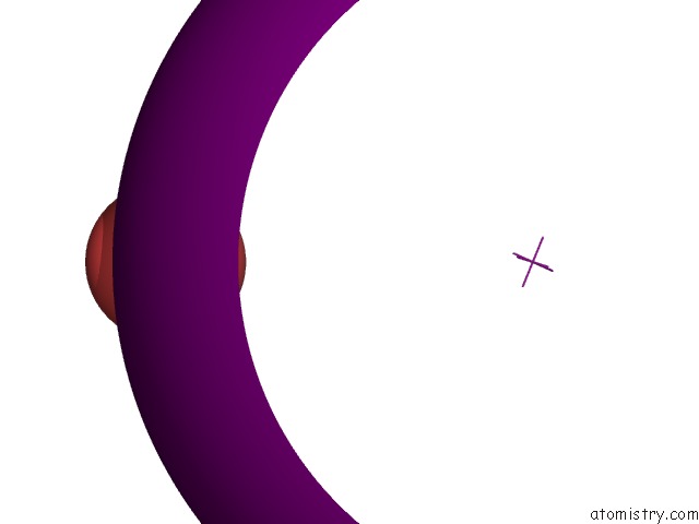
Iodine binding site 2 out of 13 in 6an0
Go back to
Iodine binding site 2 out
of 13 in the Crystal Structure of Histidinol Dehydrogenase From Elizabethkingia Anophelis
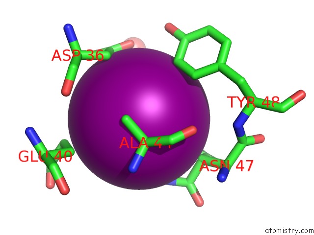
Mono view
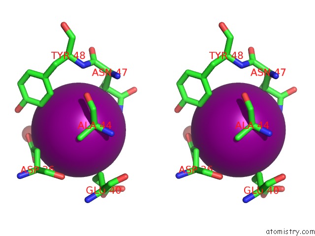
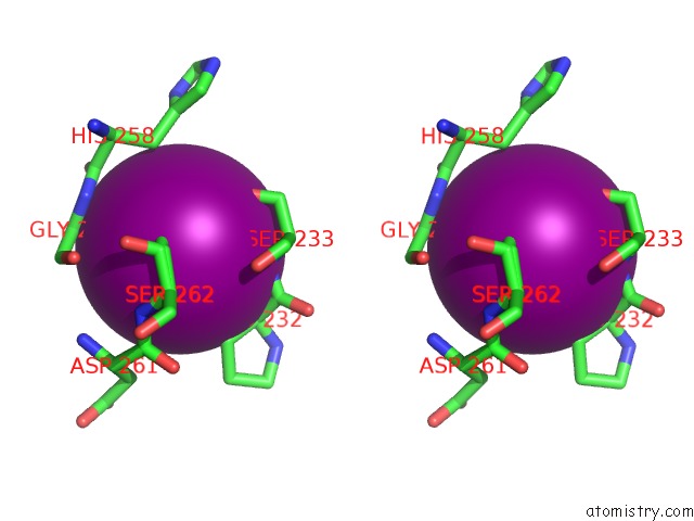
Stereo pair view
Mono view
Stereo pair view
A full contact list of Iodine with other atoms in the I binding
site number 2 of Crystal Structure of Histidinol Dehydrogenase From Elizabethkingia Anophelis within 5.0Å range:
|
Iodine binding site 3 out of 13 in 6an0
Go back to
Iodine binding site 3 out
of 13 in the Crystal Structure of Histidinol Dehydrogenase From Elizabethkingia Anophelis
Mono view
Stereo pair view
Mono view
Stereo pair view
A full contact list of Iodine with other atoms in the I binding
site number 3 of Crystal Structure of Histidinol Dehydrogenase From Elizabethkingia Anophelis within 5.0Å range:
|
Iodine binding site 4 out of 13 in 6an0
Go back to
Iodine binding site 4 out
of 13 in the Crystal Structure of Histidinol Dehydrogenase From Elizabethkingia Anophelis
Mono view
Stereo pair view
Mono view
Stereo pair view
A full contact list of Iodine with other atoms in the I binding
site number 4 of Crystal Structure of Histidinol Dehydrogenase From Elizabethkingia Anophelis within 5.0Å range:
|
Iodine binding site 5 out of 13 in 6an0
Go back to
Iodine binding site 5 out
of 13 in the Crystal Structure of Histidinol Dehydrogenase From Elizabethkingia Anophelis
Mono view
Stereo pair view
Mono view
Stereo pair view
A full contact list of Iodine with other atoms in the I binding
site number 5 of Crystal Structure of Histidinol Dehydrogenase From Elizabethkingia Anophelis within 5.0Å range:
|
Iodine binding site 6 out of 13 in 6an0
Go back to
Iodine binding site 6 out
of 13 in the Crystal Structure of Histidinol Dehydrogenase From Elizabethkingia Anophelis
Mono view
Stereo pair view
Mono view
Stereo pair view
A full contact list of Iodine with other atoms in the I binding
site number 6 of Crystal Structure of Histidinol Dehydrogenase From Elizabethkingia Anophelis within 5.0Å range:
|
Iodine binding site 7 out of 13 in 6an0
Go back to
Iodine binding site 7 out
of 13 in the Crystal Structure of Histidinol Dehydrogenase From Elizabethkingia Anophelis
Mono view
Stereo pair view
Mono view
Stereo pair view
A full contact list of Iodine with other atoms in the I binding
site number 7 of Crystal Structure of Histidinol Dehydrogenase From Elizabethkingia Anophelis within 5.0Å range:
|
Iodine binding site 8 out of 13 in 6an0
Go back to
Iodine binding site 8 out
of 13 in the Crystal Structure of Histidinol Dehydrogenase From Elizabethkingia Anophelis
Mono view
Stereo pair view
Mono view
Stereo pair view
A full contact list of Iodine with other atoms in the I binding
site number 8 of Crystal Structure of Histidinol Dehydrogenase From Elizabethkingia Anophelis within 5.0Å range:
|
Iodine binding site 9 out of 13 in 6an0
Go back to
Iodine binding site 9 out
of 13 in the Crystal Structure of Histidinol Dehydrogenase From Elizabethkingia Anophelis
Mono view
Stereo pair view
Mono view
Stereo pair view
A full contact list of Iodine with other atoms in the I binding
site number 9 of Crystal Structure of Histidinol Dehydrogenase From Elizabethkingia Anophelis within 5.0Å range:
|
Iodine binding site 10 out of 13 in 6an0
Go back to
Iodine binding site 10 out
of 13 in the Crystal Structure of Histidinol Dehydrogenase From Elizabethkingia Anophelis
Mono view
Stereo pair view
Mono view
Stereo pair view
A full contact list of Iodine with other atoms in the I binding
site number 10 of Crystal Structure of Histidinol Dehydrogenase From Elizabethkingia Anophelis within 5.0Å range:
|
Reference:
D.M.Dranow,
S.J.Mayclin,
D.D.Lorimer,
T.E.Edwards.
Crystal Structure of Histidinol Dehydrogenase From Elizabethkingia Anophelis To Be Published.
Page generated: Sun Aug 11 22:29:03 2024
Last articles
Zn in 9J0NZn in 9J0O
Zn in 9J0P
Zn in 9FJX
Zn in 9EKB
Zn in 9C0F
Zn in 9CAH
Zn in 9CH0
Zn in 9CH3
Zn in 9CH1