Iodine »
PDB 5w4i-6axx »
6axr »
Iodine in PDB 6axr: Structure of the P122A Mutant of the Hiv-1 Capsid Protein
Protein crystallography data
The structure of Structure of the P122A Mutant of the Hiv-1 Capsid Protein, PDB code: 6axr
was solved by
A.T.Gres,
K.A.Kirby,
S.G.Sarafianos,
with X-Ray Crystallography technique. A brief refinement statistics is given in the table below:
| Resolution Low / High (Å) | 46.91 / 2.30 |
| Space group | P 6 |
| Cell size a, b, c (Å), α, β, γ (°) | 92.728, 92.728, 57.791, 90.00, 90.00, 120.00 |
| R / Rfree (%) | 24 / 26.8 |
Other elements in 6axr:
The structure of Structure of the P122A Mutant of the Hiv-1 Capsid Protein also contains other interesting chemical elements:
| Chlorine | (Cl) | 5 atoms |
Iodine Binding Sites:
The binding sites of Iodine atom in the Structure of the P122A Mutant of the Hiv-1 Capsid Protein
(pdb code 6axr). This binding sites where shown within
5.0 Angstroms radius around Iodine atom.
In total 8 binding sites of Iodine where determined in the Structure of the P122A Mutant of the Hiv-1 Capsid Protein, PDB code: 6axr:
Jump to Iodine binding site number: 1; 2; 3; 4; 5; 6; 7; 8;
In total 8 binding sites of Iodine where determined in the Structure of the P122A Mutant of the Hiv-1 Capsid Protein, PDB code: 6axr:
Jump to Iodine binding site number: 1; 2; 3; 4; 5; 6; 7; 8;
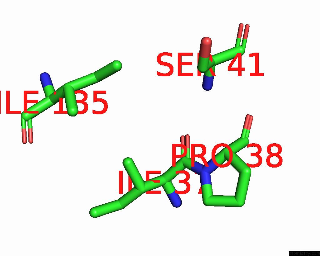
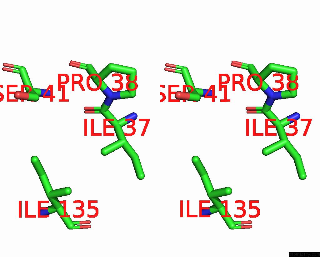
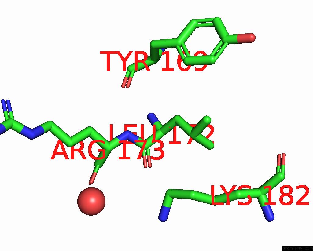
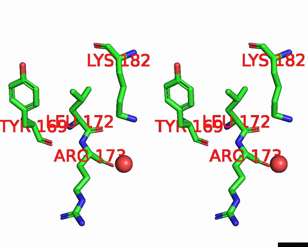
Iodine binding site 1 out of 8 in 6axr
Go back to
Iodine binding site 1 out
of 8 in the Structure of the P122A Mutant of the Hiv-1 Capsid Protein
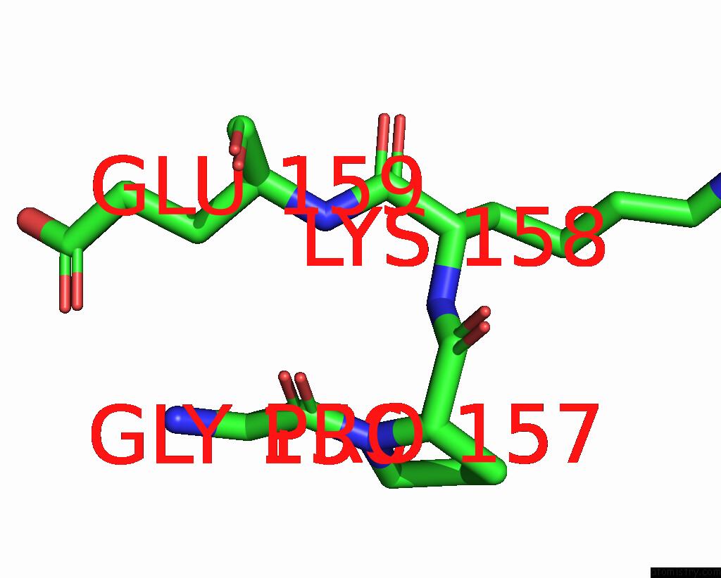
Mono view
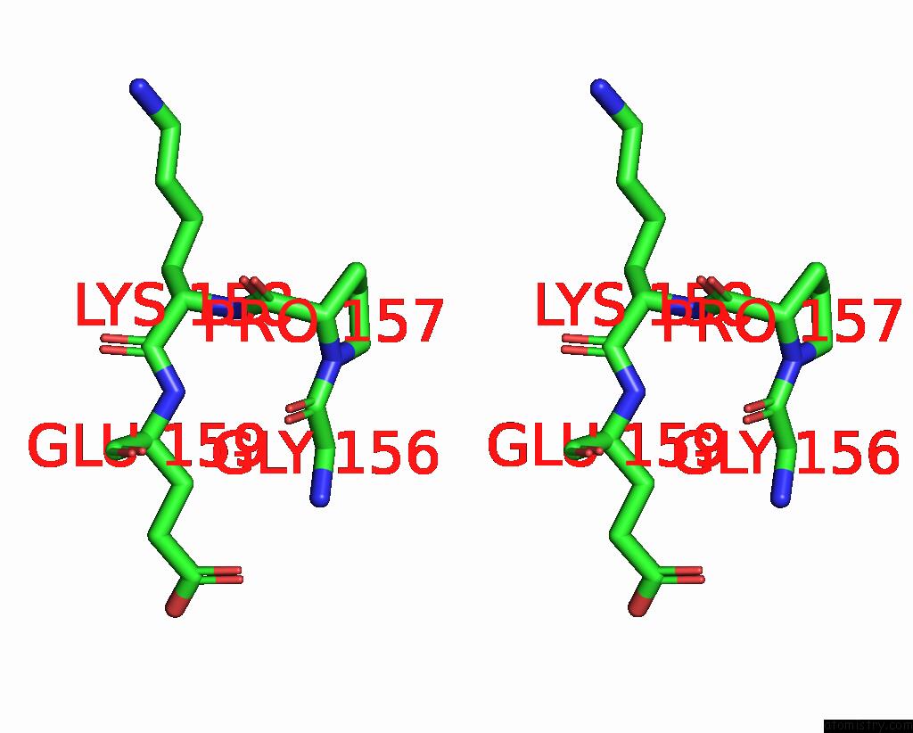
Stereo pair view
Mono view
Stereo pair view
A full contact list of Iodine with other atoms in the I binding
site number 1 of Structure of the P122A Mutant of the Hiv-1 Capsid Protein within 5.0Å range:
|
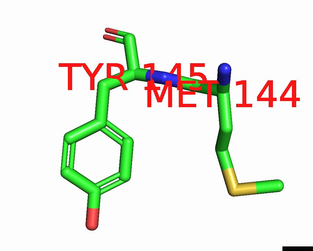
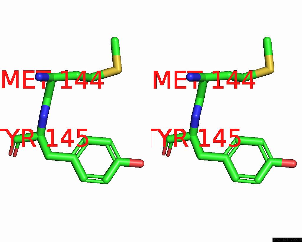
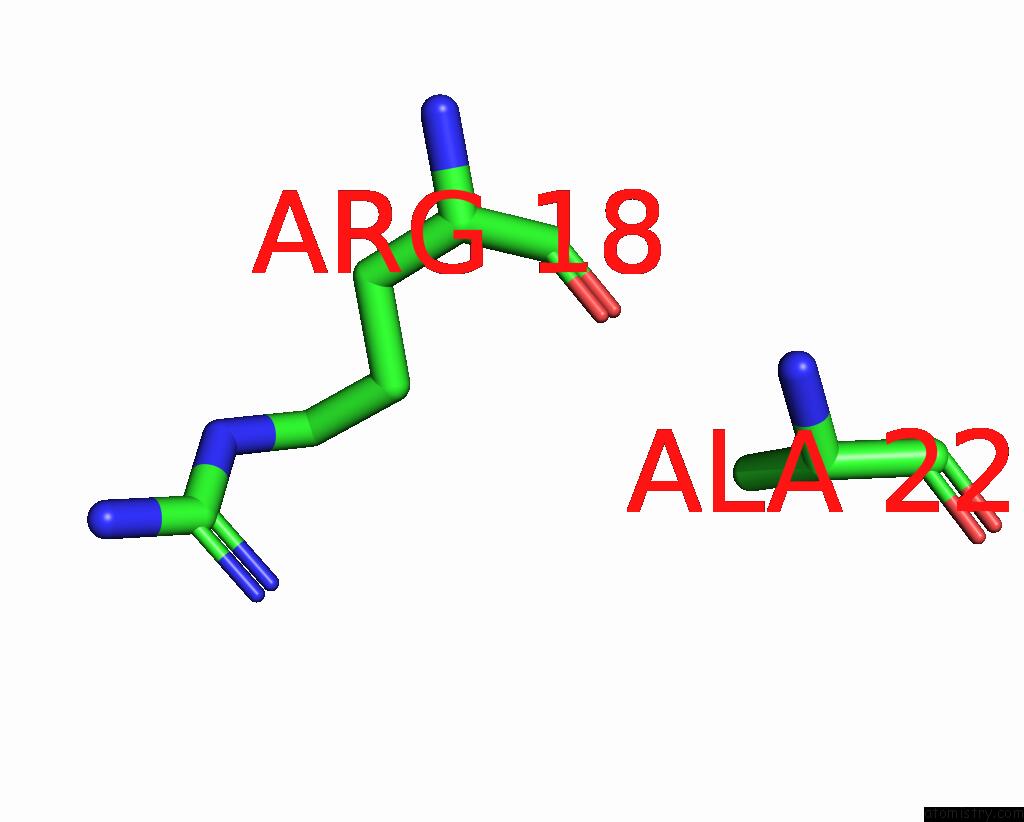
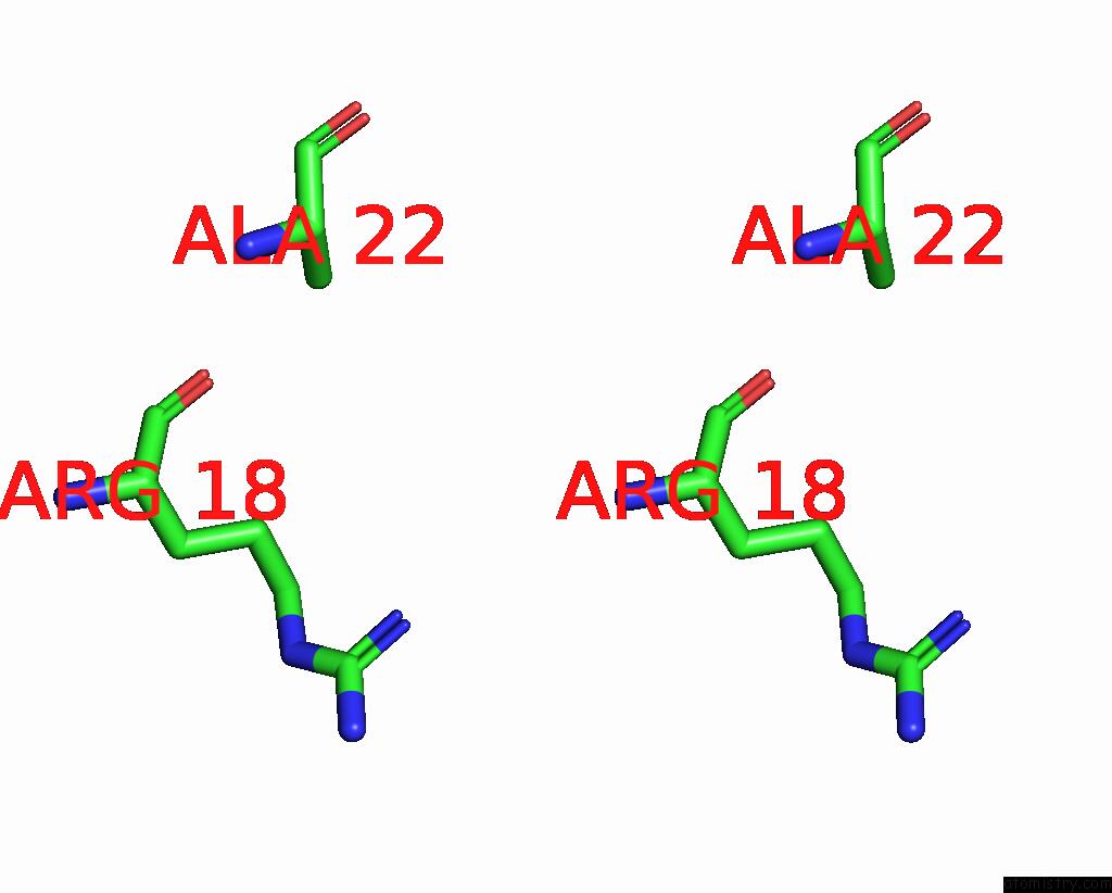
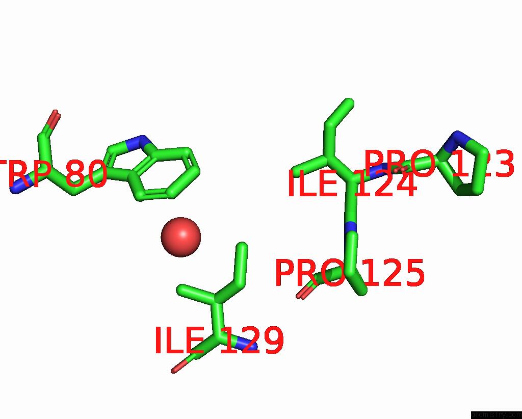
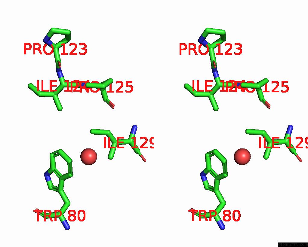
Iodine binding site 2 out of 8 in 6axr
Go back to
Iodine binding site 2 out
of 8 in the Structure of the P122A Mutant of the Hiv-1 Capsid Protein
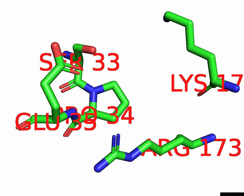
Mono view
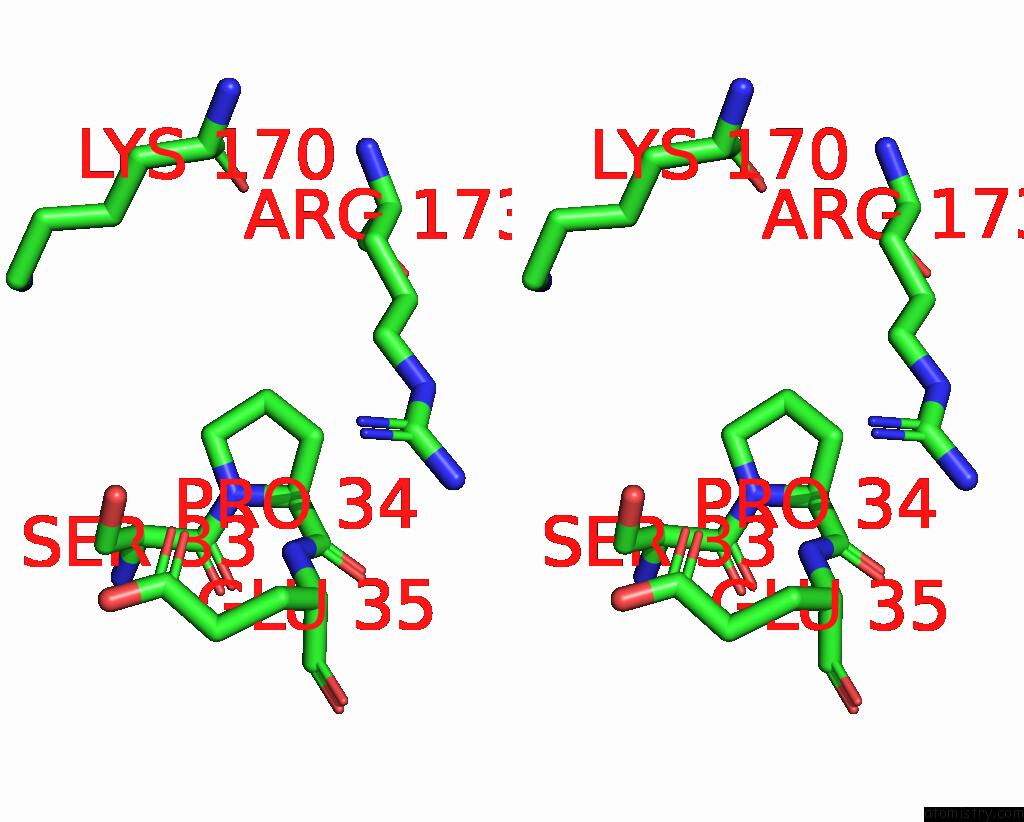
Stereo pair view
Mono view
Stereo pair view
A full contact list of Iodine with other atoms in the I binding
site number 2 of Structure of the P122A Mutant of the Hiv-1 Capsid Protein within 5.0Å range:
|
Iodine binding site 3 out of 8 in 6axr
Go back to
Iodine binding site 3 out
of 8 in the Structure of the P122A Mutant of the Hiv-1 Capsid Protein
Mono view
Stereo pair view
Mono view
Stereo pair view
A full contact list of Iodine with other atoms in the I binding
site number 3 of Structure of the P122A Mutant of the Hiv-1 Capsid Protein within 5.0Å range:
|
Iodine binding site 4 out of 8 in 6axr
Go back to
Iodine binding site 4 out
of 8 in the Structure of the P122A Mutant of the Hiv-1 Capsid Protein
Mono view
Stereo pair view
Mono view
Stereo pair view
A full contact list of Iodine with other atoms in the I binding
site number 4 of Structure of the P122A Mutant of the Hiv-1 Capsid Protein within 5.0Å range:
|
Iodine binding site 5 out of 8 in 6axr
Go back to
Iodine binding site 5 out
of 8 in the Structure of the P122A Mutant of the Hiv-1 Capsid Protein
Mono view
Stereo pair view
Mono view
Stereo pair view
A full contact list of Iodine with other atoms in the I binding
site number 5 of Structure of the P122A Mutant of the Hiv-1 Capsid Protein within 5.0Å range:
|
Iodine binding site 6 out of 8 in 6axr
Go back to
Iodine binding site 6 out
of 8 in the Structure of the P122A Mutant of the Hiv-1 Capsid Protein
Mono view
Stereo pair view
Mono view
Stereo pair view
A full contact list of Iodine with other atoms in the I binding
site number 6 of Structure of the P122A Mutant of the Hiv-1 Capsid Protein within 5.0Å range:
|
Iodine binding site 7 out of 8 in 6axr
Go back to
Iodine binding site 7 out
of 8 in the Structure of the P122A Mutant of the Hiv-1 Capsid Protein

Mono view
Stereo pair view
Mono view
Stereo pair view
A full contact list of Iodine with other atoms in the I binding
site number 7 of Structure of the P122A Mutant of the Hiv-1 Capsid Protein within 5.0Å range:
|
Iodine binding site 8 out of 8 in 6axr
Go back to
Iodine binding site 8 out
of 8 in the Structure of the P122A Mutant of the Hiv-1 Capsid Protein
Mono view
Stereo pair view
Mono view
Stereo pair view
A full contact list of Iodine with other atoms in the I binding
site number 8 of Structure of the P122A Mutant of the Hiv-1 Capsid Protein within 5.0Å range:
|
Reference:
M.Novikova,
L.J.Adams,
J.Fontana,
A.T.Gres,
M.Balasubramaniam,
D.C.Winkler,
S.B.Kudchodkar,
F.Soheilian,
S.G.Sarafianos,
A.C.Steven,
E.O.Freed.
Identification of A Structural Element in Hiv-1 Gag Required For Virus Particle Assembly and Maturation. Mbio V. 9 2018.
ISSN: ESSN 2150-7511
PubMed: 30327442
DOI: 10.1128/MBIO.01567-18
Page generated: Sun Aug 11 22:30:47 2024
ISSN: ESSN 2150-7511
PubMed: 30327442
DOI: 10.1128/MBIO.01567-18
Last articles
Zn in 9J0NZn in 9J0O
Zn in 9J0P
Zn in 9FJX
Zn in 9EKB
Zn in 9C0F
Zn in 9CAH
Zn in 9CH0
Zn in 9CH3
Zn in 9CH1