Iodine »
PDB 6axy-6doe »
6bxg »
Iodine in PDB 6bxg: 1.45 Angstrom Resolution Crystal Structure of Pdz Domain of Carboxy- Terminal Protease From Vibrio Cholerae in Complex with Peptide.
Protein crystallography data
The structure of 1.45 Angstrom Resolution Crystal Structure of Pdz Domain of Carboxy- Terminal Protease From Vibrio Cholerae in Complex with Peptide., PDB code: 6bxg
was solved by
G.Minasov,
L.Shuvalova,
E.V.Filippova,
O.Kiryukhina,
S.Grimshaw,
K.Kwon,
W.F.Anderson,
K.J.F.Satchell,
A.Joachimiak,
Center For Structuralgenomics Of Infectious Diseases (Csgid),
with X-Ray Crystallography technique. A brief refinement statistics is given in the table below:
| Resolution Low / High (Å) | 29.82 / 1.45 |
| Space group | P 65 |
| Cell size a, b, c (Å), α, β, γ (°) | 35.579, 35.579, 118.394, 90.00, 90.00, 120.00 |
| R / Rfree (%) | 13.2 / 17.8 |
Other elements in 6bxg:
The structure of 1.45 Angstrom Resolution Crystal Structure of Pdz Domain of Carboxy- Terminal Protease From Vibrio Cholerae in Complex with Peptide. also contains other interesting chemical elements:
| Chlorine | (Cl) | 2 atoms |
Iodine Binding Sites:
Pages:
>>> Page 1 <<< Page 2, Binding sites: 11 - 15;Binding sites:
The binding sites of Iodine atom in the 1.45 Angstrom Resolution Crystal Structure of Pdz Domain of Carboxy- Terminal Protease From Vibrio Cholerae in Complex with Peptide. (pdb code 6bxg). This binding sites where shown within 5.0 Angstroms radius around Iodine atom.In total 15 binding sites of Iodine where determined in the 1.45 Angstrom Resolution Crystal Structure of Pdz Domain of Carboxy- Terminal Protease From Vibrio Cholerae in Complex with Peptide., PDB code: 6bxg:
Jump to Iodine binding site number: 1; 2; 3; 4; 5; 6; 7; 8; 9; 10;
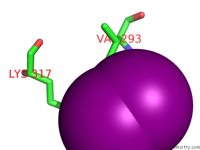
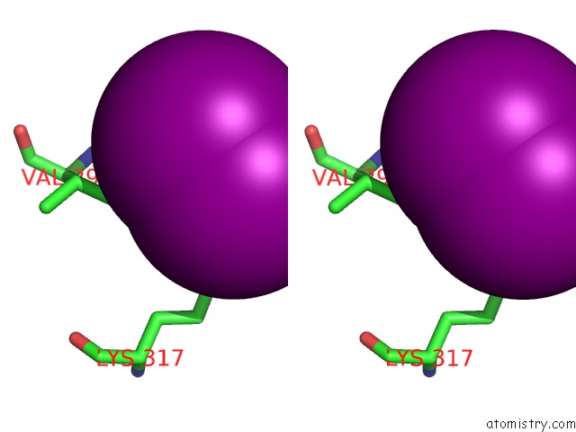
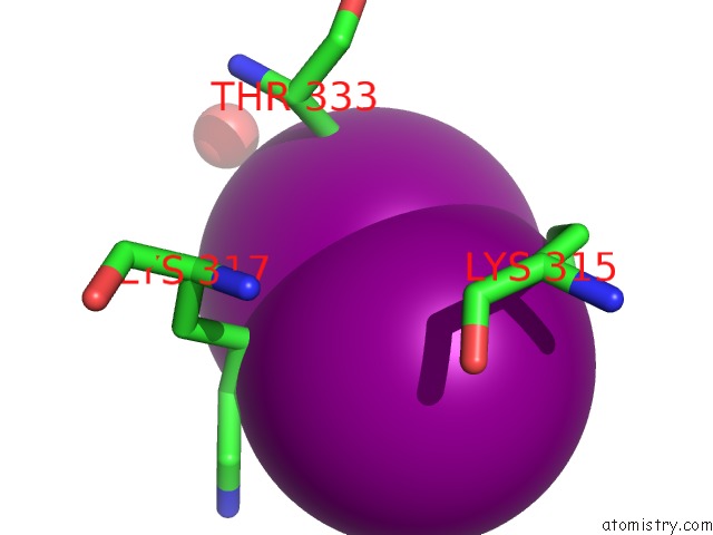
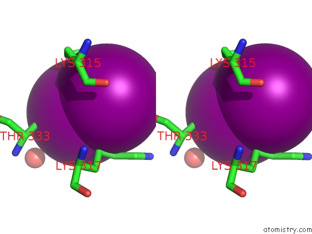
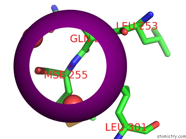
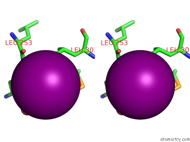
Iodine binding site 1 out of 15 in 6bxg
Go back to
Iodine binding site 1 out
of 15 in the 1.45 Angstrom Resolution Crystal Structure of Pdz Domain of Carboxy- Terminal Protease From Vibrio Cholerae in Complex with Peptide.
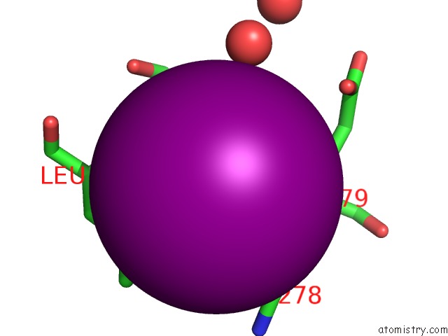
Mono view
Stereo pair view
Mono view
Stereo pair view
A full contact list of Iodine with other atoms in the I binding
site number 1 of 1.45 Angstrom Resolution Crystal Structure of Pdz Domain of Carboxy- Terminal Protease From Vibrio Cholerae in Complex with Peptide. within 5.0Å range:
|
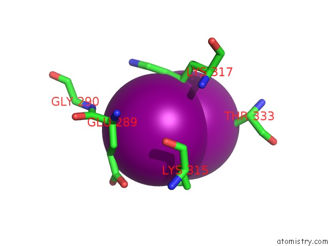
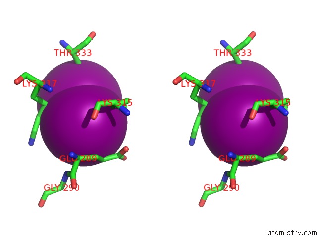
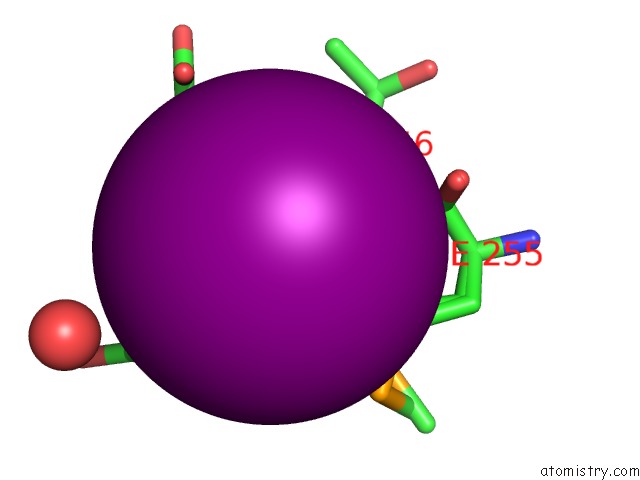
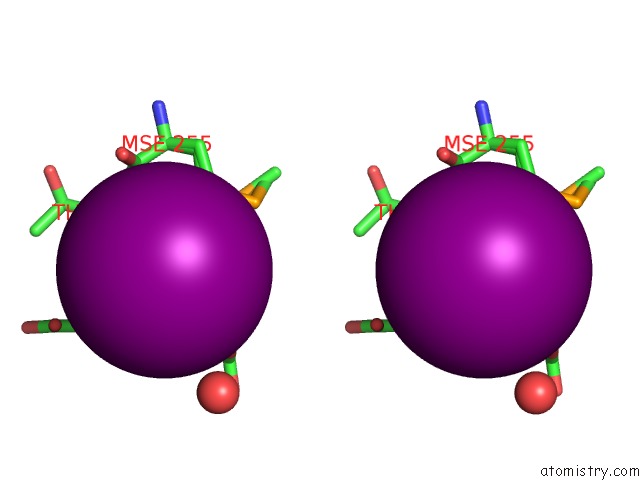
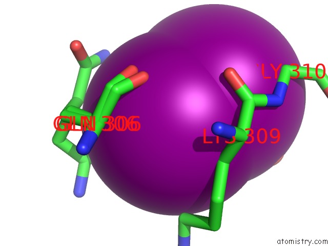
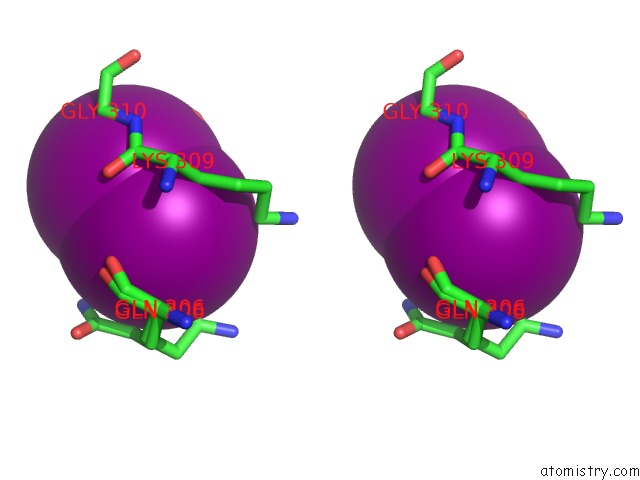
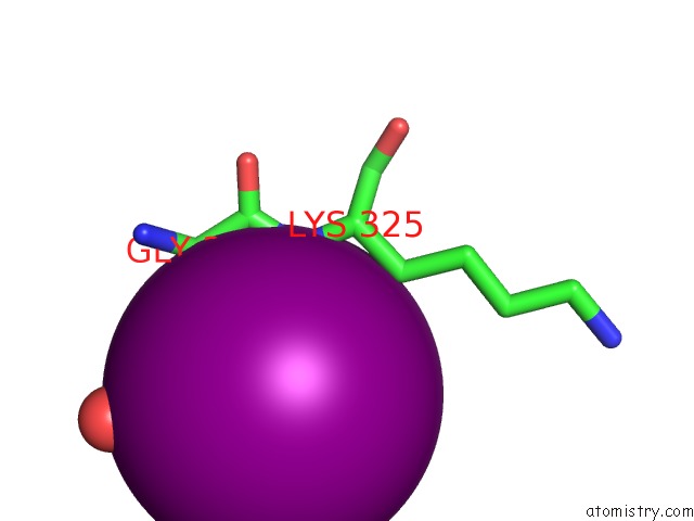
Iodine binding site 2 out of 15 in 6bxg
Go back to
Iodine binding site 2 out
of 15 in the 1.45 Angstrom Resolution Crystal Structure of Pdz Domain of Carboxy- Terminal Protease From Vibrio Cholerae in Complex with Peptide.
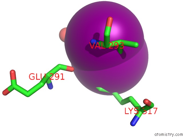
Mono view
Stereo pair view
Mono view
Stereo pair view
A full contact list of Iodine with other atoms in the I binding
site number 2 of 1.45 Angstrom Resolution Crystal Structure of Pdz Domain of Carboxy- Terminal Protease From Vibrio Cholerae in Complex with Peptide. within 5.0Å range:
|
Iodine binding site 3 out of 15 in 6bxg
Go back to
Iodine binding site 3 out
of 15 in the 1.45 Angstrom Resolution Crystal Structure of Pdz Domain of Carboxy- Terminal Protease From Vibrio Cholerae in Complex with Peptide.
Mono view
Stereo pair view
Mono view
Stereo pair view
A full contact list of Iodine with other atoms in the I binding
site number 3 of 1.45 Angstrom Resolution Crystal Structure of Pdz Domain of Carboxy- Terminal Protease From Vibrio Cholerae in Complex with Peptide. within 5.0Å range:
|
Iodine binding site 4 out of 15 in 6bxg
Go back to
Iodine binding site 4 out
of 15 in the 1.45 Angstrom Resolution Crystal Structure of Pdz Domain of Carboxy- Terminal Protease From Vibrio Cholerae in Complex with Peptide.
Mono view
Stereo pair view
Mono view
Stereo pair view
A full contact list of Iodine with other atoms in the I binding
site number 4 of 1.45 Angstrom Resolution Crystal Structure of Pdz Domain of Carboxy- Terminal Protease From Vibrio Cholerae in Complex with Peptide. within 5.0Å range:
|
Iodine binding site 5 out of 15 in 6bxg
Go back to
Iodine binding site 5 out
of 15 in the 1.45 Angstrom Resolution Crystal Structure of Pdz Domain of Carboxy- Terminal Protease From Vibrio Cholerae in Complex with Peptide.
Mono view
Stereo pair view
Mono view
Stereo pair view
A full contact list of Iodine with other atoms in the I binding
site number 5 of 1.45 Angstrom Resolution Crystal Structure of Pdz Domain of Carboxy- Terminal Protease From Vibrio Cholerae in Complex with Peptide. within 5.0Å range:
|
Iodine binding site 6 out of 15 in 6bxg
Go back to
Iodine binding site 6 out
of 15 in the 1.45 Angstrom Resolution Crystal Structure of Pdz Domain of Carboxy- Terminal Protease From Vibrio Cholerae in Complex with Peptide.
Mono view
Stereo pair view
Mono view
Stereo pair view
A full contact list of Iodine with other atoms in the I binding
site number 6 of 1.45 Angstrom Resolution Crystal Structure of Pdz Domain of Carboxy- Terminal Protease From Vibrio Cholerae in Complex with Peptide. within 5.0Å range:
|
Iodine binding site 7 out of 15 in 6bxg
Go back to
Iodine binding site 7 out
of 15 in the 1.45 Angstrom Resolution Crystal Structure of Pdz Domain of Carboxy- Terminal Protease From Vibrio Cholerae in Complex with Peptide.
Mono view
Stereo pair view
Mono view
Stereo pair view
A full contact list of Iodine with other atoms in the I binding
site number 7 of 1.45 Angstrom Resolution Crystal Structure of Pdz Domain of Carboxy- Terminal Protease From Vibrio Cholerae in Complex with Peptide. within 5.0Å range:
|
Iodine binding site 8 out of 15 in 6bxg
Go back to
Iodine binding site 8 out
of 15 in the 1.45 Angstrom Resolution Crystal Structure of Pdz Domain of Carboxy- Terminal Protease From Vibrio Cholerae in Complex with Peptide.
Mono view
Stereo pair view
Mono view
Stereo pair view
A full contact list of Iodine with other atoms in the I binding
site number 8 of 1.45 Angstrom Resolution Crystal Structure of Pdz Domain of Carboxy- Terminal Protease From Vibrio Cholerae in Complex with Peptide. within 5.0Å range:
|
Iodine binding site 9 out of 15 in 6bxg
Go back to
Iodine binding site 9 out
of 15 in the 1.45 Angstrom Resolution Crystal Structure of Pdz Domain of Carboxy- Terminal Protease From Vibrio Cholerae in Complex with Peptide.
Mono view
Stereo pair view
Mono view
Stereo pair view
A full contact list of Iodine with other atoms in the I binding
site number 9 of 1.45 Angstrom Resolution Crystal Structure of Pdz Domain of Carboxy- Terminal Protease From Vibrio Cholerae in Complex with Peptide. within 5.0Å range:
|
Iodine binding site 10 out of 15 in 6bxg
Go back to
Iodine binding site 10 out
of 15 in the 1.45 Angstrom Resolution Crystal Structure of Pdz Domain of Carboxy- Terminal Protease From Vibrio Cholerae in Complex with Peptide.
Mono view
Stereo pair view
Mono view
Stereo pair view
A full contact list of Iodine with other atoms in the I binding
site number 10 of 1.45 Angstrom Resolution Crystal Structure of Pdz Domain of Carboxy- Terminal Protease From Vibrio Cholerae in Complex with Peptide. within 5.0Å range:
|
Reference:
G.Minasov,
L.Shuvalova,
E.V.Filippova,
O.Kiryukhina,
S.Grimshaw,
K.Kwon,
W.F.Anderson,
K.J.F.Satchell,
A.Joachimiak,
Center For Structural Genomics Of Infectious Diseases(Csgid).
1.45 Angstrom Resolution Crystal Structure of Pdz Domain of Carboxy-Terminal Protease From Vibrio Cholerae in Complex with Peptide. To Be Published.
Page generated: Sun Aug 11 22:45:58 2024
Last articles
Zn in 9MJ5Zn in 9HNW
Zn in 9G0L
Zn in 9FNE
Zn in 9DZN
Zn in 9E0I
Zn in 9D32
Zn in 9DAK
Zn in 8ZXC
Zn in 8ZUF