Iodine »
PDB 6axy-6doe »
6cvl »
Iodine in PDB 6cvl: Crystal Structure of the Escherichia Coli Atpgs-Bound Metni Methionine Abc Transporter in Complex with Its Metq Binding Protein
Protein crystallography data
The structure of Crystal Structure of the Escherichia Coli Atpgs-Bound Metni Methionine Abc Transporter in Complex with Its Metq Binding Protein, PDB code: 6cvl
was solved by
P.T.Nguyen,
J.T.Kaiser,
D.C.Rees,
with X-Ray Crystallography technique. A brief refinement statistics is given in the table below:
| Resolution Low / High (Å) | 39.85 / 2.95 |
| Space group | P 32 2 1 |
| Cell size a, b, c (Å), α, β, γ (°) | 107.960, 107.960, 354.520, 90.00, 90.00, 120.00 |
| R / Rfree (%) | 20.8 / 22.6 |
Other elements in 6cvl:
The structure of Crystal Structure of the Escherichia Coli Atpgs-Bound Metni Methionine Abc Transporter in Complex with Its Metq Binding Protein also contains other interesting chemical elements:
| Mercury | (Hg) | 3 atoms |
Iodine Binding Sites:
The binding sites of Iodine atom in the Crystal Structure of the Escherichia Coli Atpgs-Bound Metni Methionine Abc Transporter in Complex with Its Metq Binding Protein
(pdb code 6cvl). This binding sites where shown within
5.0 Angstroms radius around Iodine atom.
In total 2 binding sites of Iodine where determined in the Crystal Structure of the Escherichia Coli Atpgs-Bound Metni Methionine Abc Transporter in Complex with Its Metq Binding Protein, PDB code: 6cvl:
Jump to Iodine binding site number: 1; 2;
In total 2 binding sites of Iodine where determined in the Crystal Structure of the Escherichia Coli Atpgs-Bound Metni Methionine Abc Transporter in Complex with Its Metq Binding Protein, PDB code: 6cvl:
Jump to Iodine binding site number: 1; 2;
Iodine binding site 1 out of 2 in 6cvl
Go back to
Iodine binding site 1 out
of 2 in the Crystal Structure of the Escherichia Coli Atpgs-Bound Metni Methionine Abc Transporter in Complex with Its Metq Binding Protein
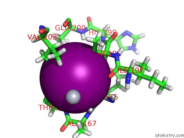
Mono view
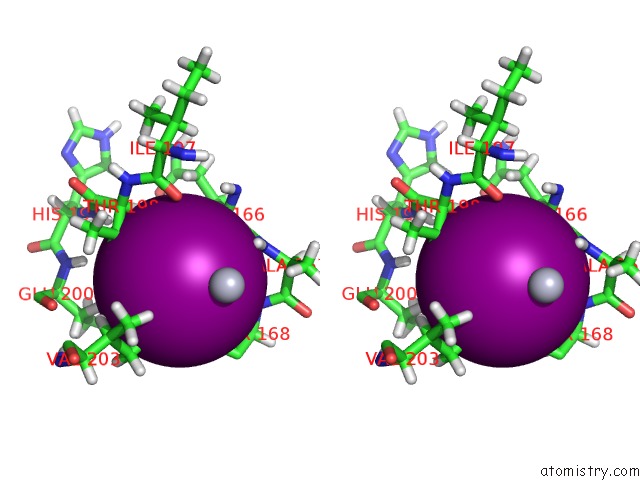
Stereo pair view
Mono view
Stereo pair view
A full contact list of Iodine with other atoms in the I binding
site number 1 of Crystal Structure of the Escherichia Coli Atpgs-Bound Metni Methionine Abc Transporter in Complex with Its Metq Binding Protein within 5.0Å range:
|
Iodine binding site 2 out of 2 in 6cvl
Go back to
Iodine binding site 2 out
of 2 in the Crystal Structure of the Escherichia Coli Atpgs-Bound Metni Methionine Abc Transporter in Complex with Its Metq Binding Protein
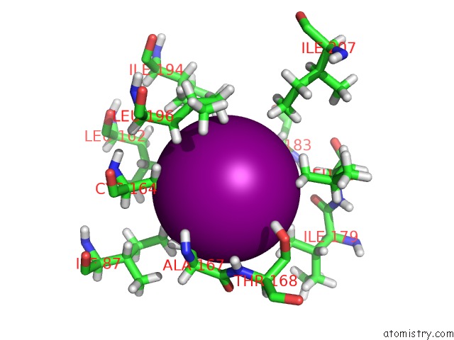
Mono view
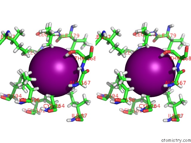
Stereo pair view
Mono view
Stereo pair view
A full contact list of Iodine with other atoms in the I binding
site number 2 of Crystal Structure of the Escherichia Coli Atpgs-Bound Metni Methionine Abc Transporter in Complex with Its Metq Binding Protein within 5.0Å range:
|
Reference:
P.T.Nguyen,
J.Y.Lai,
A.T.Lee,
J.T.Kaiser,
D.C.Rees.
Noncanonical Role For the Binding Protein in Substrate Uptake By the Metni Methionine Atp Binding Cassette (Abc) Transporter. Proc. Natl. Acad. Sci. V. 115 10596 2018U.S.A..
ISSN: ESSN 1091-6490
PubMed: 30352853
DOI: 10.1073/PNAS.1811003115
Page generated: Fri Aug 8 21:08:03 2025
ISSN: ESSN 1091-6490
PubMed: 30352853
DOI: 10.1073/PNAS.1811003115
Last articles
K in 4EVYK in 4EOU
K in 4ETM
K in 4ESK
K in 4ES8
K in 4ERT
K in 4ERD
K in 4ENC
K in 4EK1
K in 4ENB