Iodine »
PDB 6eql-6k60 »
6eql »
Iodine in PDB 6eql: Crystal Structure of Human Glycogenin-1 (GYG1) TYR195PIPHE Mutant Complexed with Manganese and Udp
Enzymatic activity of Crystal Structure of Human Glycogenin-1 (GYG1) TYR195PIPHE Mutant Complexed with Manganese and Udp
All present enzymatic activity of Crystal Structure of Human Glycogenin-1 (GYG1) TYR195PIPHE Mutant Complexed with Manganese and Udp:
2.4.1.186;
2.4.1.186;
Protein crystallography data
The structure of Crystal Structure of Human Glycogenin-1 (GYG1) TYR195PIPHE Mutant Complexed with Manganese and Udp, PDB code: 6eql
was solved by
H.J.Bailey,
J.Kopec,
M.K.Bilyard,
G.A.Bezerra,
S.Seo Lee,
C.H.Arrowsmith,
A.M.Edwards,
C.Bountra,
B.G.Davis,
W.W.Yue,
with X-Ray Crystallography technique. A brief refinement statistics is given in the table below:
| Resolution Low / High (Å) | 46.55 / 2.38 |
| Space group | P 21 21 21 |
| Cell size a, b, c (Å), α, β, γ (°) | 66.170, 81.050, 111.200, 90.00, 90.00, 90.00 |
| R / Rfree (%) | 22.5 / 28.1 |
Other elements in 6eql:
The structure of Crystal Structure of Human Glycogenin-1 (GYG1) TYR195PIPHE Mutant Complexed with Manganese and Udp also contains other interesting chemical elements:
| Manganese | (Mn) | 2 atoms |
Iodine Binding Sites:
The binding sites of Iodine atom in the Crystal Structure of Human Glycogenin-1 (GYG1) TYR195PIPHE Mutant Complexed with Manganese and Udp
(pdb code 6eql). This binding sites where shown within
5.0 Angstroms radius around Iodine atom.
In total only one binding site of Iodine was determined in the Crystal Structure of Human Glycogenin-1 (GYG1) TYR195PIPHE Mutant Complexed with Manganese and Udp, PDB code: 6eql:
In total only one binding site of Iodine was determined in the Crystal Structure of Human Glycogenin-1 (GYG1) TYR195PIPHE Mutant Complexed with Manganese and Udp, PDB code: 6eql:
Iodine binding site 1 out of 1 in 6eql
Go back to
Iodine binding site 1 out
of 1 in the Crystal Structure of Human Glycogenin-1 (GYG1) TYR195PIPHE Mutant Complexed with Manganese and Udp
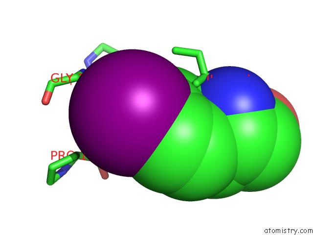
Mono view
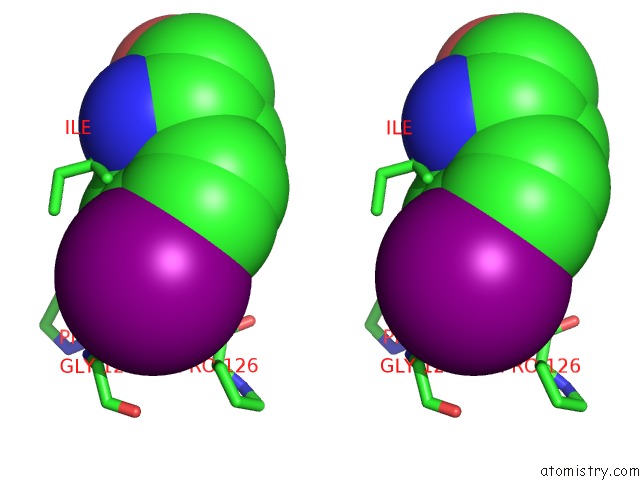
Stereo pair view
Mono view
Stereo pair view
A full contact list of Iodine with other atoms in the I binding
site number 1 of Crystal Structure of Human Glycogenin-1 (GYG1) TYR195PIPHE Mutant Complexed with Manganese and Udp within 5.0Å range:
|
Reference:
M.K.Bilyard,
H.J.Bailey,
L.Raich,
M.A.Gafitescu,
T.Machida,
J.Iglesias-Fernandez,
S.S.Lee,
C.D.Spicer,
C.Rovira,
W.W.Yue,
B.G.Davis.
Palladium-Mediated Enzyme Activation Suggests Multiphase Initiation of Glycogenesis. Nature V. 563 235 2018.
ISSN: ESSN 1476-4687
PubMed: 30356213
DOI: 10.1038/S41586-018-0644-7
Page generated: Fri Aug 8 21:24:23 2025
ISSN: ESSN 1476-4687
PubMed: 30356213
DOI: 10.1038/S41586-018-0644-7
Last articles
Mg in 1XPOMg in 1XP7
Mg in 1XPF
Mg in 1XPE
Mg in 1XP5
Mg in 1XP0
Mg in 1XOZ
Mg in 1XOR
Mg in 1XOT
Mg in 1XOQ