Iodine »
PDB 6eql-6k60 »
6gaj »
Iodine in PDB 6gaj: Crystal Structure of the T1L Reovirus SIGMA1 Coiled Coil Tail (Iodide)
Protein crystallography data
The structure of Crystal Structure of the T1L Reovirus SIGMA1 Coiled Coil Tail (Iodide), PDB code: 6gaj
was solved by
M.H.Dietrich,
T.Stehle,
with X-Ray Crystallography technique. A brief refinement statistics is given in the table below:
| Resolution Low / High (Å) | 48.73 / 1.35 |
| Space group | P 1 21 1 |
| Cell size a, b, c (Å), α, β, γ (°) | 52.700, 37.590, 89.720, 90.00, 100.53, 90.00 |
| R / Rfree (%) | 17.5 / 20.3 |
Other elements in 6gaj:
The structure of Crystal Structure of the T1L Reovirus SIGMA1 Coiled Coil Tail (Iodide) also contains other interesting chemical elements:
| Chlorine | (Cl) | 4 atoms |
Iodine Binding Sites:
The binding sites of Iodine atom in the Crystal Structure of the T1L Reovirus SIGMA1 Coiled Coil Tail (Iodide)
(pdb code 6gaj). This binding sites where shown within
5.0 Angstroms radius around Iodine atom.
In total 3 binding sites of Iodine where determined in the Crystal Structure of the T1L Reovirus SIGMA1 Coiled Coil Tail (Iodide), PDB code: 6gaj:
Jump to Iodine binding site number: 1; 2; 3;
In total 3 binding sites of Iodine where determined in the Crystal Structure of the T1L Reovirus SIGMA1 Coiled Coil Tail (Iodide), PDB code: 6gaj:
Jump to Iodine binding site number: 1; 2; 3;
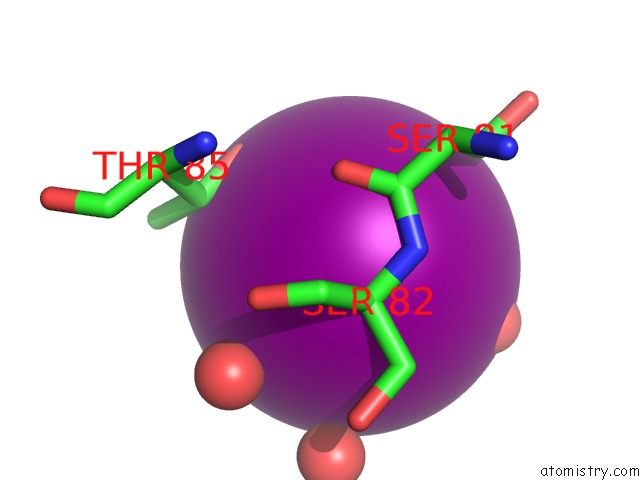
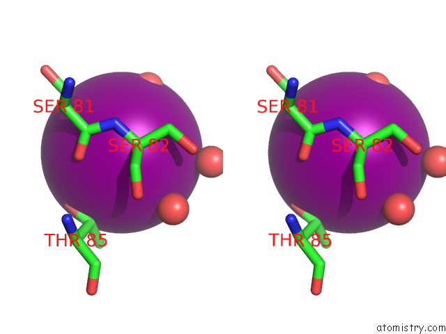
Iodine binding site 1 out of 3 in 6gaj
Go back to
Iodine binding site 1 out
of 3 in the Crystal Structure of the T1L Reovirus SIGMA1 Coiled Coil Tail (Iodide)
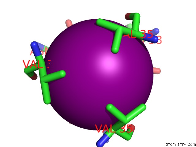
Mono view
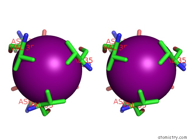
Stereo pair view
Mono view
Stereo pair view
A full contact list of Iodine with other atoms in the I binding
site number 1 of Crystal Structure of the T1L Reovirus SIGMA1 Coiled Coil Tail (Iodide) within 5.0Å range:
|
Iodine binding site 2 out of 3 in 6gaj
Go back to
Iodine binding site 2 out
of 3 in the Crystal Structure of the T1L Reovirus SIGMA1 Coiled Coil Tail (Iodide)
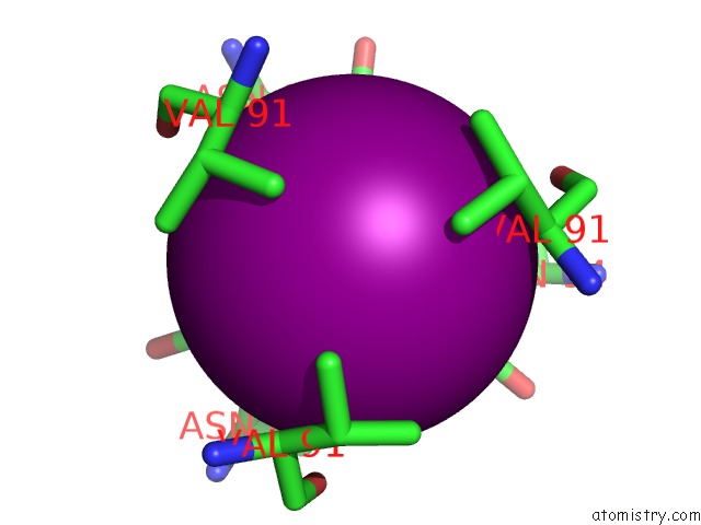
Mono view
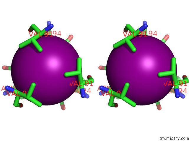
Stereo pair view
Mono view
Stereo pair view
A full contact list of Iodine with other atoms in the I binding
site number 2 of Crystal Structure of the T1L Reovirus SIGMA1 Coiled Coil Tail (Iodide) within 5.0Å range:
|
Iodine binding site 3 out of 3 in 6gaj
Go back to
Iodine binding site 3 out
of 3 in the Crystal Structure of the T1L Reovirus SIGMA1 Coiled Coil Tail (Iodide)
Mono view
Stereo pair view
Mono view
Stereo pair view
A full contact list of Iodine with other atoms in the I binding
site number 3 of Crystal Structure of the T1L Reovirus SIGMA1 Coiled Coil Tail (Iodide) within 5.0Å range:
|
Reference:
M.H.Dietrich,
K.M.Ogden,
J.M.Long,
R.Ebenhoch,
A.Thor,
T.S.Dermody,
T.Stehle.
Structural and Functional Features of the Reovirus Sigma 1 Tail. J. Virol. V. 92 2018.
ISSN: ESSN 1098-5514
PubMed: 29695426
DOI: 10.1128/JVI.00336-18
Page generated: Sun Aug 11 23:15:02 2024
ISSN: ESSN 1098-5514
PubMed: 29695426
DOI: 10.1128/JVI.00336-18
Last articles
Zn in 9J0NZn in 9J0O
Zn in 9J0P
Zn in 9FJX
Zn in 9EKB
Zn in 9C0F
Zn in 9CAH
Zn in 9CH0
Zn in 9CH3
Zn in 9CH1