Iodine »
PDB 6eql-6k60 »
6iuh »
Iodine in PDB 6iuh: Crystal Structure of GIT1 Pbd Domain in Complex with Liprin-ALPHA2
Protein crystallography data
The structure of Crystal Structure of GIT1 Pbd Domain in Complex with Liprin-ALPHA2, PDB code: 6iuh
was solved by
M.Liang,
Z.Wei,
with X-Ray Crystallography technique. A brief refinement statistics is given in the table below:
| Resolution Low / High (Å) | 49.57 / 1.80 |
| Space group | P 21 21 21 |
| Cell size a, b, c (Å), α, β, γ (°) | 88.200, 38.617, 99.134, 90.00, 90.00, 90.00 |
| R / Rfree (%) | 22.7 / 26.7 |
Iodine Binding Sites:
The binding sites of Iodine atom in the Crystal Structure of GIT1 Pbd Domain in Complex with Liprin-ALPHA2
(pdb code 6iuh). This binding sites where shown within
5.0 Angstroms radius around Iodine atom.
In total 3 binding sites of Iodine where determined in the Crystal Structure of GIT1 Pbd Domain in Complex with Liprin-ALPHA2, PDB code: 6iuh:
Jump to Iodine binding site number: 1; 2; 3;
In total 3 binding sites of Iodine where determined in the Crystal Structure of GIT1 Pbd Domain in Complex with Liprin-ALPHA2, PDB code: 6iuh:
Jump to Iodine binding site number: 1; 2; 3;
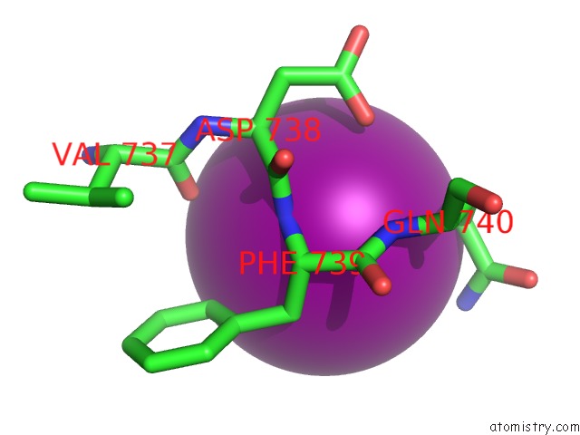
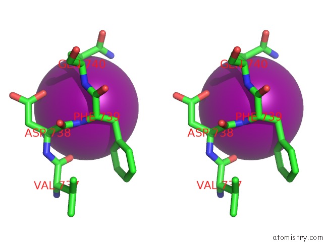
Iodine binding site 1 out of 3 in 6iuh
Go back to
Iodine binding site 1 out
of 3 in the Crystal Structure of GIT1 Pbd Domain in Complex with Liprin-ALPHA2
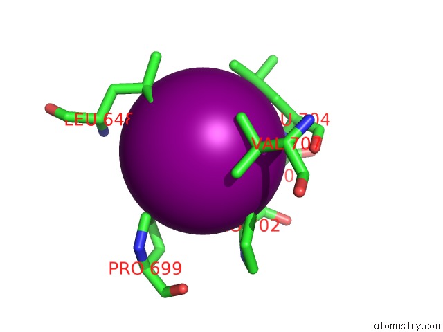
Mono view
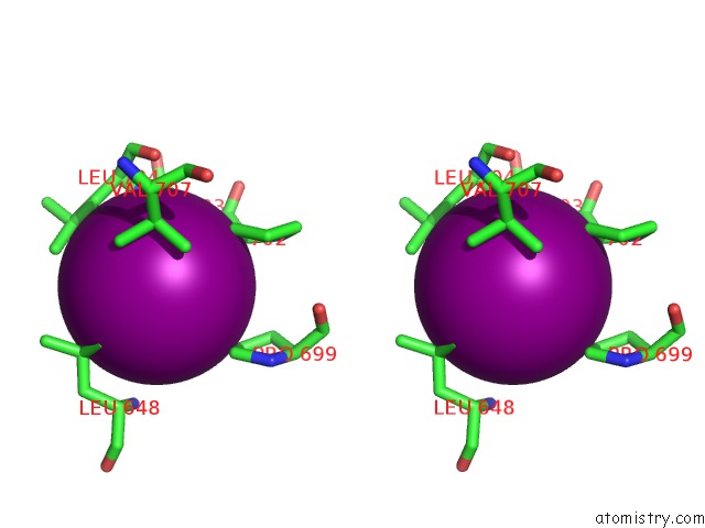
Stereo pair view
Mono view
Stereo pair view
A full contact list of Iodine with other atoms in the I binding
site number 1 of Crystal Structure of GIT1 Pbd Domain in Complex with Liprin-ALPHA2 within 5.0Å range:
|
Iodine binding site 2 out of 3 in 6iuh
Go back to
Iodine binding site 2 out
of 3 in the Crystal Structure of GIT1 Pbd Domain in Complex with Liprin-ALPHA2
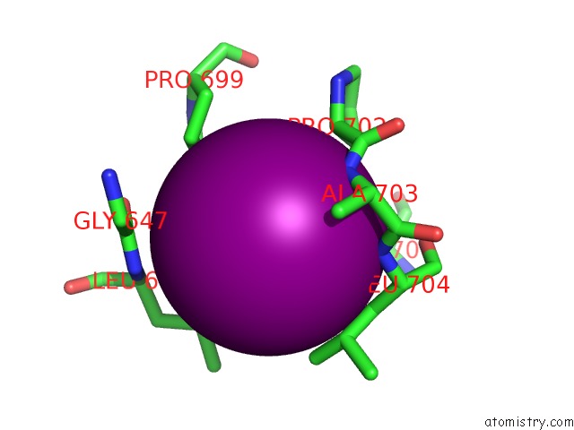
Mono view
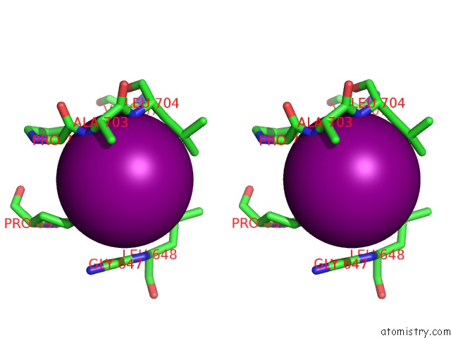
Stereo pair view
Mono view
Stereo pair view
A full contact list of Iodine with other atoms in the I binding
site number 2 of Crystal Structure of GIT1 Pbd Domain in Complex with Liprin-ALPHA2 within 5.0Å range:
|
Iodine binding site 3 out of 3 in 6iuh
Go back to
Iodine binding site 3 out
of 3 in the Crystal Structure of GIT1 Pbd Domain in Complex with Liprin-ALPHA2
Mono view
Stereo pair view
Mono view
Stereo pair view
A full contact list of Iodine with other atoms in the I binding
site number 3 of Crystal Structure of GIT1 Pbd Domain in Complex with Liprin-ALPHA2 within 5.0Å range:
|
Reference:
M.Liang,
X.Xie,
J.Pan,
G.Jin,
C.Yu,
Z.Wei.
Structural Basis of the Target-Binding Mode of the G Protein-Coupled Receptor Kinase-Interacting Protein in the Regulation of Focal Adhesion Dynamics. J. Biol. Chem. V. 294 5827 2019.
ISSN: ESSN 1083-351X
PubMed: 30737283
DOI: 10.1074/JBC.RA118.006915
Page generated: Sun Aug 11 23:16:28 2024
ISSN: ESSN 1083-351X
PubMed: 30737283
DOI: 10.1074/JBC.RA118.006915
Last articles
Zn in 9J0NZn in 9J0O
Zn in 9J0P
Zn in 9FJX
Zn in 9EKB
Zn in 9C0F
Zn in 9CAH
Zn in 9CH0
Zn in 9CH3
Zn in 9CH1