Iodine »
PDB 6k9o-6nfk »
6n7o »
Iodine in PDB 6n7o: Crystal Structure of GIL01 GP7
Protein crystallography data
The structure of Crystal Structure of GIL01 GP7, PDB code: 6n7o
was solved by
N.A.Caveney,
N.C.J.Strynadka,
with X-Ray Crystallography technique. A brief refinement statistics is given in the table below:
| Resolution Low / High (Å) | 32.16 / 1.70 |
| Space group | P 21 21 21 |
| Cell size a, b, c (Å), α, β, γ (°) | 39.690, 44.700, 46.300, 90.00, 90.00, 90.00 |
| R / Rfree (%) | 19.3 / 23 |
Iodine Binding Sites:
The binding sites of Iodine atom in the Crystal Structure of GIL01 GP7
(pdb code 6n7o). This binding sites where shown within
5.0 Angstroms radius around Iodine atom.
In total 3 binding sites of Iodine where determined in the Crystal Structure of GIL01 GP7, PDB code: 6n7o:
Jump to Iodine binding site number: 1; 2; 3;
In total 3 binding sites of Iodine where determined in the Crystal Structure of GIL01 GP7, PDB code: 6n7o:
Jump to Iodine binding site number: 1; 2; 3;
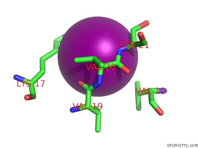
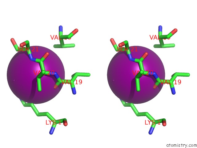
Iodine binding site 1 out of 3 in 6n7o
Go back to
Iodine binding site 1 out
of 3 in the Crystal Structure of GIL01 GP7
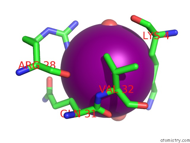
Mono view
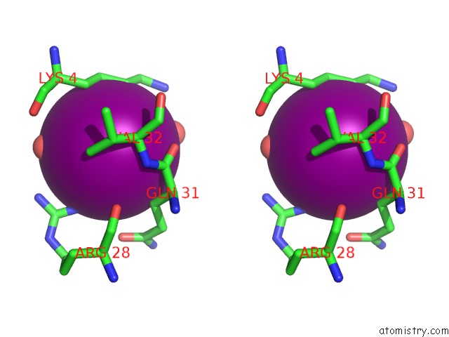
Stereo pair view
Mono view
Stereo pair view
A full contact list of Iodine with other atoms in the I binding
site number 1 of Crystal Structure of GIL01 GP7 within 5.0Å range:
|
Iodine binding site 2 out of 3 in 6n7o
Go back to
Iodine binding site 2 out
of 3 in the Crystal Structure of GIL01 GP7
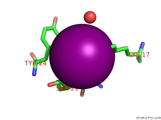
Mono view
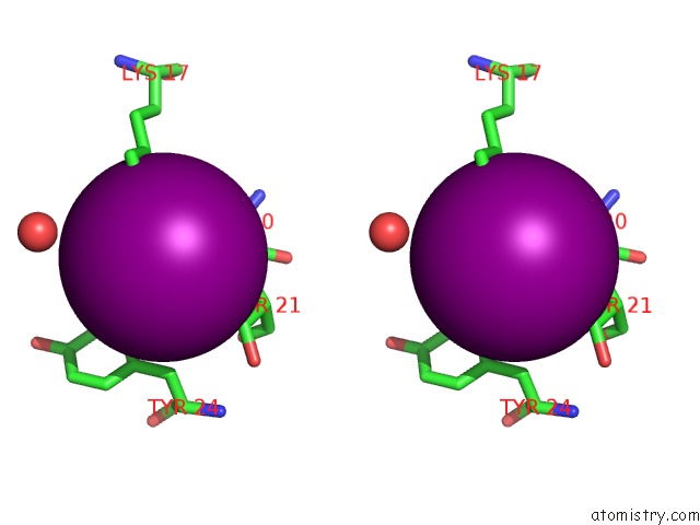
Stereo pair view
Mono view
Stereo pair view
A full contact list of Iodine with other atoms in the I binding
site number 2 of Crystal Structure of GIL01 GP7 within 5.0Å range:
|
Iodine binding site 3 out of 3 in 6n7o
Go back to
Iodine binding site 3 out
of 3 in the Crystal Structure of GIL01 GP7
Mono view
Stereo pair view
Mono view
Stereo pair view
A full contact list of Iodine with other atoms in the I binding
site number 3 of Crystal Structure of GIL01 GP7 within 5.0Å range:
|
Reference:
N.A.Caveney,
A.Pavlin,
G.Caballero,
M.Bahun,
V.Hodnik,
L.De Castro,
N.Fornelos,
M.Butala,
N.C.J.Strynadka.
Structural Insights Into Bacteriophage GIL01 GP7 Inhibition of Host Lexa Repressor. Structure V. 27 1094 2019.
ISSN: ISSN 0969-2126
PubMed: 31056420
DOI: 10.1016/J.STR.2019.03.019
Page generated: Sun Aug 11 23:40:32 2024
ISSN: ISSN 0969-2126
PubMed: 31056420
DOI: 10.1016/J.STR.2019.03.019
Last articles
F in 7FUIF in 7FU0
F in 7FU4
F in 7FTT
F in 7FU2
F in 7FTM
F in 7FTS
F in 7FTQ
F in 7FQL
F in 7FTO