Iodine »
PDB 6w0u-7ben »
6wgy »
Iodine in PDB 6wgy: Crystal Structure of A Putative Citrate Synthase 2 From Mycobacterium Bovis in Complex with Citrate
Enzymatic activity of Crystal Structure of A Putative Citrate Synthase 2 From Mycobacterium Bovis in Complex with Citrate
All present enzymatic activity of Crystal Structure of A Putative Citrate Synthase 2 From Mycobacterium Bovis in Complex with Citrate:
2.3.3.16;
2.3.3.16;
Protein crystallography data
The structure of Crystal Structure of A Putative Citrate Synthase 2 From Mycobacterium Bovis in Complex with Citrate, PDB code: 6wgy
was solved by
Seattle Structural Genomics Center For Infectious Disease (Ssgcid),
with X-Ray Crystallography technique. A brief refinement statistics is given in the table below:
| Resolution Low / High (Å) | 47.43 / 2.30 |
| Space group | P 21 21 2 |
| Cell size a, b, c (Å), α, β, γ (°) | 127.140, 153.370, 100.580, 90.00, 90.00, 90.00 |
| R / Rfree (%) | 17.4 / 22.5 |
Iodine Binding Sites:
Pages:
>>> Page 1 <<< Page 2, Binding sites: 11 - 20; Page 3, Binding sites: 21 - 30; Page 4, Binding sites: 31 - 40; Page 5, Binding sites: 41 - 50; Page 6, Binding sites: 51 - 60; Page 7, Binding sites: 61 - 70; Page 8, Binding sites: 71 - 80; Page 9, Binding sites: 81 - 90; Page 10, Binding sites: 91 - 100; Page 11, Binding sites: 101 - 110; Page 12, Binding sites: 111 - 112;Binding sites:
The binding sites of Iodine atom in the Crystal Structure of A Putative Citrate Synthase 2 From Mycobacterium Bovis in Complex with Citrate (pdb code 6wgy). This binding sites where shown within 5.0 Angstroms radius around Iodine atom.In total 112 binding sites of Iodine where determined in the Crystal Structure of A Putative Citrate Synthase 2 From Mycobacterium Bovis in Complex with Citrate, PDB code: 6wgy:
Jump to Iodine binding site number: 1; 2; 3; 4; 5; 6; 7; 8; 9; 10;
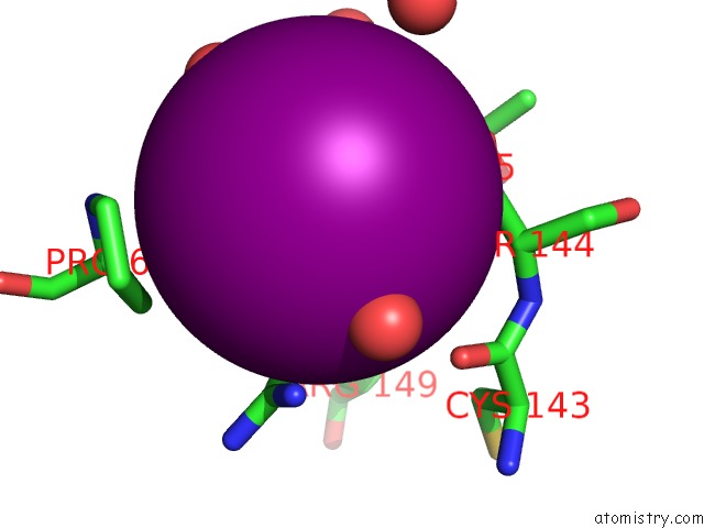
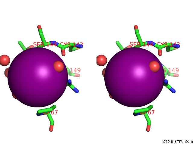
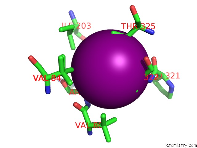
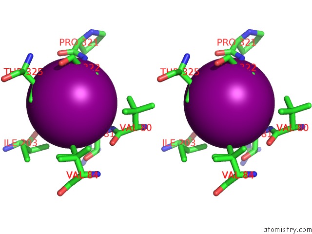
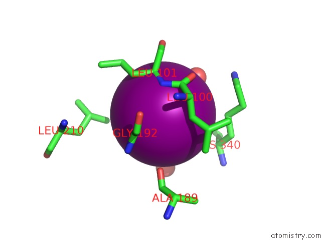
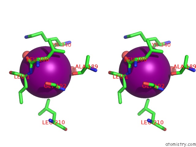
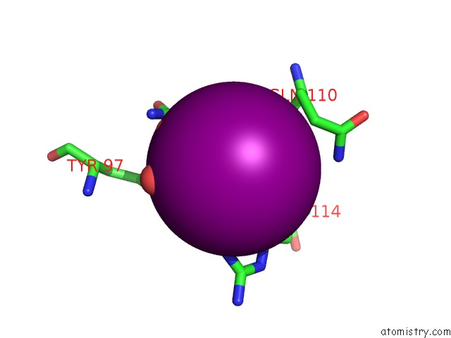
Iodine binding site 1 out of 112 in 6wgy
Go back to
Iodine binding site 1 out
of 112 in the Crystal Structure of A Putative Citrate Synthase 2 From Mycobacterium Bovis in Complex with Citrate
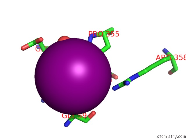
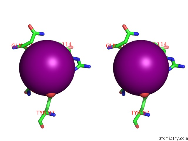
Mono view
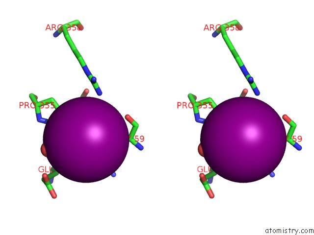
Stereo pair view
Mono view
Stereo pair view
A full contact list of Iodine with other atoms in the I binding
site number 1 of Crystal Structure of A Putative Citrate Synthase 2 From Mycobacterium Bovis in Complex with Citrate within 5.0Å range:
|
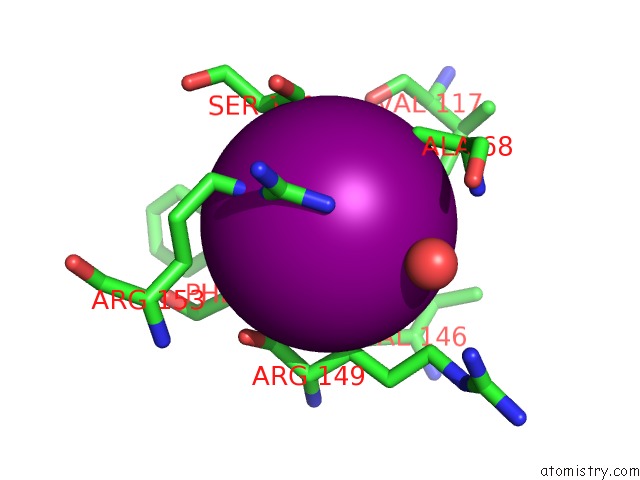
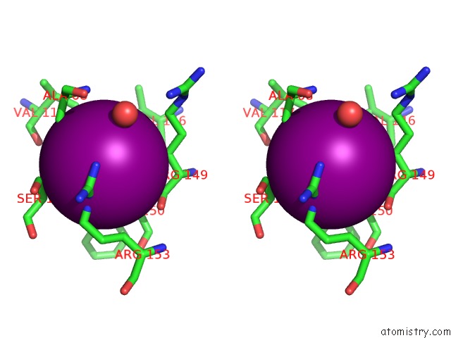
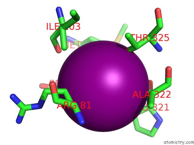
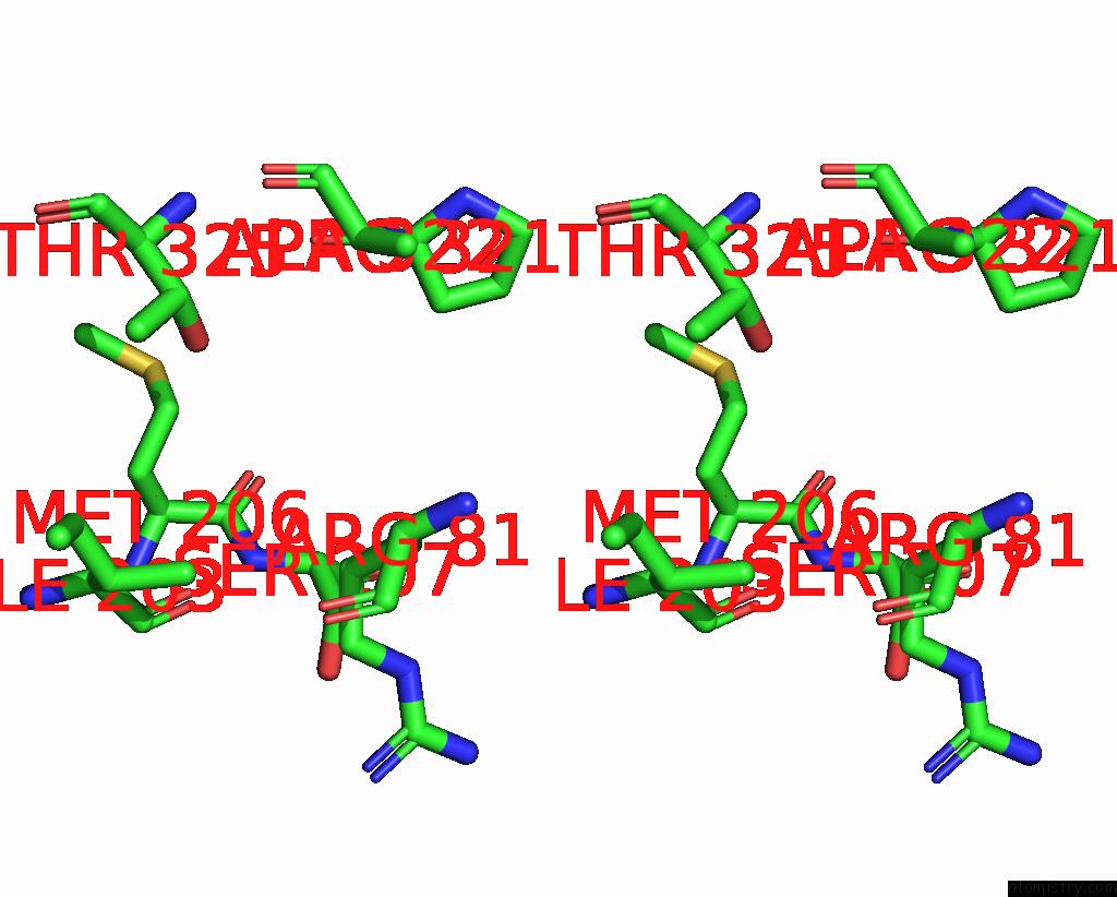
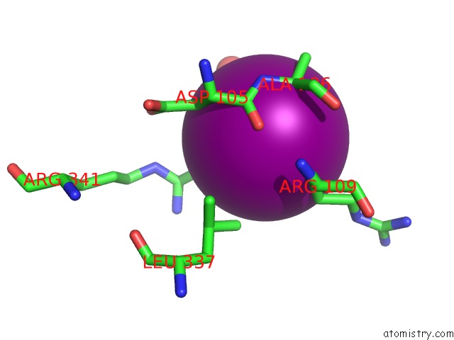
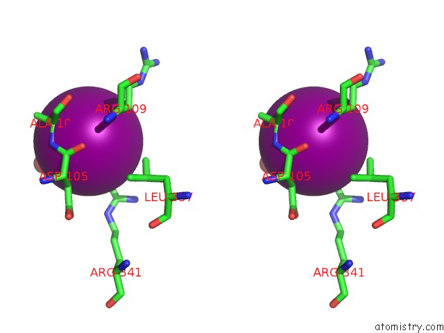
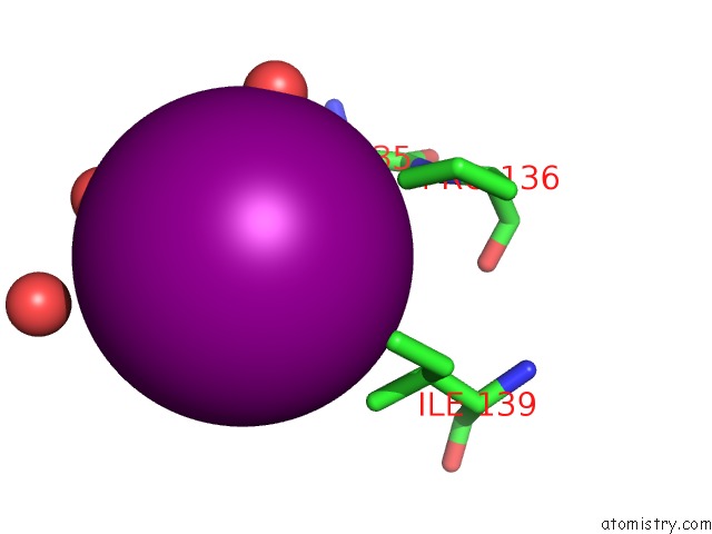
Iodine binding site 2 out of 112 in 6wgy
Go back to
Iodine binding site 2 out
of 112 in the Crystal Structure of A Putative Citrate Synthase 2 From Mycobacterium Bovis in Complex with Citrate
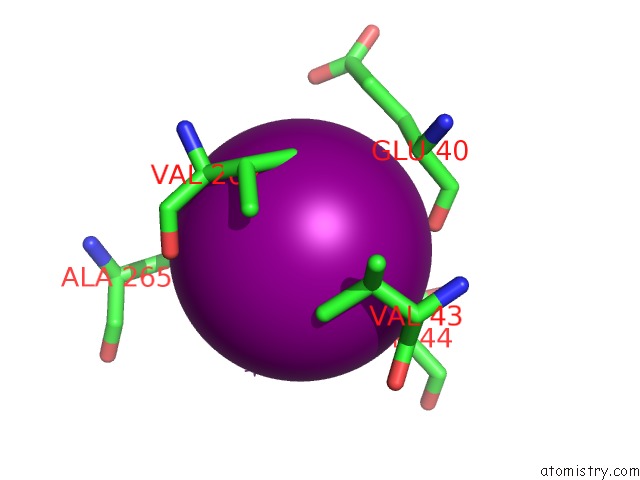
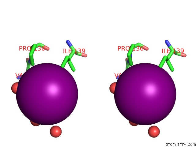
Mono view
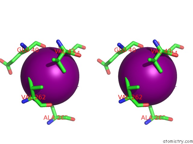
Stereo pair view
Mono view
Stereo pair view
A full contact list of Iodine with other atoms in the I binding
site number 2 of Crystal Structure of A Putative Citrate Synthase 2 From Mycobacterium Bovis in Complex with Citrate within 5.0Å range:
|
Iodine binding site 3 out of 112 in 6wgy
Go back to
Iodine binding site 3 out
of 112 in the Crystal Structure of A Putative Citrate Synthase 2 From Mycobacterium Bovis in Complex with Citrate
Mono view
Stereo pair view
Mono view
Stereo pair view
A full contact list of Iodine with other atoms in the I binding
site number 3 of Crystal Structure of A Putative Citrate Synthase 2 From Mycobacterium Bovis in Complex with Citrate within 5.0Å range:
|
Iodine binding site 4 out of 112 in 6wgy
Go back to
Iodine binding site 4 out
of 112 in the Crystal Structure of A Putative Citrate Synthase 2 From Mycobacterium Bovis in Complex with Citrate
Mono view
Stereo pair view
Mono view
Stereo pair view
A full contact list of Iodine with other atoms in the I binding
site number 4 of Crystal Structure of A Putative Citrate Synthase 2 From Mycobacterium Bovis in Complex with Citrate within 5.0Å range:
|
Iodine binding site 5 out of 112 in 6wgy
Go back to
Iodine binding site 5 out
of 112 in the Crystal Structure of A Putative Citrate Synthase 2 From Mycobacterium Bovis in Complex with Citrate
Mono view
Stereo pair view
Mono view
Stereo pair view
A full contact list of Iodine with other atoms in the I binding
site number 5 of Crystal Structure of A Putative Citrate Synthase 2 From Mycobacterium Bovis in Complex with Citrate within 5.0Å range:
|
Iodine binding site 6 out of 112 in 6wgy
Go back to
Iodine binding site 6 out
of 112 in the Crystal Structure of A Putative Citrate Synthase 2 From Mycobacterium Bovis in Complex with Citrate
Mono view
Stereo pair view
Mono view
Stereo pair view
A full contact list of Iodine with other atoms in the I binding
site number 6 of Crystal Structure of A Putative Citrate Synthase 2 From Mycobacterium Bovis in Complex with Citrate within 5.0Å range:
|
Iodine binding site 7 out of 112 in 6wgy
Go back to
Iodine binding site 7 out
of 112 in the Crystal Structure of A Putative Citrate Synthase 2 From Mycobacterium Bovis in Complex with Citrate
Mono view
Stereo pair view
Mono view
Stereo pair view
A full contact list of Iodine with other atoms in the I binding
site number 7 of Crystal Structure of A Putative Citrate Synthase 2 From Mycobacterium Bovis in Complex with Citrate within 5.0Å range:
|
Iodine binding site 8 out of 112 in 6wgy
Go back to
Iodine binding site 8 out
of 112 in the Crystal Structure of A Putative Citrate Synthase 2 From Mycobacterium Bovis in Complex with Citrate
Mono view
Stereo pair view
Mono view
Stereo pair view
A full contact list of Iodine with other atoms in the I binding
site number 8 of Crystal Structure of A Putative Citrate Synthase 2 From Mycobacterium Bovis in Complex with Citrate within 5.0Å range:
|
Iodine binding site 9 out of 112 in 6wgy
Go back to
Iodine binding site 9 out
of 112 in the Crystal Structure of A Putative Citrate Synthase 2 From Mycobacterium Bovis in Complex with Citrate
Mono view
Stereo pair view
Mono view
Stereo pair view
A full contact list of Iodine with other atoms in the I binding
site number 9 of Crystal Structure of A Putative Citrate Synthase 2 From Mycobacterium Bovis in Complex with Citrate within 5.0Å range:
|
Iodine binding site 10 out of 112 in 6wgy
Go back to
Iodine binding site 10 out
of 112 in the Crystal Structure of A Putative Citrate Synthase 2 From Mycobacterium Bovis in Complex with Citrate
Mono view
Stereo pair view
Mono view
Stereo pair view
A full contact list of Iodine with other atoms in the I binding
site number 10 of Crystal Structure of A Putative Citrate Synthase 2 From Mycobacterium Bovis in Complex with Citrate within 5.0Å range:
|
Reference:
J.Abendroth,
J.K.Yano,
P.S.Horanyi,
D.D.Lorimer,
T.E.Edwards.
Crystal Structure of A Putative Citrate Synthase 2 From Mycobacterium Bovis in Complex with Citrate To Be Published.
Page generated: Fri Aug 8 22:26:28 2025
Last articles
Mg in 3B9BMg in 3B8E
Mg in 3B7L
Mg in 3B6B
Mg in 3B7W
Mg in 3B6V
Mg in 3B6U
Mg in 3B5I
Mg in 3B4A
Mg in 3B6R