Iodine »
PDB 6w0u-7ben »
6xnl »
Iodine in PDB 6xnl: GCN4-P1 Peptide Trimer with Iodo-Phenylalanine Residue at Position 16 (Ipf-F16)
Protein crystallography data
The structure of GCN4-P1 Peptide Trimer with Iodo-Phenylalanine Residue at Position 16 (Ipf-F16), PDB code: 6xnl
was solved by
R.K.Rowe Hartje,
R.S.Czarny,
A.Ho,
with X-Ray Crystallography technique. A brief refinement statistics is given in the table below:
| Resolution Low / High (Å) | 19.85 / 2.20 |
| Space group | C 1 2 1 |
| Cell size a, b, c (Å), α, β, γ (°) | 59.069, 34.848, 46.712, 90, 100.44, 90 |
| R / Rfree (%) | 22.2 / 32.1 |
Other elements in 6xnl:
The structure of GCN4-P1 Peptide Trimer with Iodo-Phenylalanine Residue at Position 16 (Ipf-F16) also contains other interesting chemical elements:
| Sodium | (Na) | 2 atoms |
Iodine Binding Sites:
The binding sites of Iodine atom in the GCN4-P1 Peptide Trimer with Iodo-Phenylalanine Residue at Position 16 (Ipf-F16)
(pdb code 6xnl). This binding sites where shown within
5.0 Angstroms radius around Iodine atom.
In total only one binding site of Iodine was determined in the GCN4-P1 Peptide Trimer with Iodo-Phenylalanine Residue at Position 16 (Ipf-F16), PDB code: 6xnl:
In total only one binding site of Iodine was determined in the GCN4-P1 Peptide Trimer with Iodo-Phenylalanine Residue at Position 16 (Ipf-F16), PDB code: 6xnl:
Iodine binding site 1 out of 1 in 6xnl
Go back to
Iodine binding site 1 out
of 1 in the GCN4-P1 Peptide Trimer with Iodo-Phenylalanine Residue at Position 16 (Ipf-F16)

Mono view
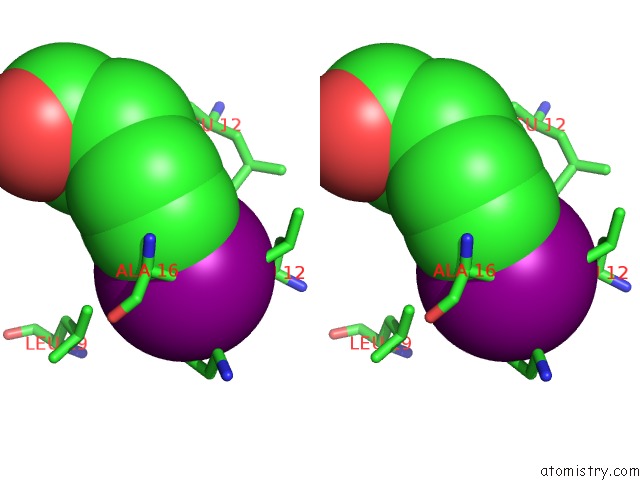
Stereo pair view
Mono view
Stereo pair view
A full contact list of Iodine with other atoms in the I binding
site number 1 of GCN4-P1 Peptide Trimer with Iodo-Phenylalanine Residue at Position 16 (Ipf-F16) within 5.0Å range:
|
Reference:
R.K.Rowe Hartje,
M.Ferrero,
G.Cavallo,
P.Metrangolo,
A.Ho,
R.Czarny,
P.S.Ho.
Engineering Specific Protein-Protein Interactions Through Halogen and Hydrogen Bonds To Be Published.
Page generated: Fri Aug 8 22:31:55 2025
Last articles
K in 5HHKK in 5HDK
K in 5G3W
K in 5HHJ
K in 5HAF
K in 5H5A
K in 5H6G
K in 5GZK
K in 5GEP
K in 5GUF