Iodine »
PDB 7ben-7fgj »
7ccy »
Iodine in PDB 7ccy: Crystal Structure of the 2-Iodoporphobilinogen-Bound Holo Form of Human Hydroxymethylbilane Synthase
Enzymatic activity of Crystal Structure of the 2-Iodoporphobilinogen-Bound Holo Form of Human Hydroxymethylbilane Synthase
All present enzymatic activity of Crystal Structure of the 2-Iodoporphobilinogen-Bound Holo Form of Human Hydroxymethylbilane Synthase:
2.5.1.61;
2.5.1.61;
Protein crystallography data
The structure of Crystal Structure of the 2-Iodoporphobilinogen-Bound Holo Form of Human Hydroxymethylbilane Synthase, PDB code: 7ccy
was solved by
H.Sato,
M.Sugishima,
K.Wada,
K.Hirabayashi,
M.Tsukaguchi,
with X-Ray Crystallography technique. A brief refinement statistics is given in the table below:
| Resolution Low / High (Å) | 48.91 / 2.40 |
| Space group | P 21 21 21 |
| Cell size a, b, c (Å), α, β, γ (°) | 74.011, 81.237, 109.097, 90, 90, 90 |
| R / Rfree (%) | 20.2 / 27.3 |
Iodine Binding Sites:
The binding sites of Iodine atom in the Crystal Structure of the 2-Iodoporphobilinogen-Bound Holo Form of Human Hydroxymethylbilane Synthase
(pdb code 7ccy). This binding sites where shown within
5.0 Angstroms radius around Iodine atom.
In total only one binding site of Iodine was determined in the Crystal Structure of the 2-Iodoporphobilinogen-Bound Holo Form of Human Hydroxymethylbilane Synthase, PDB code: 7ccy:
In total only one binding site of Iodine was determined in the Crystal Structure of the 2-Iodoporphobilinogen-Bound Holo Form of Human Hydroxymethylbilane Synthase, PDB code: 7ccy:
Iodine binding site 1 out of 1 in 7ccy
Go back to
Iodine binding site 1 out
of 1 in the Crystal Structure of the 2-Iodoporphobilinogen-Bound Holo Form of Human Hydroxymethylbilane Synthase
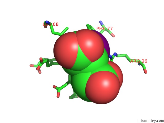
Mono view
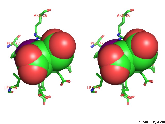
Stereo pair view
Mono view
Stereo pair view
A full contact list of Iodine with other atoms in the I binding
site number 1 of Crystal Structure of the 2-Iodoporphobilinogen-Bound Holo Form of Human Hydroxymethylbilane Synthase within 5.0Å range:
|
Reference:
H.Sato,
M.Sugishima,
M.Tsukaguchi,
T.Masuko,
M.Iijima,
M.Takano,
Y.Omata,
K.Hirabayashi,
K.Wada,
Y.Hisaeda,
K.Yamamoto.
Crystal Structures of Hydroxymethylbilane Synthase Complexed with A Substrate Analog: A Single Substrate-Binding Site For Four Consecutive Condensation Steps. Biochem.J. V. 478 1023 2021.
ISSN: ESSN 1470-8728
PubMed: 33600566
DOI: 10.1042/BCJ20200996
Page generated: Mon Aug 12 00:54:51 2024
ISSN: ESSN 1470-8728
PubMed: 33600566
DOI: 10.1042/BCJ20200996
Last articles
Zn in 9JYWZn in 9IR4
Zn in 9IR3
Zn in 9GMX
Zn in 9GMW
Zn in 9JEJ
Zn in 9ERF
Zn in 9ERE
Zn in 9EGV
Zn in 9EGW