Iodine »
PDB 7fgk-7lod »
7juy »
Iodine in PDB 7juy: Crystal Structure of KSR1:MEK1 in Complex with Amp-Pnp, and Allosteric Mek Inhibitor Cobimetinib
Enzymatic activity of Crystal Structure of KSR1:MEK1 in Complex with Amp-Pnp, and Allosteric Mek Inhibitor Cobimetinib
All present enzymatic activity of Crystal Structure of KSR1:MEK1 in Complex with Amp-Pnp, and Allosteric Mek Inhibitor Cobimetinib:
2.7.11.1; 2.7.12.2;
2.7.11.1; 2.7.12.2;
Protein crystallography data
The structure of Crystal Structure of KSR1:MEK1 in Complex with Amp-Pnp, and Allosteric Mek Inhibitor Cobimetinib, PDB code: 7juy
was solved by
Z.M.Khan,
A.C.Dar,
with X-Ray Crystallography technique. A brief refinement statistics is given in the table below:
| Resolution Low / High (Å) | 49.65 / 3.10 |
| Space group | P 61 2 2 |
| Cell size a, b, c (Å), α, β, γ (°) | 136.000, 136.000, 219.000, 90.00, 90.00, 120.00 |
| R / Rfree (%) | 24.7 / 25.7 |
Other elements in 7juy:
The structure of Crystal Structure of KSR1:MEK1 in Complex with Amp-Pnp, and Allosteric Mek Inhibitor Cobimetinib also contains other interesting chemical elements:
| Fluorine | (F) | 3 atoms |
| Magnesium | (Mg) | 2 atoms |
Iodine Binding Sites:
The binding sites of Iodine atom in the Crystal Structure of KSR1:MEK1 in Complex with Amp-Pnp, and Allosteric Mek Inhibitor Cobimetinib
(pdb code 7juy). This binding sites where shown within
5.0 Angstroms radius around Iodine atom.
In total only one binding site of Iodine was determined in the Crystal Structure of KSR1:MEK1 in Complex with Amp-Pnp, and Allosteric Mek Inhibitor Cobimetinib, PDB code: 7juy:
In total only one binding site of Iodine was determined in the Crystal Structure of KSR1:MEK1 in Complex with Amp-Pnp, and Allosteric Mek Inhibitor Cobimetinib, PDB code: 7juy:
Iodine binding site 1 out of 1 in 7juy
Go back to
Iodine binding site 1 out
of 1 in the Crystal Structure of KSR1:MEK1 in Complex with Amp-Pnp, and Allosteric Mek Inhibitor Cobimetinib
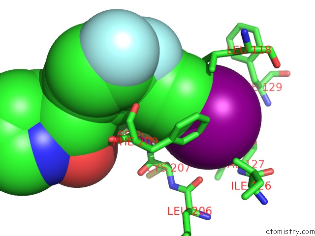
Mono view
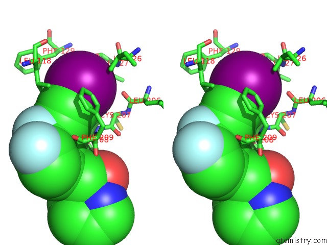
Stereo pair view
Mono view
Stereo pair view
A full contact list of Iodine with other atoms in the I binding
site number 1 of Crystal Structure of KSR1:MEK1 in Complex with Amp-Pnp, and Allosteric Mek Inhibitor Cobimetinib within 5.0Å range:
|
Reference:
Z.M.Khan,
A.M.Real,
W.M.Marsiglia,
A.Chow,
M.E.Duffy,
J.R.Yerabolu,
A.P.Scopton,
A.C.Dar.
Structural Basis For the Action of the Drug Trametinib at Ksr-Bound Mek. Nature 2020.
ISSN: ESSN 1476-4687
PubMed: 32927473
DOI: 10.1038/S41586-020-2760-4
Page generated: Mon Aug 12 01:13:32 2024
ISSN: ESSN 1476-4687
PubMed: 32927473
DOI: 10.1038/S41586-020-2760-4
Last articles
Zn in 9MJ5Zn in 9HNW
Zn in 9G0L
Zn in 9FNE
Zn in 9DZN
Zn in 9E0I
Zn in 9D32
Zn in 9DAK
Zn in 8ZXC
Zn in 8ZUF