Iodine »
PDB 7px1-7se4 »
7qdt »
Iodine in PDB 7qdt: Crystal Structure of A Mutant (P393GX) Thyroid Receptor Alpha Ligand Binding Domain Designed to Model Dominant Negative Human Mutations.
Protein crystallography data
The structure of Crystal Structure of A Mutant (P393GX) Thyroid Receptor Alpha Ligand Binding Domain Designed to Model Dominant Negative Human Mutations., PDB code: 7qdt
was solved by
B.Romartinez-Alonso,
L.Fairall,
M.Agostini,
K.Chatterjee,
J.Schwabe,
with X-Ray Crystallography technique. A brief refinement statistics is given in the table below:
| Resolution Low / High (Å) | 72.06 / 3.00 |
| Space group | P 64 2 2 |
| Cell size a, b, c (Å), α, β, γ (°) | 143.328, 143.328, 88.502, 90, 90, 120 |
| R / Rfree (%) | 18.3 / 22.8 |
Iodine Binding Sites:
The binding sites of Iodine atom in the Crystal Structure of A Mutant (P393GX) Thyroid Receptor Alpha Ligand Binding Domain Designed to Model Dominant Negative Human Mutations.
(pdb code 7qdt). This binding sites where shown within
5.0 Angstroms radius around Iodine atom.
In total 3 binding sites of Iodine where determined in the Crystal Structure of A Mutant (P393GX) Thyroid Receptor Alpha Ligand Binding Domain Designed to Model Dominant Negative Human Mutations., PDB code: 7qdt:
Jump to Iodine binding site number: 1; 2; 3;
In total 3 binding sites of Iodine where determined in the Crystal Structure of A Mutant (P393GX) Thyroid Receptor Alpha Ligand Binding Domain Designed to Model Dominant Negative Human Mutations., PDB code: 7qdt:
Jump to Iodine binding site number: 1; 2; 3;
Iodine binding site 1 out of 3 in 7qdt
Go back to
Iodine binding site 1 out
of 3 in the Crystal Structure of A Mutant (P393GX) Thyroid Receptor Alpha Ligand Binding Domain Designed to Model Dominant Negative Human Mutations.

Mono view
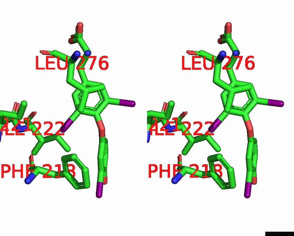
Stereo pair view
Mono view
Stereo pair view
A full contact list of Iodine with other atoms in the I binding
site number 1 of Crystal Structure of A Mutant (P393GX) Thyroid Receptor Alpha Ligand Binding Domain Designed to Model Dominant Negative Human Mutations. within 5.0Å range:
|
Iodine binding site 2 out of 3 in 7qdt
Go back to
Iodine binding site 2 out
of 3 in the Crystal Structure of A Mutant (P393GX) Thyroid Receptor Alpha Ligand Binding Domain Designed to Model Dominant Negative Human Mutations.
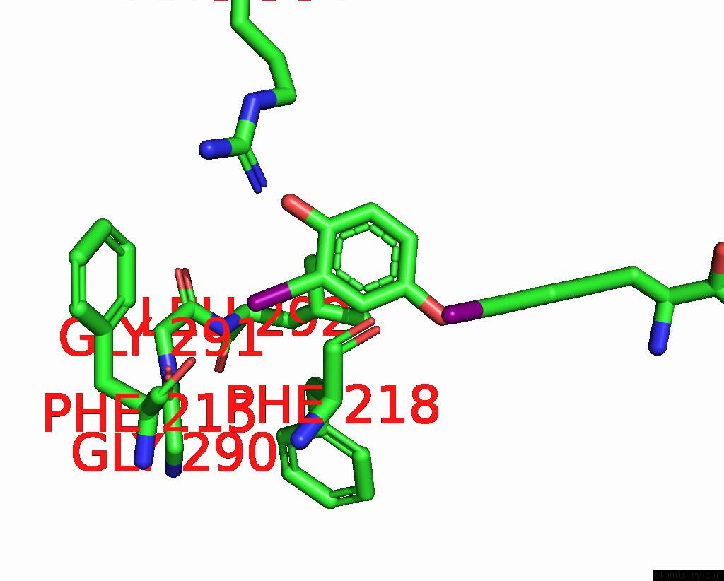
Mono view

Stereo pair view
Mono view
Stereo pair view
A full contact list of Iodine with other atoms in the I binding
site number 2 of Crystal Structure of A Mutant (P393GX) Thyroid Receptor Alpha Ligand Binding Domain Designed to Model Dominant Negative Human Mutations. within 5.0Å range:
|
Iodine binding site 3 out of 3 in 7qdt
Go back to
Iodine binding site 3 out
of 3 in the Crystal Structure of A Mutant (P393GX) Thyroid Receptor Alpha Ligand Binding Domain Designed to Model Dominant Negative Human Mutations.

Mono view

Stereo pair view
Mono view
Stereo pair view
A full contact list of Iodine with other atoms in the I binding
site number 3 of Crystal Structure of A Mutant (P393GX) Thyroid Receptor Alpha Ligand Binding Domain Designed to Model Dominant Negative Human Mutations. within 5.0Å range:
|
Reference:
B.Romartinez-Alonso,
M.Agostini,
H.Jones,
J.Mclellan,
D.E.Sood,
N.Tomkinson,
F.Marelli,
I.Gentile,
W.E.Visser,
E.Schoenmakers,
L.Fairall,
M.Privalsky,
C.Moran,
L.Persani,
K.Chatterjee,
J.W.R.Schwabe.
Structure-Guided Approach to Relieving Transcriptional Repression in Resistance to Thyroid Hormone Alpha. Mol.Cell.Biol. V. 42 36321 2022.
ISSN: ESSN 1098-5549
PubMed: 34871063
DOI: 10.1128/MCB.00363-21
Page generated: Mon Aug 12 01:59:16 2024
ISSN: ESSN 1098-5549
PubMed: 34871063
DOI: 10.1128/MCB.00363-21
Last articles
Zn in 9J0NZn in 9J0O
Zn in 9J0P
Zn in 9FJX
Zn in 9EKB
Zn in 9C0F
Zn in 9CAH
Zn in 9CH0
Zn in 9CH3
Zn in 9CH1