Iodine »
PDB 7px1-7se4 »
7qs2 »
Iodine in PDB 7qs2: Crystal Structure of B30.2 Pryspry Domain of TRIM15
Protein crystallography data
The structure of Crystal Structure of B30.2 Pryspry Domain of TRIM15, PDB code: 7qs2
was solved by
A.Chaikuad,
R.Zhubi,
S.Knapp,
Structural Genomics Consortium (Sgc),
with X-Ray Crystallography technique. A brief refinement statistics is given in the table below:
| Resolution Low / High (Å) | 41.23 / 1.70 |
| Space group | P 41 21 2 |
| Cell size a, b, c (Å), α, β, γ (°) | 54.764, 54.764, 125.265, 90, 90, 90 |
| R / Rfree (%) | 16.9 / 19.9 |
Iodine Binding Sites:
The binding sites of Iodine atom in the Crystal Structure of B30.2 Pryspry Domain of TRIM15
(pdb code 7qs2). This binding sites where shown within
5.0 Angstroms radius around Iodine atom.
In total 4 binding sites of Iodine where determined in the Crystal Structure of B30.2 Pryspry Domain of TRIM15, PDB code: 7qs2:
Jump to Iodine binding site number: 1; 2; 3; 4;
In total 4 binding sites of Iodine where determined in the Crystal Structure of B30.2 Pryspry Domain of TRIM15, PDB code: 7qs2:
Jump to Iodine binding site number: 1; 2; 3; 4;
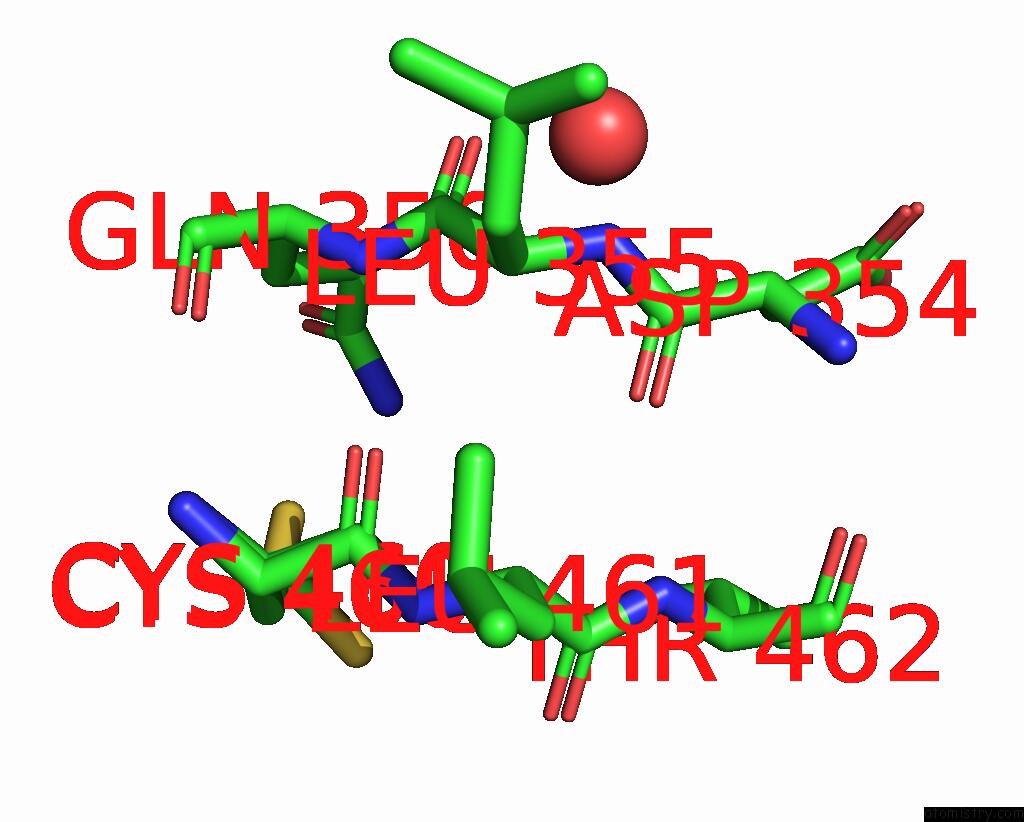
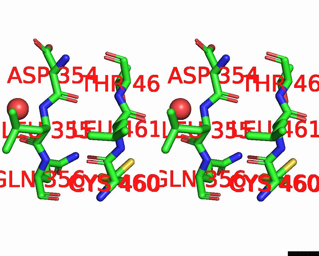
Iodine binding site 1 out of 4 in 7qs2
Go back to
Iodine binding site 1 out
of 4 in the Crystal Structure of B30.2 Pryspry Domain of TRIM15
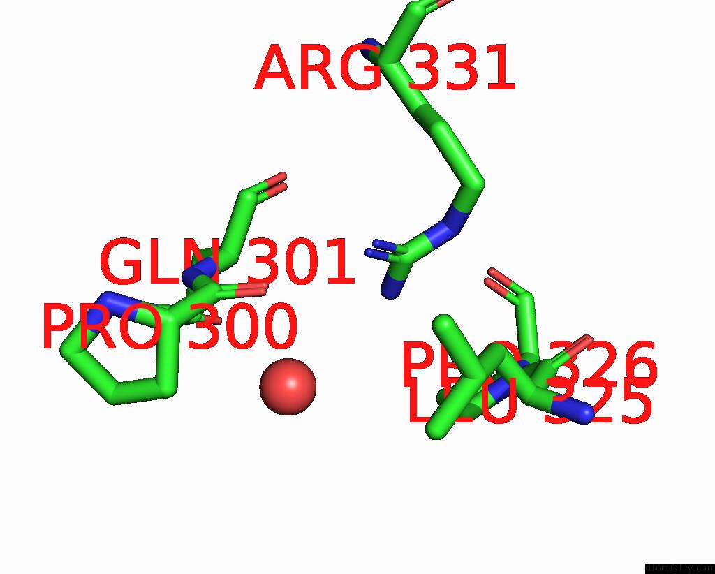
Mono view
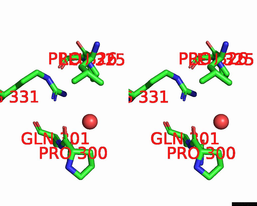
Stereo pair view
Mono view
Stereo pair view
A full contact list of Iodine with other atoms in the I binding
site number 1 of Crystal Structure of B30.2 Pryspry Domain of TRIM15 within 5.0Å range:
|
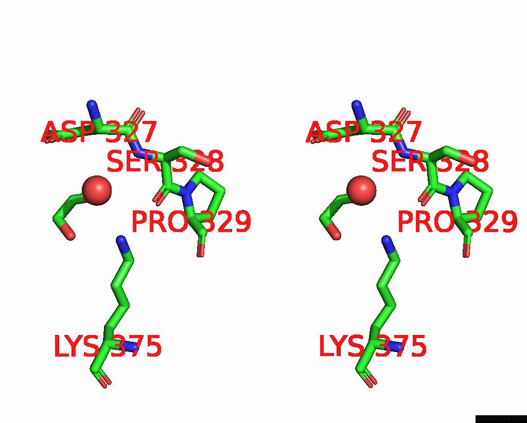
Iodine binding site 2 out of 4 in 7qs2
Go back to
Iodine binding site 2 out
of 4 in the Crystal Structure of B30.2 Pryspry Domain of TRIM15

Mono view
Stereo pair view
Mono view
Stereo pair view
A full contact list of Iodine with other atoms in the I binding
site number 2 of Crystal Structure of B30.2 Pryspry Domain of TRIM15 within 5.0Å range:
|
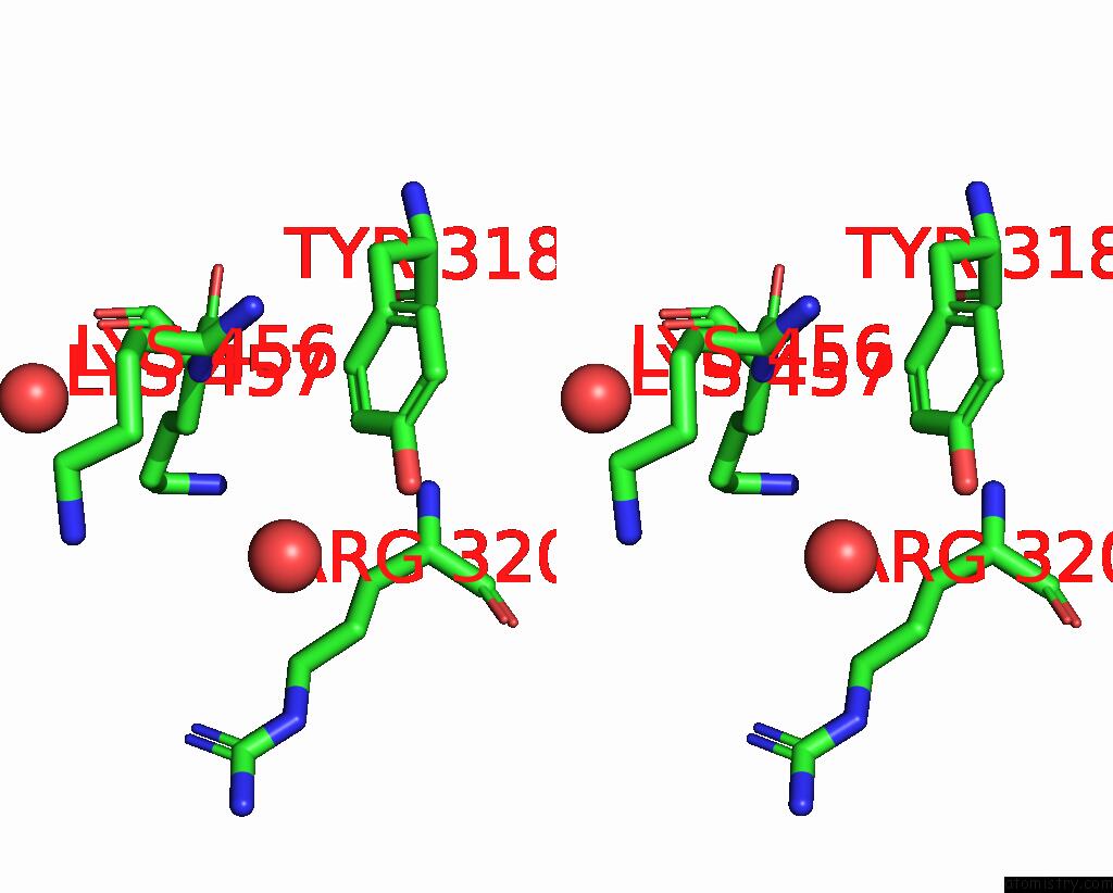
Iodine binding site 3 out of 4 in 7qs2
Go back to
Iodine binding site 3 out
of 4 in the Crystal Structure of B30.2 Pryspry Domain of TRIM15

Mono view
Stereo pair view
Mono view
Stereo pair view
A full contact list of Iodine with other atoms in the I binding
site number 3 of Crystal Structure of B30.2 Pryspry Domain of TRIM15 within 5.0Å range:
|
Iodine binding site 4 out of 4 in 7qs2
Go back to
Iodine binding site 4 out
of 4 in the Crystal Structure of B30.2 Pryspry Domain of TRIM15
Mono view
Stereo pair view
Mono view
Stereo pair view
A full contact list of Iodine with other atoms in the I binding
site number 4 of Crystal Structure of B30.2 Pryspry Domain of TRIM15 within 5.0Å range:
|
Reference:
A.Chaikuad,
R.Zhubi,
S.Knapp,
Structural Genomics Consortium (Sgc).
Crystal Structure of B30.2 Pryspry Domain of TRIM15 To Be Published.
Page generated: Mon Aug 12 01:59:16 2024
Last articles
Ca in 2ODICa in 2OF1
Ca in 2OEE
Ca in 2OBH
Ca in 2OEO
Ca in 2OAA
Ca in 2OAN
Ca in 2OBM
Ca in 2OBL
Ca in 2O8O