Iodine »
PDB 7px1-7se4 »
7rar »
Iodine in PDB 7rar: Structure of Q67H Mutant of Disulfide Stabilized Hiv-1 Ca Hexamer
Protein crystallography data
The structure of Structure of Q67H Mutant of Disulfide Stabilized Hiv-1 Ca Hexamer, PDB code: 7rar
was solved by
S.M.Bester,
M.Kvaratskhelia,
with X-Ray Crystallography technique. A brief refinement statistics is given in the table below:
| Resolution Low / High (Å) | 46.75 / 2.15 |
| Space group | P 6 |
| Cell size a, b, c (Å), α, β, γ (°) | 91.849, 91.849, 57.775, 90, 90, 120 |
| R / Rfree (%) | 20.8 / 24 |
Other elements in 7rar:
The structure of Structure of Q67H Mutant of Disulfide Stabilized Hiv-1 Ca Hexamer also contains other interesting chemical elements:
| Chlorine | (Cl) | 4 atoms |
Iodine Binding Sites:
The binding sites of Iodine atom in the Structure of Q67H Mutant of Disulfide Stabilized Hiv-1 Ca Hexamer
(pdb code 7rar). This binding sites where shown within
5.0 Angstroms radius around Iodine atom.
In total 6 binding sites of Iodine where determined in the Structure of Q67H Mutant of Disulfide Stabilized Hiv-1 Ca Hexamer, PDB code: 7rar:
Jump to Iodine binding site number: 1; 2; 3; 4; 5; 6;
In total 6 binding sites of Iodine where determined in the Structure of Q67H Mutant of Disulfide Stabilized Hiv-1 Ca Hexamer, PDB code: 7rar:
Jump to Iodine binding site number: 1; 2; 3; 4; 5; 6;
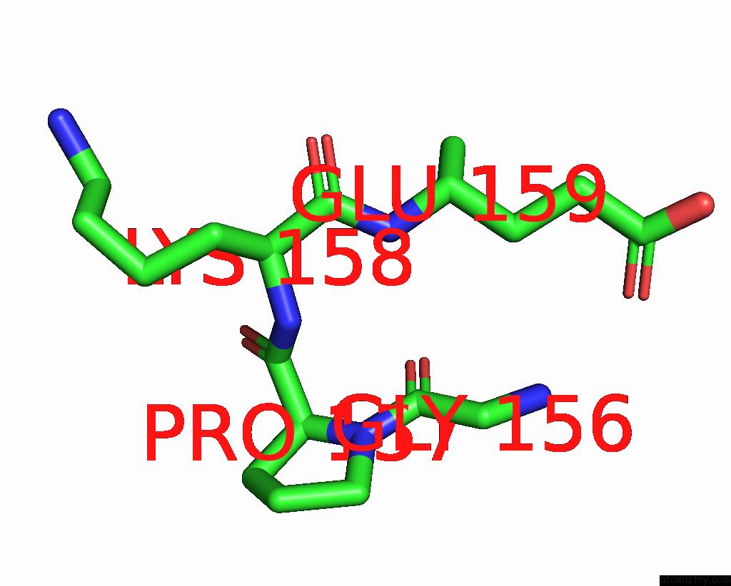
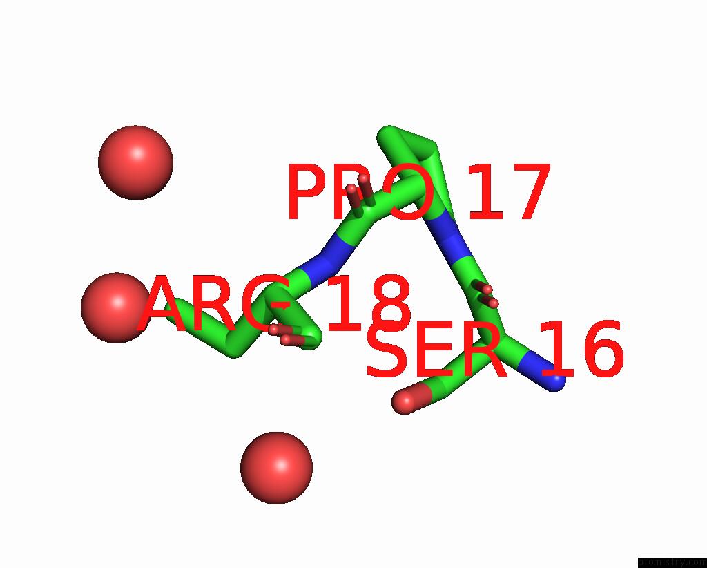
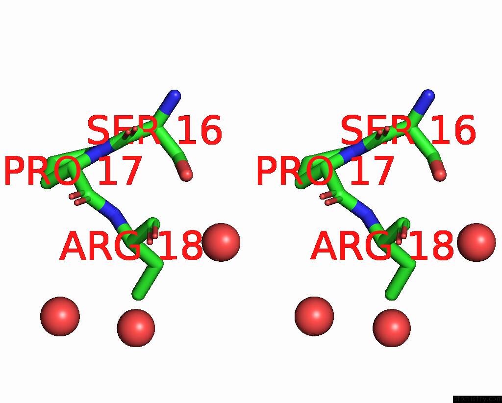
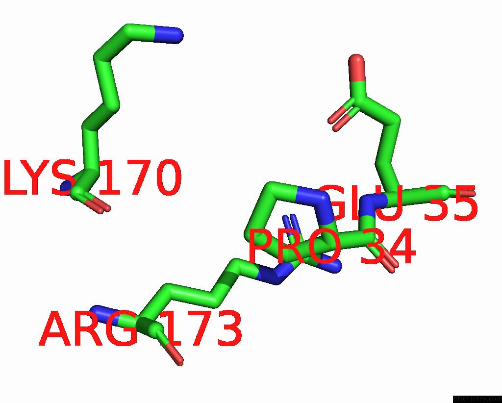
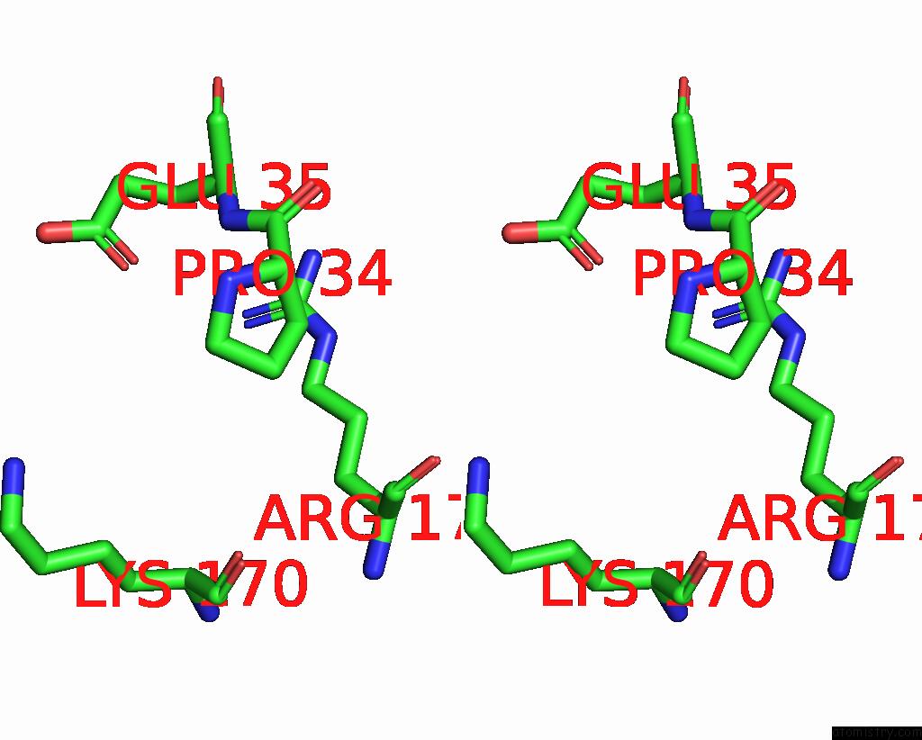
Iodine binding site 1 out of 6 in 7rar
Go back to
Iodine binding site 1 out
of 6 in the Structure of Q67H Mutant of Disulfide Stabilized Hiv-1 Ca Hexamer
Mono view

Stereo pair view
Mono view
Stereo pair view
A full contact list of Iodine with other atoms in the I binding
site number 1 of Structure of Q67H Mutant of Disulfide Stabilized Hiv-1 Ca Hexamer within 5.0Å range:
|
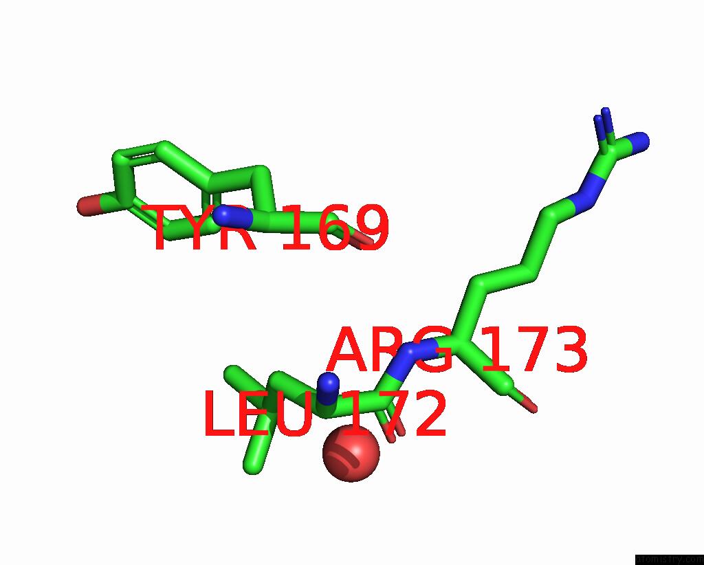
Iodine binding site 2 out of 6 in 7rar
Go back to
Iodine binding site 2 out
of 6 in the Structure of Q67H Mutant of Disulfide Stabilized Hiv-1 Ca Hexamer
Mono view

Stereo pair view
Mono view
Stereo pair view
A full contact list of Iodine with other atoms in the I binding
site number 2 of Structure of Q67H Mutant of Disulfide Stabilized Hiv-1 Ca Hexamer within 5.0Å range:
|
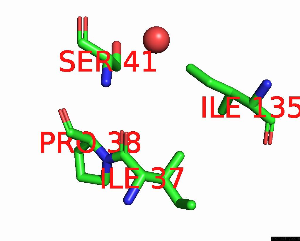
Iodine binding site 3 out of 6 in 7rar
Go back to
Iodine binding site 3 out
of 6 in the Structure of Q67H Mutant of Disulfide Stabilized Hiv-1 Ca Hexamer
Mono view
Stereo pair view
Mono view
Stereo pair view
A full contact list of Iodine with other atoms in the I binding
site number 3 of Structure of Q67H Mutant of Disulfide Stabilized Hiv-1 Ca Hexamer within 5.0Å range:
|
Iodine binding site 4 out of 6 in 7rar
Go back to
Iodine binding site 4 out
of 6 in the Structure of Q67H Mutant of Disulfide Stabilized Hiv-1 Ca Hexamer
Mono view
Stereo pair view
Mono view
Stereo pair view
A full contact list of Iodine with other atoms in the I binding
site number 4 of Structure of Q67H Mutant of Disulfide Stabilized Hiv-1 Ca Hexamer within 5.0Å range:
|
Iodine binding site 5 out of 6 in 7rar
Go back to
Iodine binding site 5 out
of 6 in the Structure of Q67H Mutant of Disulfide Stabilized Hiv-1 Ca Hexamer

Mono view

Stereo pair view
Mono view
Stereo pair view
A full contact list of Iodine with other atoms in the I binding
site number 5 of Structure of Q67H Mutant of Disulfide Stabilized Hiv-1 Ca Hexamer within 5.0Å range:
|
Iodine binding site 6 out of 6 in 7rar
Go back to
Iodine binding site 6 out
of 6 in the Structure of Q67H Mutant of Disulfide Stabilized Hiv-1 Ca Hexamer
Mono view

Stereo pair view
Mono view
Stereo pair view
A full contact list of Iodine with other atoms in the I binding
site number 6 of Structure of Q67H Mutant of Disulfide Stabilized Hiv-1 Ca Hexamer within 5.0Å range:
|
Reference:
S.M.Bester,
D.Adu-Ampratwum,
A.S.Annamalai,
G.Wei,
L.Briganti,
B.C.Murphy,
R.Haney,
J.R.Fuchs,
M.Kvaratskhelia.
Structural and Mechanistic Bases of Viral Resistance to Hiv-1 Capsid Inhibitor Lenacapavir. Mbio V. 13 80422 2022.
ISSN: ESSN 2150-7511
PubMed: 36190128
DOI: 10.1128/MBIO.01804-22
Page generated: Mon Aug 12 02:04:04 2024
ISSN: ESSN 2150-7511
PubMed: 36190128
DOI: 10.1128/MBIO.01804-22
Last articles
Zn in 9MJ5Zn in 9HNW
Zn in 9G0L
Zn in 9FNE
Zn in 9DZN
Zn in 9E0I
Zn in 9D32
Zn in 9DAK
Zn in 8ZXC
Zn in 8ZUF