Iodine in PDB 7skx: Ab Initio Structure of Proteinase K From Electron-Counted Microed Data
Enzymatic activity of Ab Initio Structure of Proteinase K From Electron-Counted Microed Data
All present enzymatic activity of Ab Initio Structure of Proteinase K From Electron-Counted Microed Data:
3.4.21.64;
Other elements in 7skx:
The structure of Ab Initio Structure of Proteinase K From Electron-Counted Microed Data also contains other interesting chemical elements:
Iodine Binding Sites:
The binding sites of Iodine atom in the Ab Initio Structure of Proteinase K From Electron-Counted Microed Data
(pdb code 7skx). This binding sites where shown within
5.0 Angstroms radius around Iodine atom.
In total 6 binding sites of Iodine where determined in the
Ab Initio Structure of Proteinase K From Electron-Counted Microed Data, PDB code: 7skx:
Jump to Iodine binding site number:
1;
2;
3;
4;
5;
6;
Iodine binding site 1 out
of 6 in 7skx
Go back to
Iodine Binding Sites List in 7skx
Iodine binding site 1 out
of 6 in the Ab Initio Structure of Proteinase K From Electron-Counted Microed Data
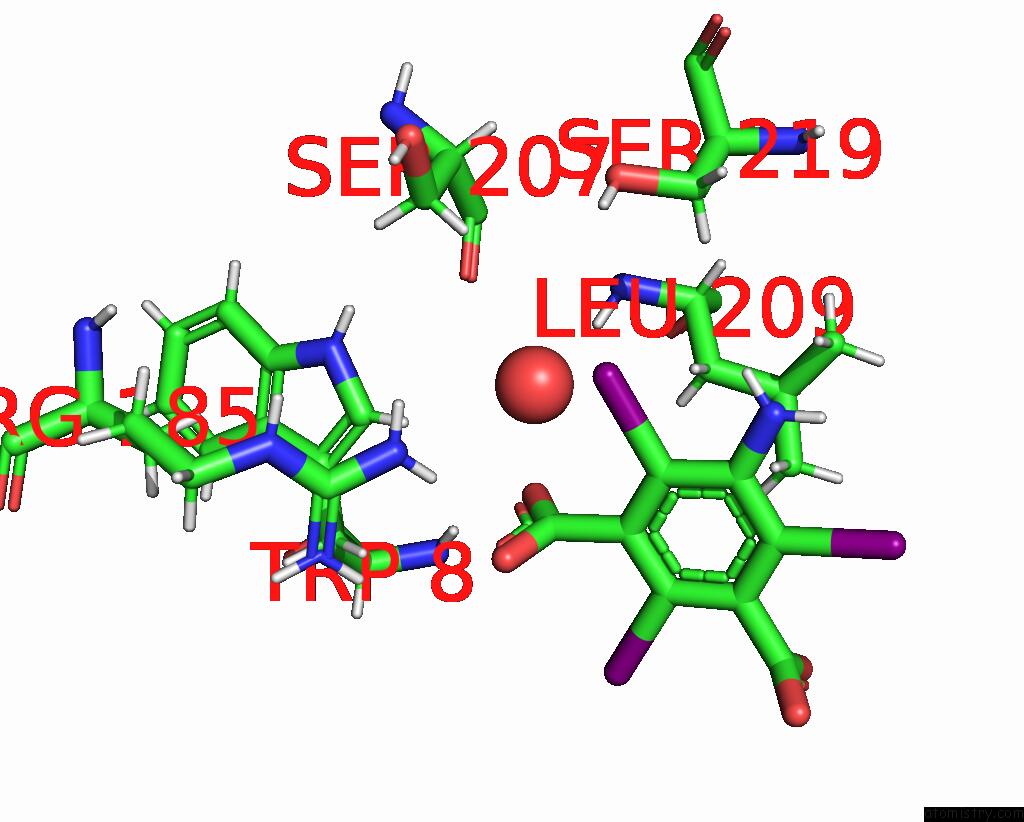
Mono view
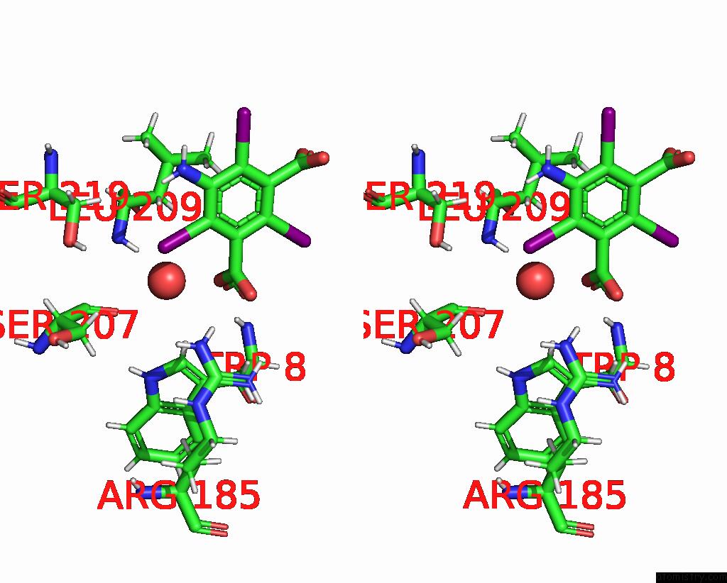
Stereo pair view
|
A full contact list of Iodine with other atoms in the I binding
site number 1 of Ab Initio Structure of Proteinase K From Electron-Counted Microed Data within 5.0Å range:
probe
|
atom
|
residue
|
distance (Å)
|
B
|
Occ
|
C:I301
b:42.6
occ:0.77
|
I3
|
C:I3C301
|
0.0
|
42.6
|
0.8
|
C6
|
C:I3C301
|
2.1
|
40.0
|
0.8
|
HN1A
|
C:I3C301
|
2.6
|
34.0
|
0.8
|
C5
|
C:I3C301
|
3.0
|
38.4
|
0.8
|
C1
|
C:I3C301
|
3.0
|
43.0
|
0.8
|
N13
|
C:I3C301
|
3.1
|
33.6
|
0.8
|
HG
|
C:SER219
|
3.2
|
16.9
|
0.0
|
C7
|
C:I3C301
|
3.3
|
32.6
|
0.8
|
HB2
|
C:SER219
|
3.4
|
14.2
|
1.0
|
HB2
|
C:LEU209
|
3.5
|
9.5
|
1.0
|
O8
|
C:I3C301
|
3.6
|
34.5
|
0.8
|
O
|
C:HOH403
|
3.7
|
9.6
|
1.0
|
HH22
|
C:ARG185
|
3.7
|
26.9
|
1.0
|
HB3
|
C:SER207
|
3.7
|
11.1
|
1.0
|
OG
|
C:SER219
|
3.8
|
17.2
|
1.0
|
O
|
C:SER207
|
3.8
|
10.1
|
1.0
|
HH21
|
C:ARG185
|
3.9
|
27.8
|
1.0
|
O9
|
C:I3C301
|
3.9
|
30.5
|
0.8
|
CB
|
C:SER219
|
4.0
|
13.9
|
1.0
|
HN13
|
C:I3C301
|
4.0
|
35.0
|
0.8
|
NH2
|
C:ARG185
|
4.2
|
27.1
|
1.0
|
HB3
|
C:SER219
|
4.3
|
13.4
|
1.0
|
C4
|
C:I3C301
|
4.3
|
40.7
|
0.8
|
C2
|
C:I3C301
|
4.3
|
48.1
|
0.8
|
H
|
C:LEU209
|
4.4
|
8.4
|
1.0
|
HD1
|
C:TRP8
|
4.4
|
10.3
|
1.0
|
CB
|
C:LEU209
|
4.4
|
9.3
|
1.0
|
HD21
|
C:LEU209
|
4.5
|
12.6
|
1.0
|
C
|
C:SER207
|
4.6
|
8.1
|
1.0
|
HD13
|
C:LEU209
|
4.7
|
9.9
|
1.0
|
CB
|
C:SER207
|
4.7
|
10.4
|
1.0
|
HB3
|
C:LEU209
|
4.7
|
9.5
|
1.0
|
N
|
C:LEU209
|
4.8
|
8.4
|
1.0
|
C3
|
C:I3C301
|
4.9
|
47.3
|
0.8
|
HB2
|
C:SER207
|
5.0
|
11.0
|
1.0
|
|
Iodine binding site 2 out
of 6 in 7skx
Go back to
Iodine Binding Sites List in 7skx
Iodine binding site 2 out
of 6 in the Ab Initio Structure of Proteinase K From Electron-Counted Microed Data
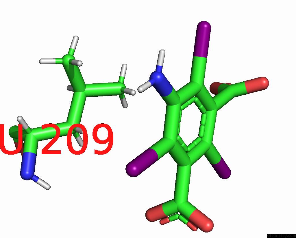
Mono view
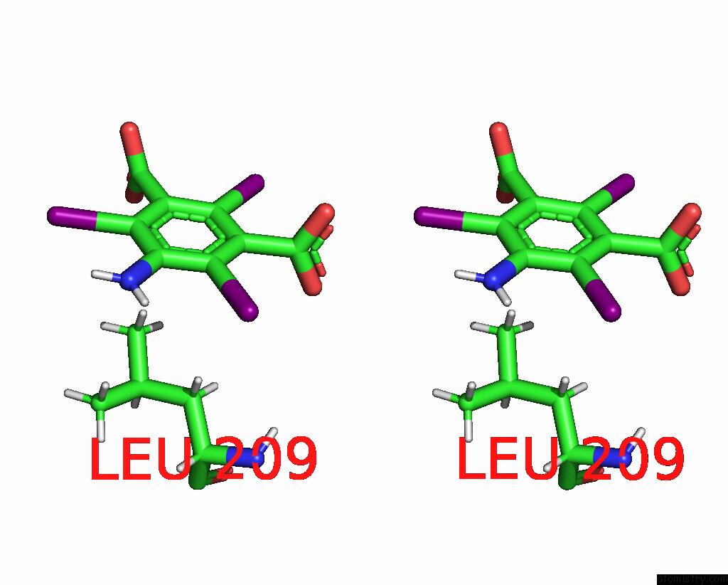
Stereo pair view
|
A full contact list of Iodine with other atoms in the I binding
site number 2 of Ab Initio Structure of Proteinase K From Electron-Counted Microed Data within 5.0Å range:
probe
|
atom
|
residue
|
distance (Å)
|
B
|
Occ
|
C:I301
b:79.8
occ:0.77
|
I2
|
C:I3C301
|
0.0
|
79.8
|
0.8
|
C4
|
C:I3C301
|
2.1
|
40.7
|
0.8
|
HN13
|
C:I3C301
|
2.7
|
35.0
|
0.8
|
C5
|
C:I3C301
|
3.0
|
38.4
|
0.8
|
C3
|
C:I3C301
|
3.1
|
47.3
|
0.8
|
N13
|
C:I3C301
|
3.2
|
33.6
|
0.8
|
C10
|
C:I3C301
|
3.3
|
47.0
|
0.8
|
O11
|
C:I3C301
|
3.5
|
63.4
|
0.8
|
HD21
|
C:LEU209
|
3.7
|
12.6
|
1.0
|
O12
|
C:I3C301
|
3.9
|
46.0
|
0.8
|
HD22
|
C:LEU209
|
4.0
|
11.8
|
1.0
|
HN1A
|
C:I3C301
|
4.1
|
34.0
|
0.8
|
CD2
|
C:LEU209
|
4.3
|
12.4
|
1.0
|
C6
|
C:I3C301
|
4.3
|
40.0
|
0.8
|
C2
|
C:I3C301
|
4.4
|
48.1
|
0.8
|
HD13
|
C:LEU209
|
4.7
|
9.9
|
1.0
|
HD12
|
C:LEU209
|
4.9
|
9.8
|
1.0
|
HD23
|
C:LEU209
|
4.9
|
12.1
|
1.0
|
C1
|
C:I3C301
|
4.9
|
43.0
|
0.8
|
|
Iodine binding site 3 out
of 6 in 7skx
Go back to
Iodine Binding Sites List in 7skx
Iodine binding site 3 out
of 6 in the Ab Initio Structure of Proteinase K From Electron-Counted Microed Data
Mono view
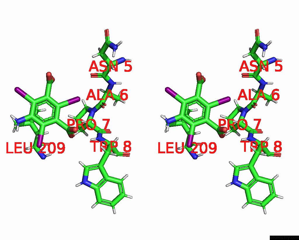
Stereo pair view
|
A full contact list of Iodine with other atoms in the I binding
site number 3 of Ab Initio Structure of Proteinase K From Electron-Counted Microed Data within 5.0Å range:
probe
|
atom
|
residue
|
distance (Å)
|
B
|
Occ
|
C:I301
b:49.8
occ:0.77
|
I1
|
C:I3C301
|
0.0
|
49.8
|
0.8
|
C2
|
C:I3C301
|
2.1
|
48.1
|
0.8
|
C1
|
C:I3C301
|
3.0
|
43.0
|
0.8
|
C3
|
C:I3C301
|
3.1
|
47.3
|
0.8
|
HA
|
C:PRO7
|
3.1
|
11.3
|
1.0
|
C7
|
C:I3C301
|
3.2
|
32.6
|
0.8
|
O
|
C:ALA6
|
3.3
|
12.3
|
1.0
|
C10
|
C:I3C301
|
3.3
|
47.0
|
0.8
|
O9
|
C:I3C301
|
3.4
|
30.5
|
0.8
|
O12
|
C:I3C301
|
3.5
|
46.0
|
0.8
|
O
|
C:ASN5
|
3.6
|
15.8
|
1.0
|
O8
|
C:I3C301
|
3.7
|
34.5
|
0.8
|
C
|
C:ALA6
|
3.8
|
11.0
|
1.0
|
O11
|
C:I3C301
|
3.9
|
63.4
|
0.8
|
CA
|
C:PRO7
|
3.9
|
11.0
|
1.0
|
N
|
C:PRO7
|
4.0
|
11.8
|
1.0
|
C
|
C:ASN5
|
4.2
|
16.3
|
1.0
|
C6
|
C:I3C301
|
4.3
|
40.0
|
0.8
|
C4
|
C:I3C301
|
4.4
|
40.7
|
0.8
|
H
|
C:TRP8
|
4.4
|
9.5
|
1.0
|
HB3
|
C:ASN5
|
4.6
|
18.7
|
1.0
|
C
|
C:PRO7
|
4.7
|
10.6
|
1.0
|
HA
|
C:ASN5
|
4.7
|
16.2
|
1.0
|
N
|
C:TRP8
|
4.7
|
9.4
|
1.0
|
N
|
C:ALA6
|
4.7
|
13.6
|
1.0
|
HD3
|
C:PRO7
|
4.8
|
13.0
|
1.0
|
CA
|
C:ALA6
|
4.8
|
12.9
|
1.0
|
HD23
|
C:LEU209
|
4.8
|
12.1
|
1.0
|
CD
|
C:PRO7
|
4.9
|
12.6
|
1.0
|
C5
|
C:I3C301
|
4.9
|
38.4
|
0.8
|
CA
|
C:ASN5
|
5.0
|
16.4
|
1.0
|
HG3
|
C:PRO7
|
5.0
|
13.0
|
1.0
|
HD21
|
C:LEU209
|
5.0
|
12.6
|
1.0
|
HA
|
C:ALA6
|
5.0
|
12.9
|
1.0
|
|
Iodine binding site 4 out
of 6 in 7skx
Go back to
Iodine Binding Sites List in 7skx
Iodine binding site 4 out
of 6 in the Ab Initio Structure of Proteinase K From Electron-Counted Microed Data
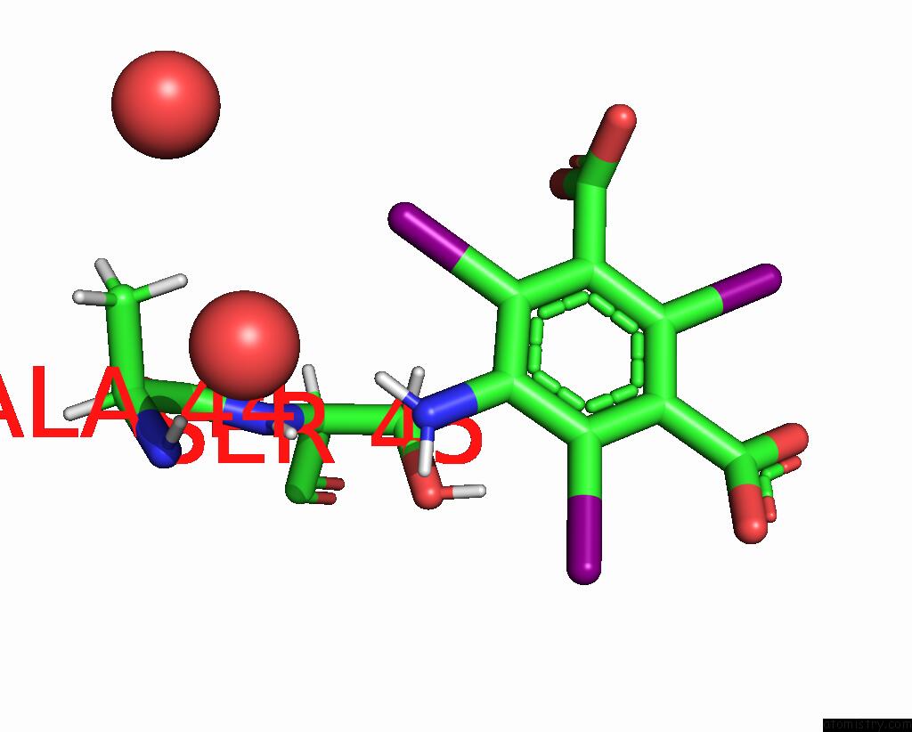
Mono view
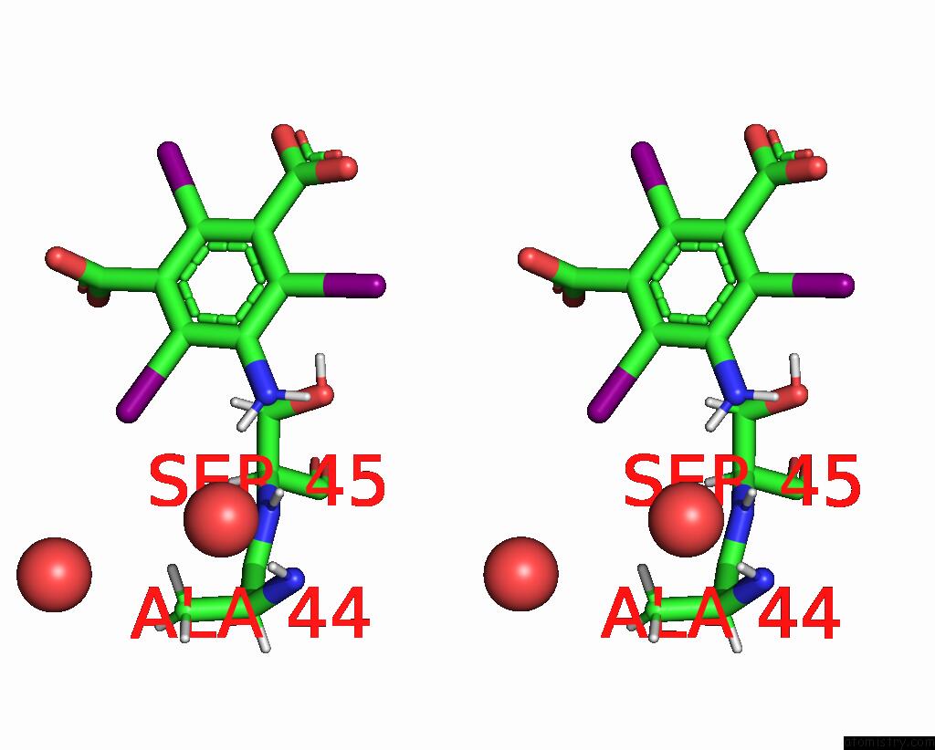
Stereo pair view
|
A full contact list of Iodine with other atoms in the I binding
site number 4 of Ab Initio Structure of Proteinase K From Electron-Counted Microed Data within 5.0Å range:
probe
|
atom
|
residue
|
distance (Å)
|
B
|
Occ
|
C:I302
b:40.0
occ:0.73
|
I3
|
C:I3C302
|
0.0
|
40.0
|
0.7
|
C6
|
C:I3C302
|
2.1
|
37.4
|
0.7
|
HN1A
|
C:I3C302
|
2.7
|
26.7
|
0.7
|
C5
|
C:I3C302
|
3.0
|
32.4
|
0.7
|
C1
|
C:I3C302
|
3.0
|
51.1
|
0.7
|
N13
|
C:I3C302
|
3.2
|
26.7
|
0.7
|
HB3
|
C:SER45
|
3.2
|
14.3
|
1.0
|
C7
|
C:I3C302
|
3.3
|
56.9
|
0.7
|
HB3
|
C:ALA44
|
3.6
|
15.0
|
1.0
|
O8
|
C:I3C302
|
3.7
|
55.7
|
0.7
|
O9
|
C:I3C302
|
3.7
|
57.4
|
0.7
|
H
|
C:SER45
|
3.9
|
12.6
|
1.0
|
HN13
|
C:I3C302
|
4.1
|
26.5
|
0.7
|
CB
|
C:SER45
|
4.2
|
14.4
|
1.0
|
O
|
C:HOH476
|
4.2
|
28.0
|
1.0
|
N
|
C:SER45
|
4.3
|
12.7
|
1.0
|
C4
|
C:I3C302
|
4.3
|
32.6
|
0.7
|
O
|
C:HOH512
|
4.4
|
25.7
|
1.0
|
C2
|
C:I3C302
|
4.4
|
53.3
|
0.7
|
CB
|
C:ALA44
|
4.5
|
14.9
|
1.0
|
HA
|
C:SER45
|
4.6
|
11.8
|
1.0
|
CA
|
C:SER45
|
4.6
|
11.8
|
1.0
|
H
|
C:ALA44
|
4.7
|
11.1
|
1.0
|
HB2
|
C:SER45
|
4.7
|
14.1
|
1.0
|
HB1
|
C:ALA44
|
4.8
|
14.3
|
1.0
|
OG
|
C:SER45
|
4.8
|
16.7
|
1.0
|
HG
|
C:SER45
|
4.8
|
16.2
|
0.0
|
C3
|
C:I3C302
|
4.9
|
49.7
|
0.7
|
C
|
C:ALA44
|
4.9
|
12.2
|
1.0
|
|
Iodine binding site 5 out
of 6 in 7skx
Go back to
Iodine Binding Sites List in 7skx
Iodine binding site 5 out
of 6 in the Ab Initio Structure of Proteinase K From Electron-Counted Microed Data
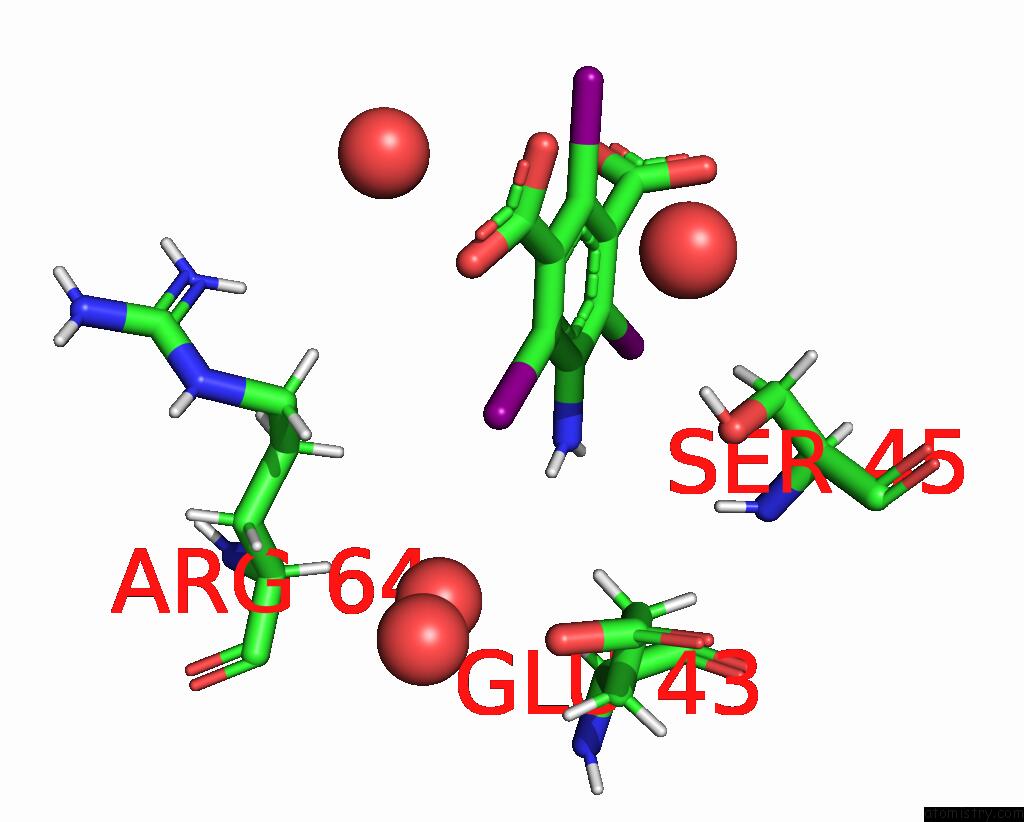
Mono view
Stereo pair view
|
A full contact list of Iodine with other atoms in the I binding
site number 5 of Ab Initio Structure of Proteinase K From Electron-Counted Microed Data within 5.0Å range:
probe
|
atom
|
residue
|
distance (Å)
|
B
|
Occ
|
C:I302
b:36.0
occ:0.73
|
I2
|
C:I3C302
|
0.0
|
36.0
|
0.7
|
C4
|
C:I3C302
|
2.1
|
32.6
|
0.7
|
O11
|
C:I3C302
|
2.7
|
73.9
|
0.7
|
HN13
|
C:I3C302
|
2.8
|
26.5
|
0.7
|
C5
|
C:I3C302
|
3.0
|
32.4
|
0.7
|
C3
|
C:I3C302
|
3.0
|
49.7
|
0.7
|
N13
|
C:I3C302
|
3.2
|
26.7
|
0.7
|
C10
|
C:I3C302
|
3.2
|
61.3
|
0.7
|
HG3
|
C:GLU43
|
3.3
|
13.0
|
1.0
|
HD2
|
C:ARG64
|
3.4
|
39.6
|
1.0
|
HG
|
C:SER45
|
3.6
|
16.2
|
0.0
|
HD3
|
C:ARG64
|
3.8
|
41.3
|
1.0
|
O
|
C:HOH442
|
4.0
|
23.5
|
1.0
|
CD
|
C:ARG64
|
4.1
|
41.2
|
1.0
|
OE2
|
C:GLU43
|
4.1
|
20.1
|
1.0
|
HN1A
|
C:I3C302
|
4.1
|
26.7
|
0.7
|
O
|
C:HOH517
|
4.2
|
19.2
|
1.0
|
OG
|
C:SER45
|
4.2
|
16.7
|
1.0
|
CG
|
C:GLU43
|
4.2
|
12.8
|
1.0
|
CD
|
C:GLU43
|
4.3
|
15.2
|
1.0
|
C6
|
C:I3C302
|
4.3
|
37.4
|
0.7
|
HG2
|
C:ARG64
|
4.4
|
26.5
|
1.0
|
C2
|
C:I3C302
|
4.4
|
53.3
|
0.7
|
O
|
C:HOH556
|
4.4
|
43.7
|
1.0
|
O12
|
C:I3C302
|
4.5
|
66.0
|
0.7
|
HG2
|
C:GLU43
|
4.5
|
13.1
|
1.0
|
CG
|
C:ARG64
|
4.8
|
27.0
|
1.0
|
O
|
C:HOH520
|
4.8
|
45.2
|
1.0
|
C1
|
C:I3C302
|
4.9
|
51.1
|
0.7
|
|
Iodine binding site 6 out
of 6 in 7skx
Go back to
Iodine Binding Sites List in 7skx
Iodine binding site 6 out
of 6 in the Ab Initio Structure of Proteinase K From Electron-Counted Microed Data
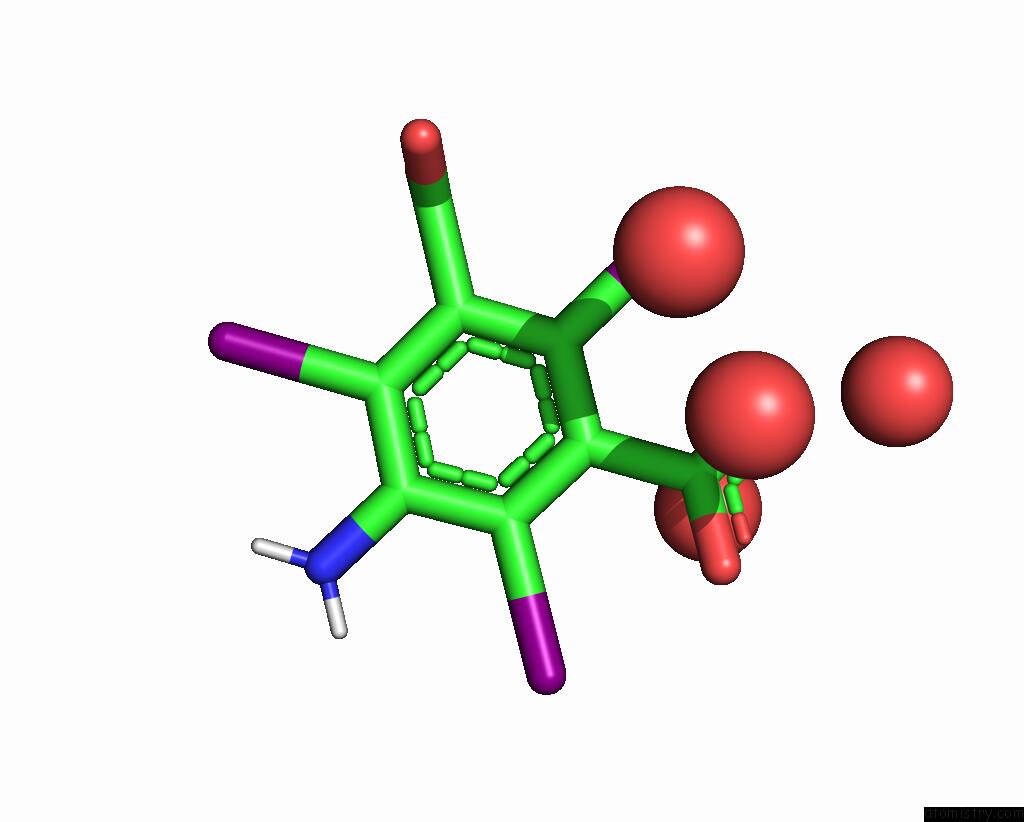
Mono view
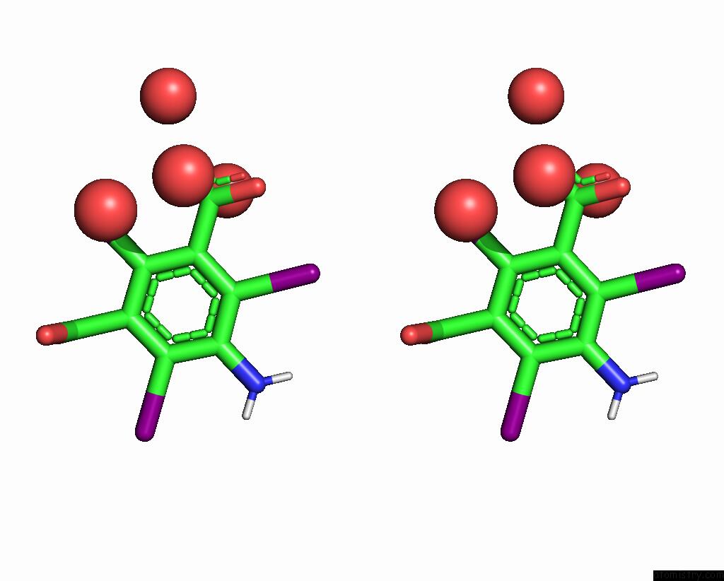
Stereo pair view
|
A full contact list of Iodine with other atoms in the I binding
site number 6 of Ab Initio Structure of Proteinase K From Electron-Counted Microed Data within 5.0Å range:
probe
|
atom
|
residue
|
distance (Å)
|
B
|
Occ
|
C:I302
b:89.6
occ:0.73
|
I1
|
C:I3C302
|
0.0
|
89.6
|
0.7
|
C2
|
C:I3C302
|
2.1
|
53.3
|
0.7
|
O12
|
C:I3C302
|
2.7
|
66.0
|
0.7
|
C1
|
C:I3C302
|
3.1
|
51.1
|
0.7
|
C3
|
C:I3C302
|
3.1
|
49.7
|
0.7
|
C7
|
C:I3C302
|
3.2
|
56.9
|
0.7
|
C10
|
C:I3C302
|
3.3
|
61.3
|
0.7
|
O
|
C:HOH601
|
3.3
|
38.6
|
1.0
|
O9
|
C:I3C302
|
3.6
|
57.4
|
0.7
|
O8
|
C:I3C302
|
3.7
|
55.7
|
0.7
|
O
|
C:HOH520
|
3.8
|
45.2
|
1.0
|
O
|
C:HOH444
|
4.2
|
38.5
|
1.0
|
C6
|
C:I3C302
|
4.4
|
37.4
|
0.7
|
C4
|
C:I3C302
|
4.4
|
32.6
|
0.7
|
O11
|
C:I3C302
|
4.5
|
73.9
|
0.7
|
O
|
C:HOH556
|
4.8
|
43.7
|
1.0
|
C5
|
C:I3C302
|
5.0
|
32.4
|
0.7
|
|
Reference:
M.W.Martynowycz,
M.T.B.Clabbers,
J.Hattne,
T.Gonen.
Ab Initio Phasing Macromolecular Structures Using Electron-Counted Microed Data. Nat.Methods V. 19 724 2022.
ISSN: ESSN 1548-7105
PubMed: 35637302
DOI: 10.1038/S41592-022-01485-4
Page generated: Mon Aug 12 02:10:52 2024
|