Iodine »
PDB 8eg4-8qwj »
8ikb »
Iodine in PDB 8ikb: Cryo-Em Structure of HNRAC1-2I Fibril.
Iodine Binding Sites:
Pages:
>>> Page 1 <<< Page 2, Binding sites: 11 - 20; Page 3, Binding sites: 21 - 28;Binding sites:
The binding sites of Iodine atom in the Cryo-Em Structure of HNRAC1-2I Fibril. (pdb code 8ikb). This binding sites where shown within 5.0 Angstroms radius around Iodine atom.In total 28 binding sites of Iodine where determined in the Cryo-Em Structure of HNRAC1-2I Fibril., PDB code: 8ikb:
Jump to Iodine binding site number: 1; 2; 3; 4; 5; 6; 7; 8; 9; 10;
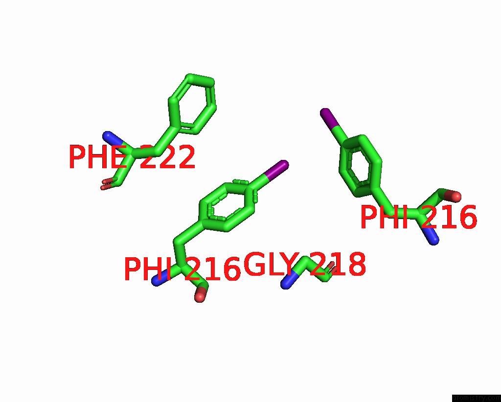
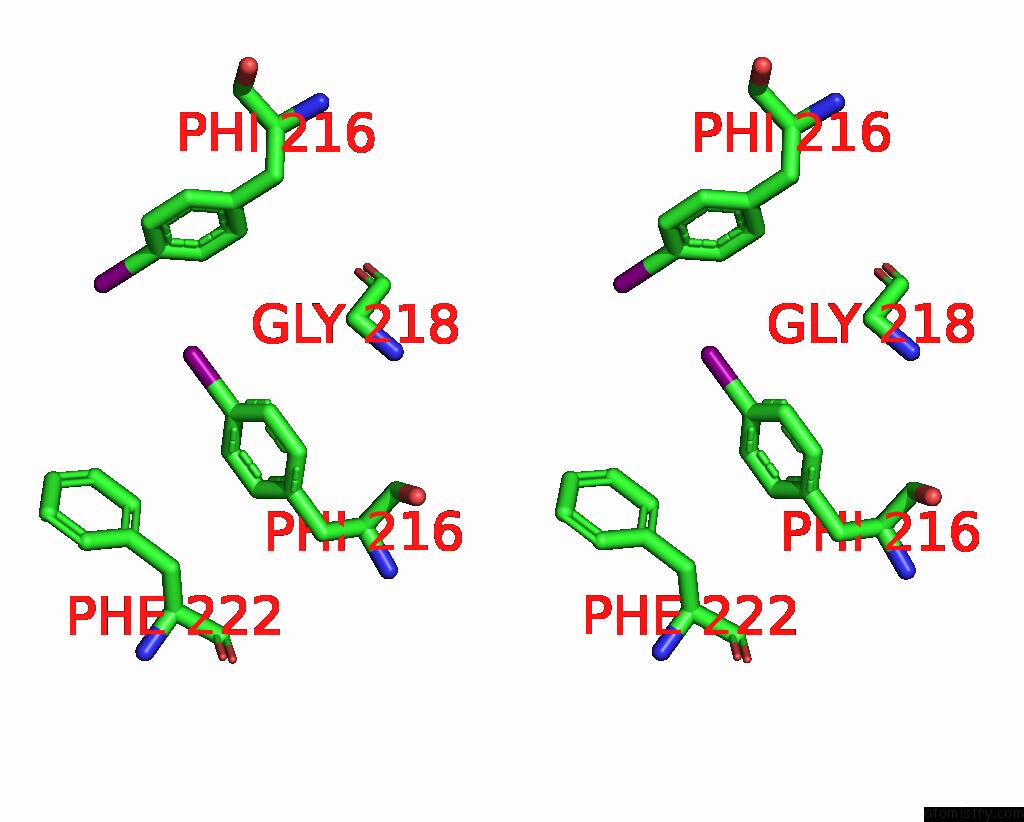
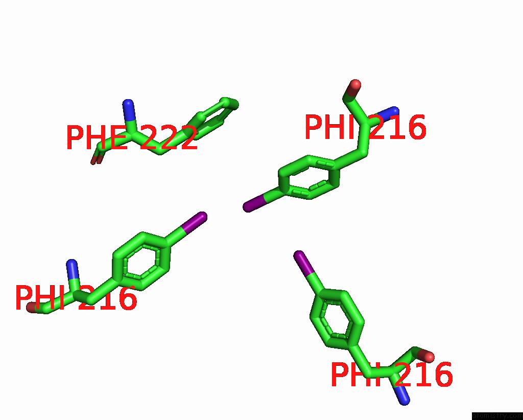
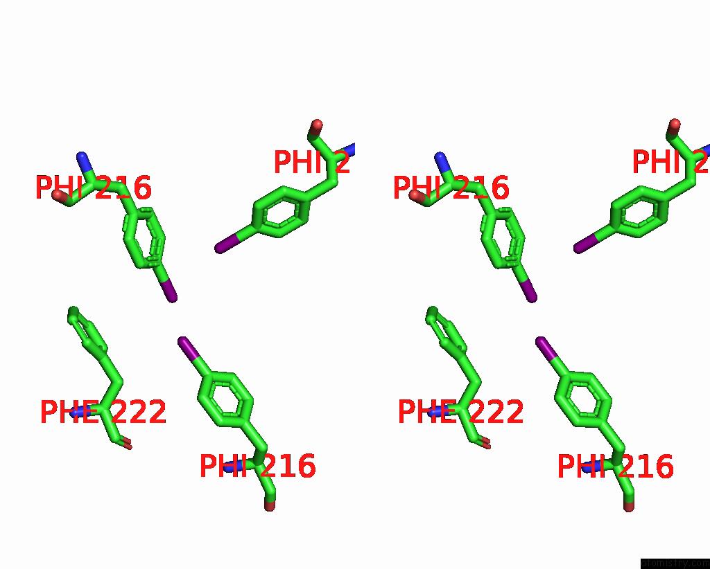
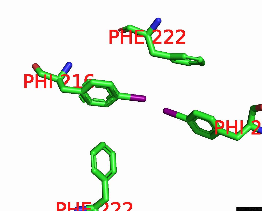
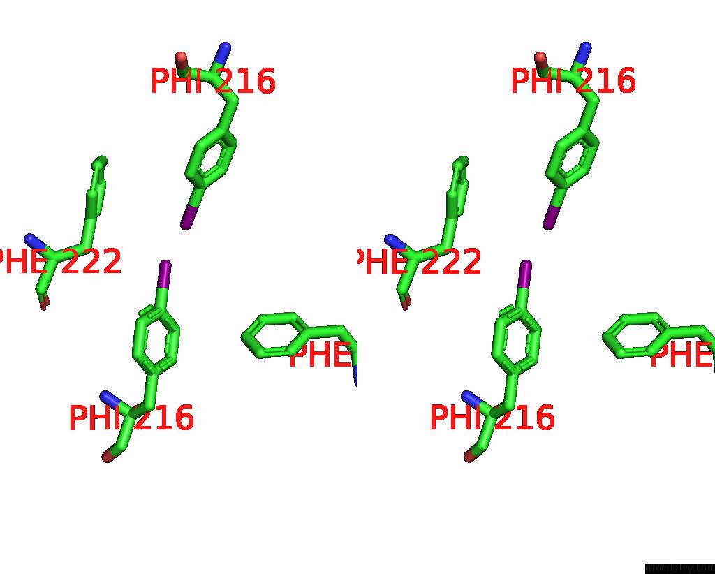
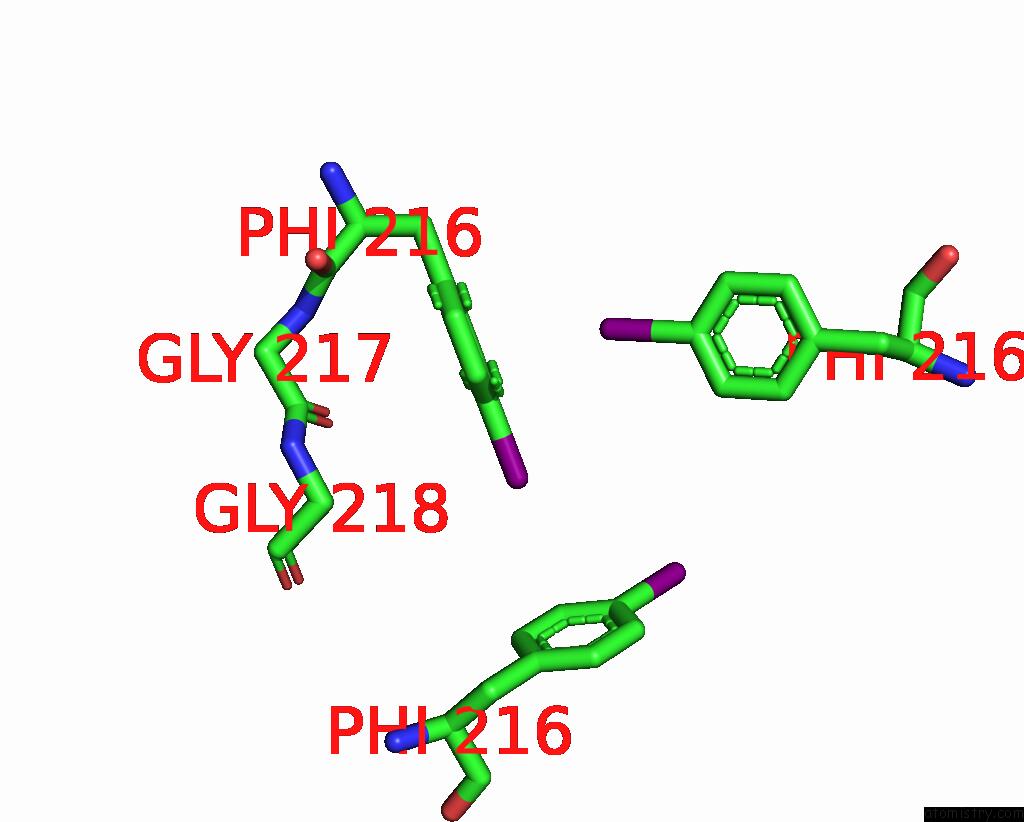
Iodine binding site 1 out of 28 in 8ikb
Go back to
Iodine binding site 1 out
of 28 in the Cryo-Em Structure of HNRAC1-2I Fibril.
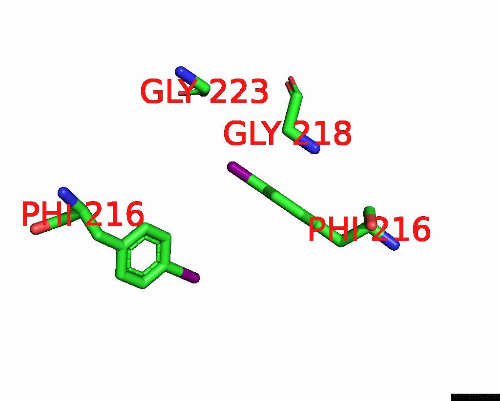
Mono view
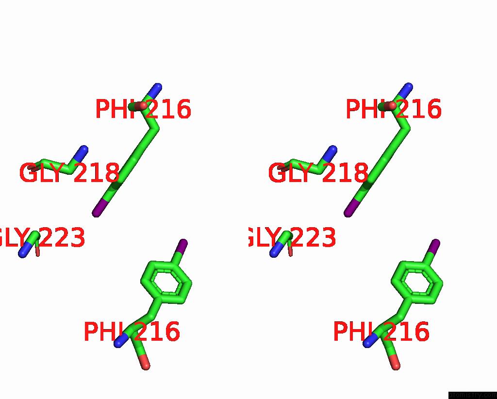
Stereo pair view
Mono view
Stereo pair view
A full contact list of Iodine with other atoms in the I binding
site number 1 of Cryo-Em Structure of HNRAC1-2I Fibril. within 5.0Å range:
|
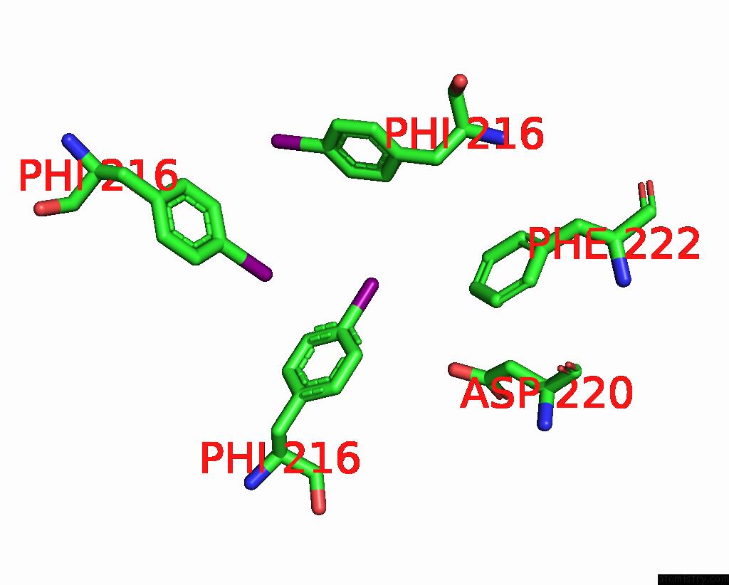
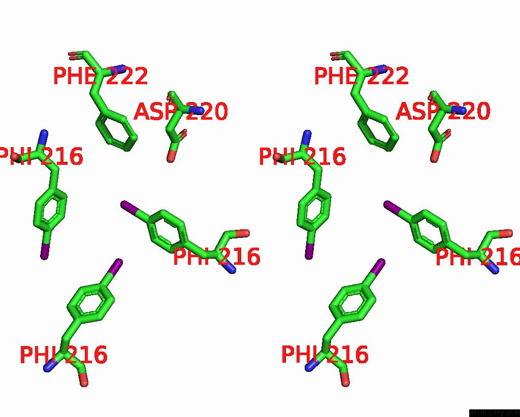
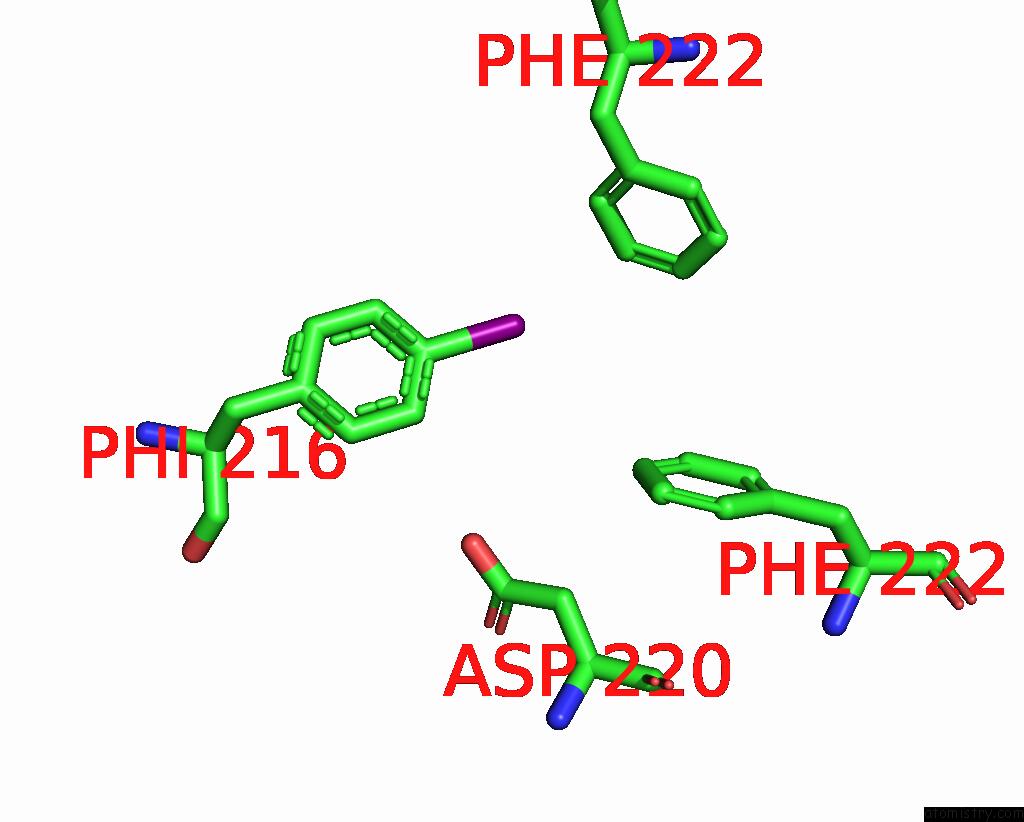
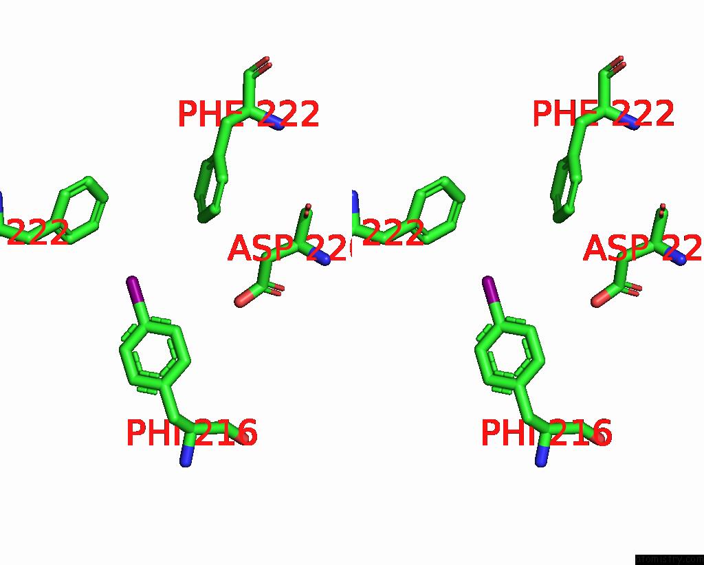
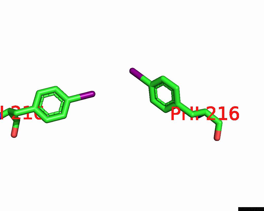
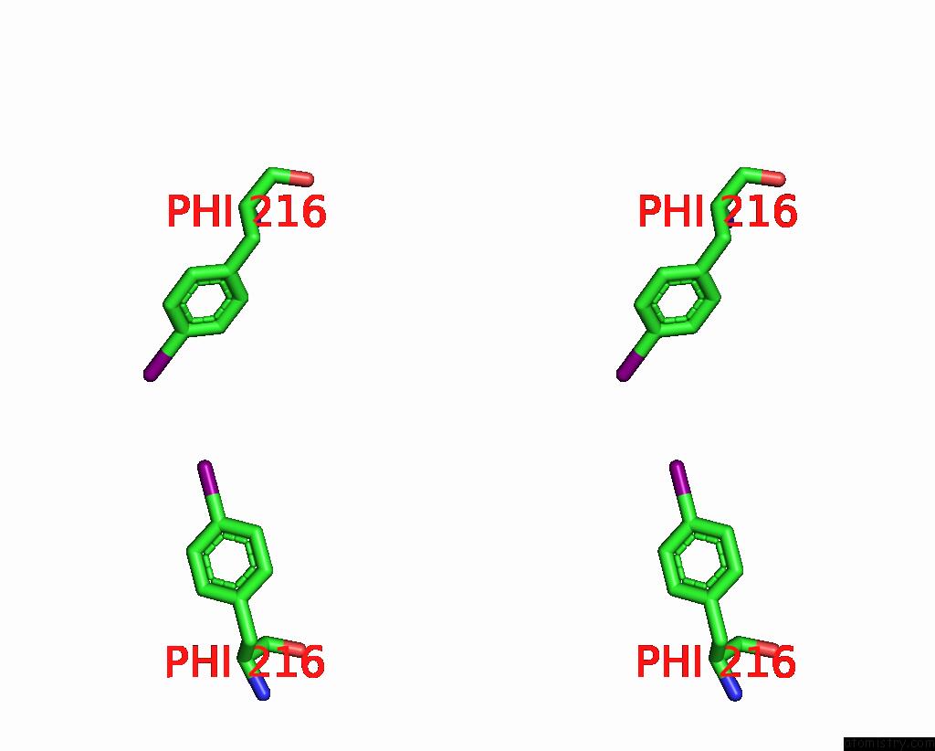
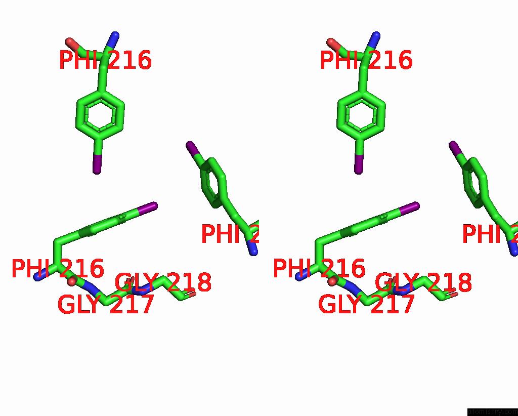
Iodine binding site 2 out of 28 in 8ikb
Go back to
Iodine binding site 2 out
of 28 in the Cryo-Em Structure of HNRAC1-2I Fibril.
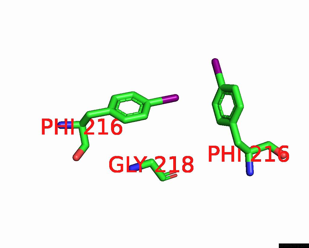
Mono view
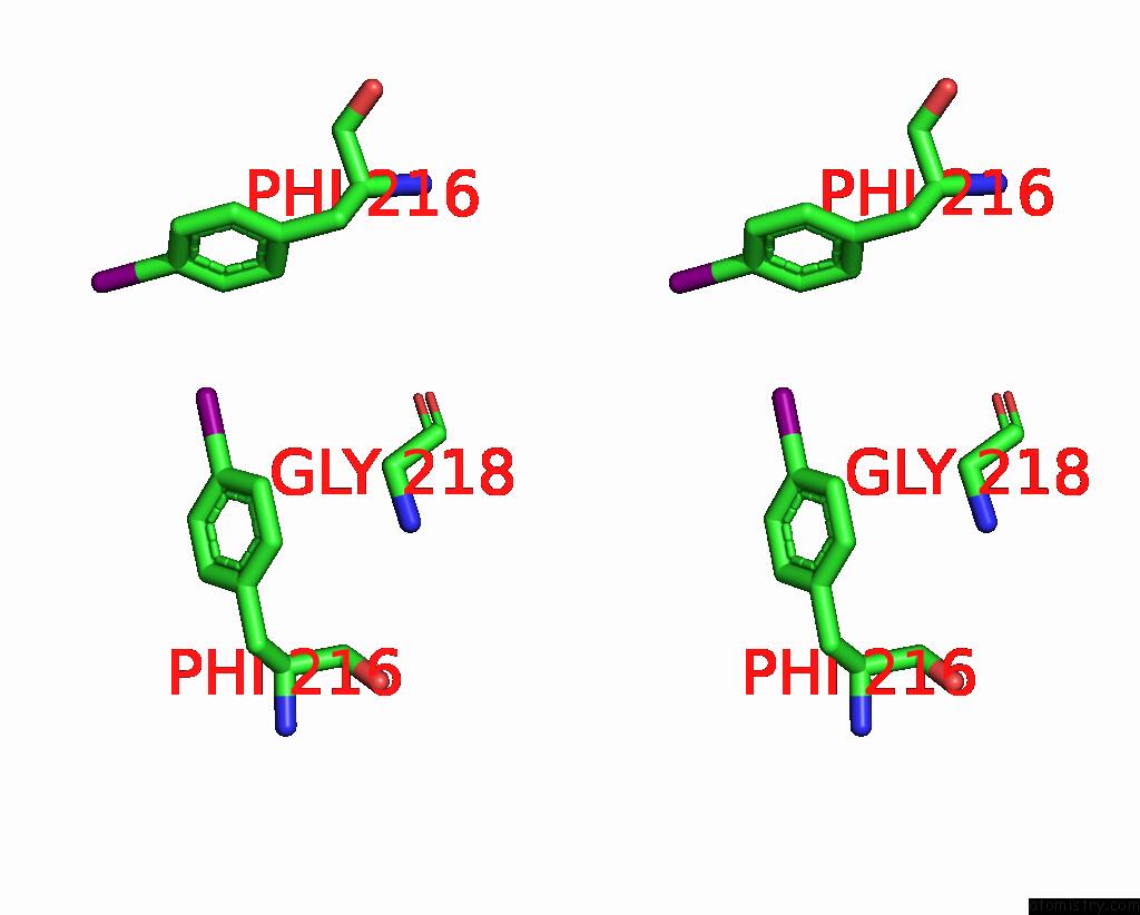
Stereo pair view
Mono view
Stereo pair view
A full contact list of Iodine with other atoms in the I binding
site number 2 of Cryo-Em Structure of HNRAC1-2I Fibril. within 5.0Å range:
|
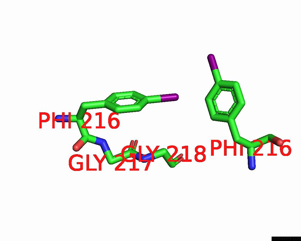
Iodine binding site 3 out of 28 in 8ikb
Go back to
Iodine binding site 3 out
of 28 in the Cryo-Em Structure of HNRAC1-2I Fibril.
Mono view
Stereo pair view
Mono view
Stereo pair view
A full contact list of Iodine with other atoms in the I binding
site number 3 of Cryo-Em Structure of HNRAC1-2I Fibril. within 5.0Å range:
|
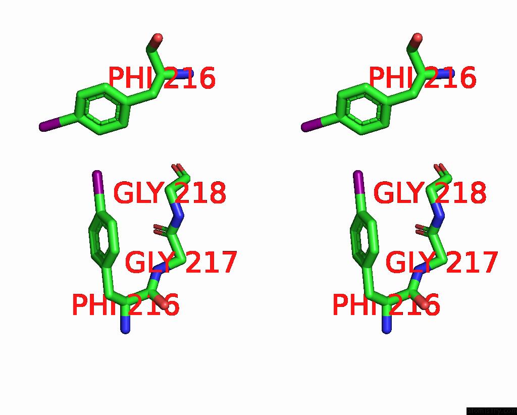
Iodine binding site 4 out of 28 in 8ikb
Go back to
Iodine binding site 4 out
of 28 in the Cryo-Em Structure of HNRAC1-2I Fibril.
Mono view
Stereo pair view
Mono view
Stereo pair view
A full contact list of Iodine with other atoms in the I binding
site number 4 of Cryo-Em Structure of HNRAC1-2I Fibril. within 5.0Å range:
|
Iodine binding site 5 out of 28 in 8ikb
Go back to
Iodine binding site 5 out
of 28 in the Cryo-Em Structure of HNRAC1-2I Fibril.
Mono view
Stereo pair view
Mono view
Stereo pair view
A full contact list of Iodine with other atoms in the I binding
site number 5 of Cryo-Em Structure of HNRAC1-2I Fibril. within 5.0Å range:
|
Iodine binding site 6 out of 28 in 8ikb
Go back to
Iodine binding site 6 out
of 28 in the Cryo-Em Structure of HNRAC1-2I Fibril.
Mono view
Stereo pair view
Mono view
Stereo pair view
A full contact list of Iodine with other atoms in the I binding
site number 6 of Cryo-Em Structure of HNRAC1-2I Fibril. within 5.0Å range:
|
Iodine binding site 7 out of 28 in 8ikb
Go back to
Iodine binding site 7 out
of 28 in the Cryo-Em Structure of HNRAC1-2I Fibril.
Mono view
Stereo pair view
Mono view
Stereo pair view
A full contact list of Iodine with other atoms in the I binding
site number 7 of Cryo-Em Structure of HNRAC1-2I Fibril. within 5.0Å range:
|
Iodine binding site 8 out of 28 in 8ikb
Go back to
Iodine binding site 8 out
of 28 in the Cryo-Em Structure of HNRAC1-2I Fibril.
Mono view
Stereo pair view
Mono view
Stereo pair view
A full contact list of Iodine with other atoms in the I binding
site number 8 of Cryo-Em Structure of HNRAC1-2I Fibril. within 5.0Å range:
|
Iodine binding site 9 out of 28 in 8ikb
Go back to
Iodine binding site 9 out
of 28 in the Cryo-Em Structure of HNRAC1-2I Fibril.
Mono view
Stereo pair view
Mono view
Stereo pair view
A full contact list of Iodine with other atoms in the I binding
site number 9 of Cryo-Em Structure of HNRAC1-2I Fibril. within 5.0Å range:
|
Iodine binding site 10 out of 28 in 8ikb
Go back to
Iodine binding site 10 out
of 28 in the Cryo-Em Structure of HNRAC1-2I Fibril.
Mono view
Stereo pair view
Mono view
Stereo pair view
A full contact list of Iodine with other atoms in the I binding
site number 10 of Cryo-Em Structure of HNRAC1-2I Fibril. within 5.0Å range:
|
Reference:
D.N.Li,
Y.Y.Ma,
D.Li,
B.Dai,
C.Liu.
Cryo-Em Structure of HNRAC1-2I Fibril. To Be Published.
Page generated: Sat Aug 9 00:13:13 2025
Last articles
K in 6PHRK in 6PHZ
K in 6PCD
K in 6P45
K in 6PC3
K in 6P9V
K in 6P0Y
K in 6OZI
K in 6P1M
K in 6OZJ