Iodine »
PDB 8e7f-8sh3 »
8iy7 »
Iodine in PDB 8iy7: Cryo-Em Structure of Dip-2I8I Fibril POLYMORPH1
Iodine Binding Sites:
Pages:
>>> Page 1 <<< Page 2, Binding sites: 11 - 20; Page 3, Binding sites: 21 - 30; Page 4, Binding sites: 31 - 40; Page 5, Binding sites: 41 - 48;Binding sites:
The binding sites of Iodine atom in the Cryo-Em Structure of Dip-2I8I Fibril POLYMORPH1 (pdb code 8iy7). This binding sites where shown within 5.0 Angstroms radius around Iodine atom.In total 48 binding sites of Iodine where determined in the Cryo-Em Structure of Dip-2I8I Fibril POLYMORPH1, PDB code: 8iy7:
Jump to Iodine binding site number: 1; 2; 3; 4; 5; 6; 7; 8; 9; 10;
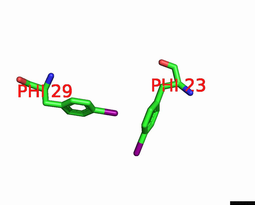
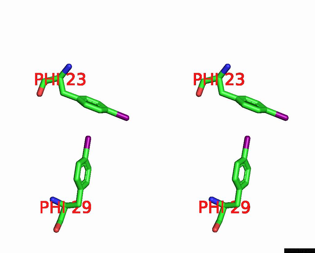
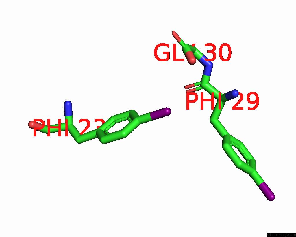
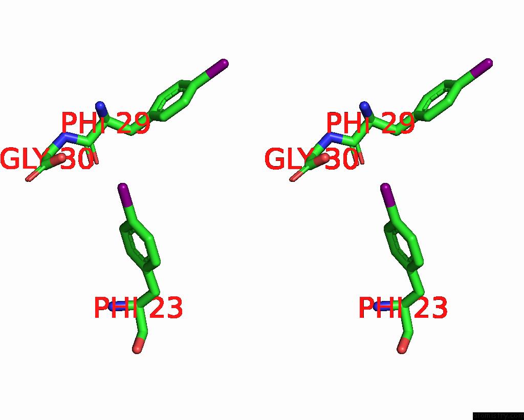
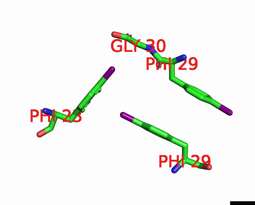
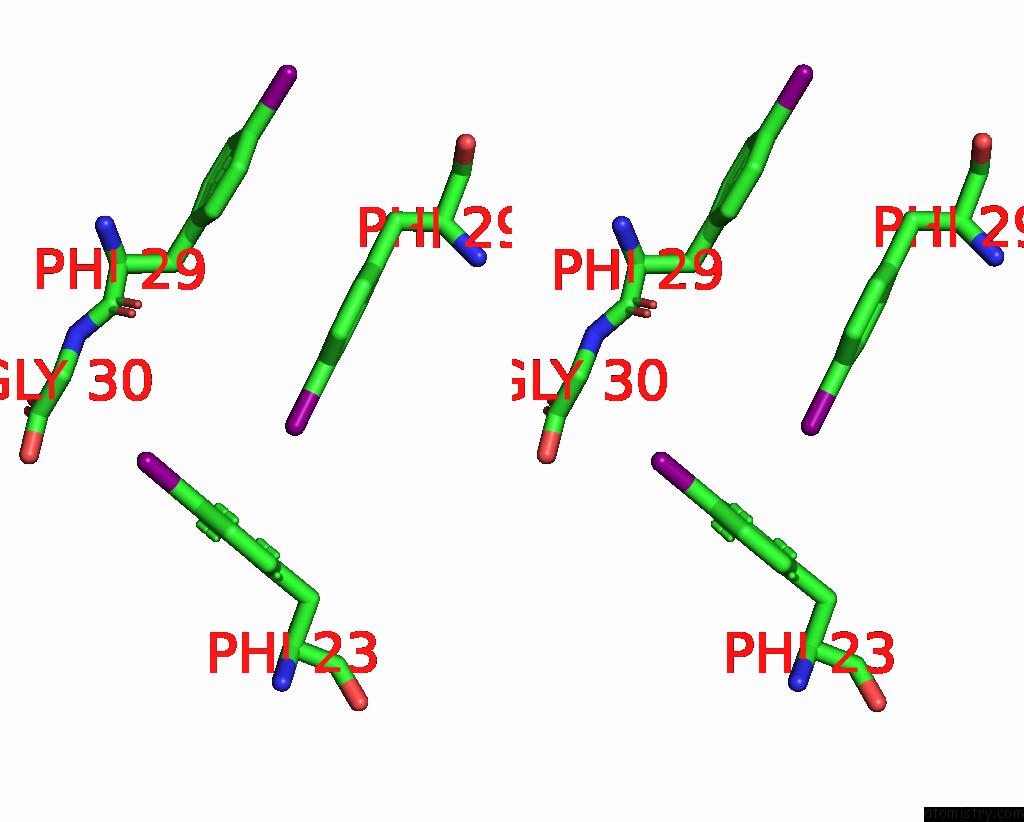
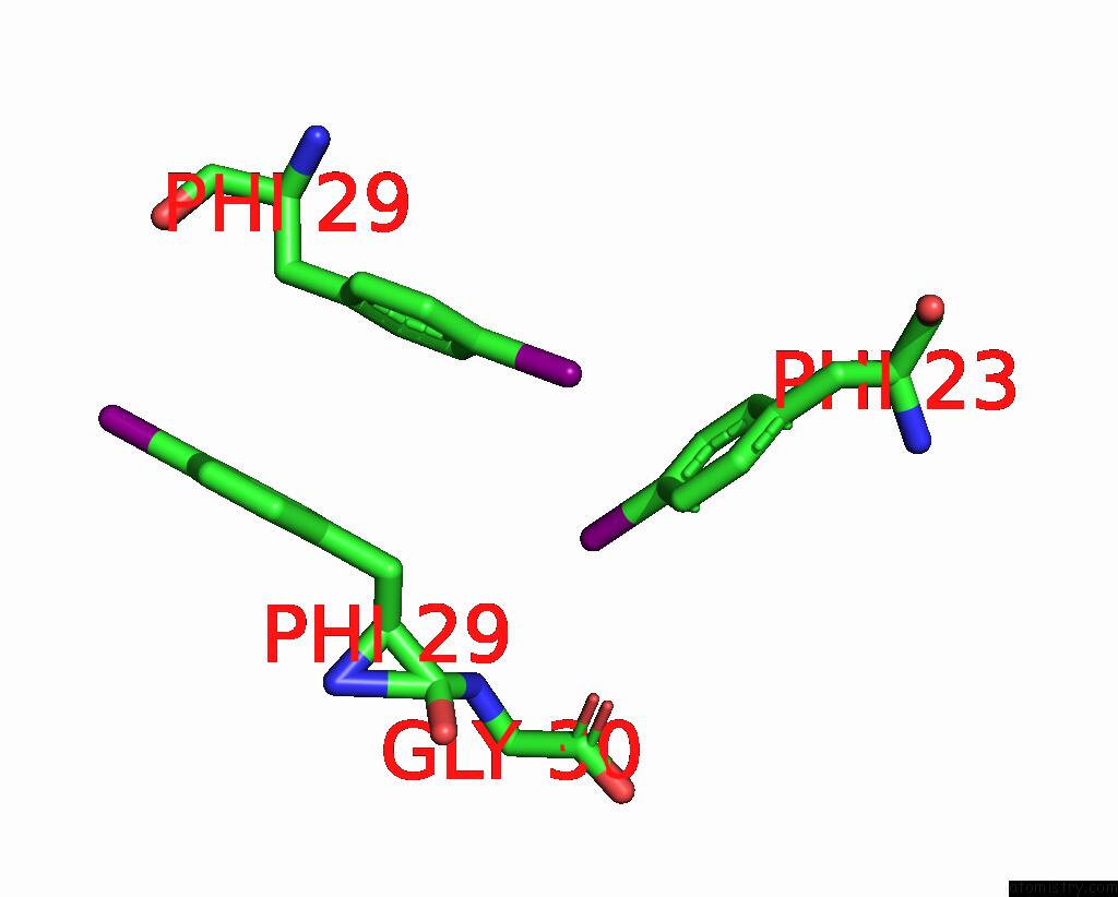
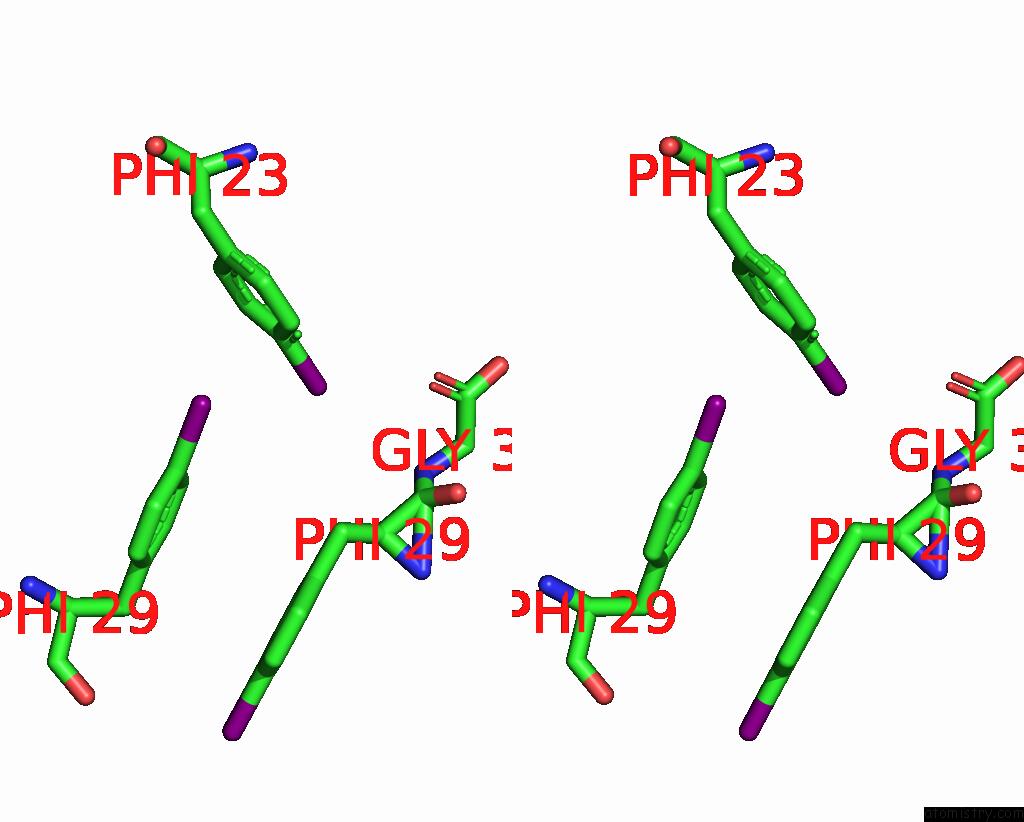
Iodine binding site 1 out of 48 in 8iy7
Go back to
Iodine binding site 1 out
of 48 in the Cryo-Em Structure of Dip-2I8I Fibril POLYMORPH1
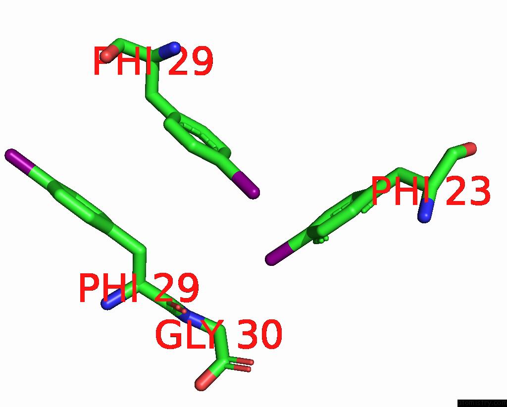
Mono view
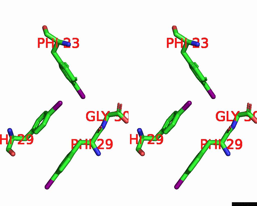
Stereo pair view
Mono view
Stereo pair view
A full contact list of Iodine with other atoms in the I binding
site number 1 of Cryo-Em Structure of Dip-2I8I Fibril POLYMORPH1 within 5.0Å range:
|
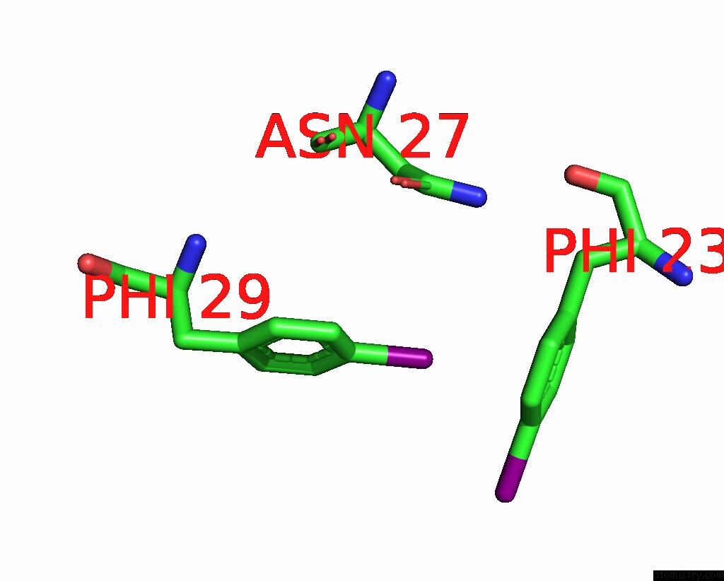
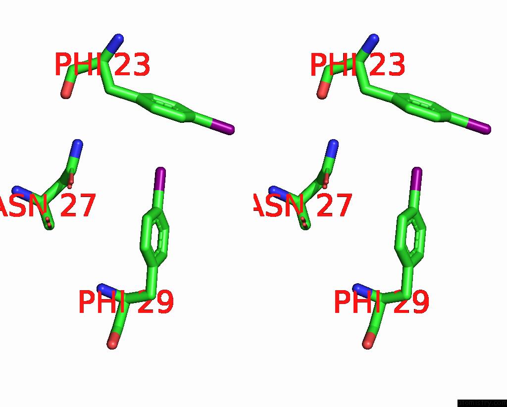
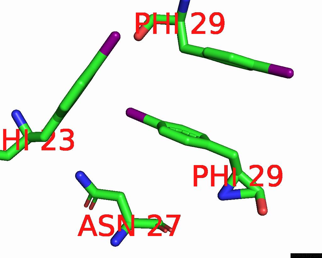
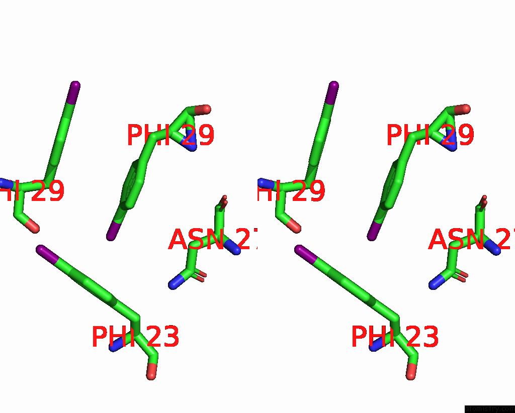
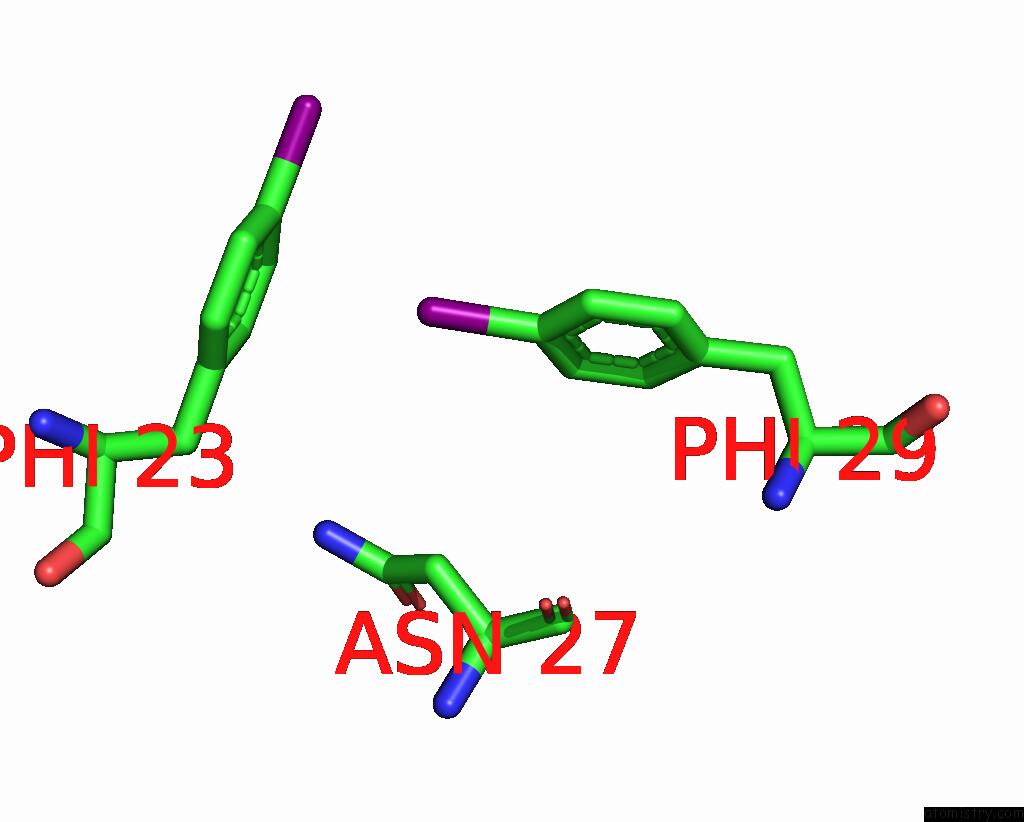
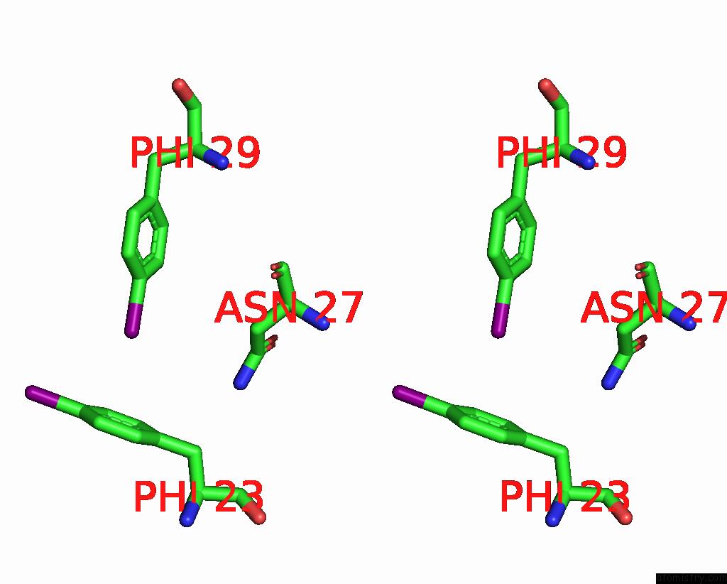
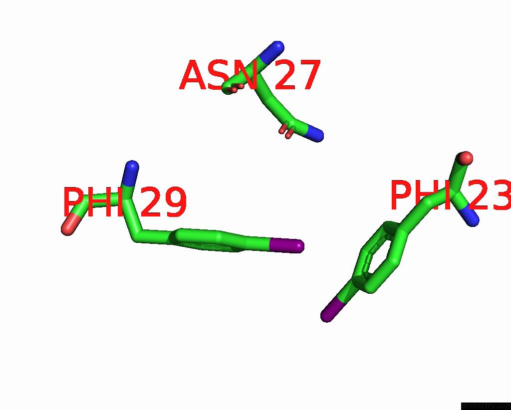
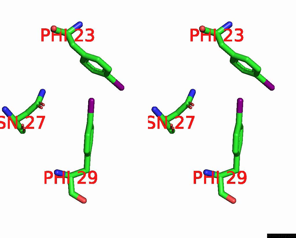
Iodine binding site 2 out of 48 in 8iy7
Go back to
Iodine binding site 2 out
of 48 in the Cryo-Em Structure of Dip-2I8I Fibril POLYMORPH1
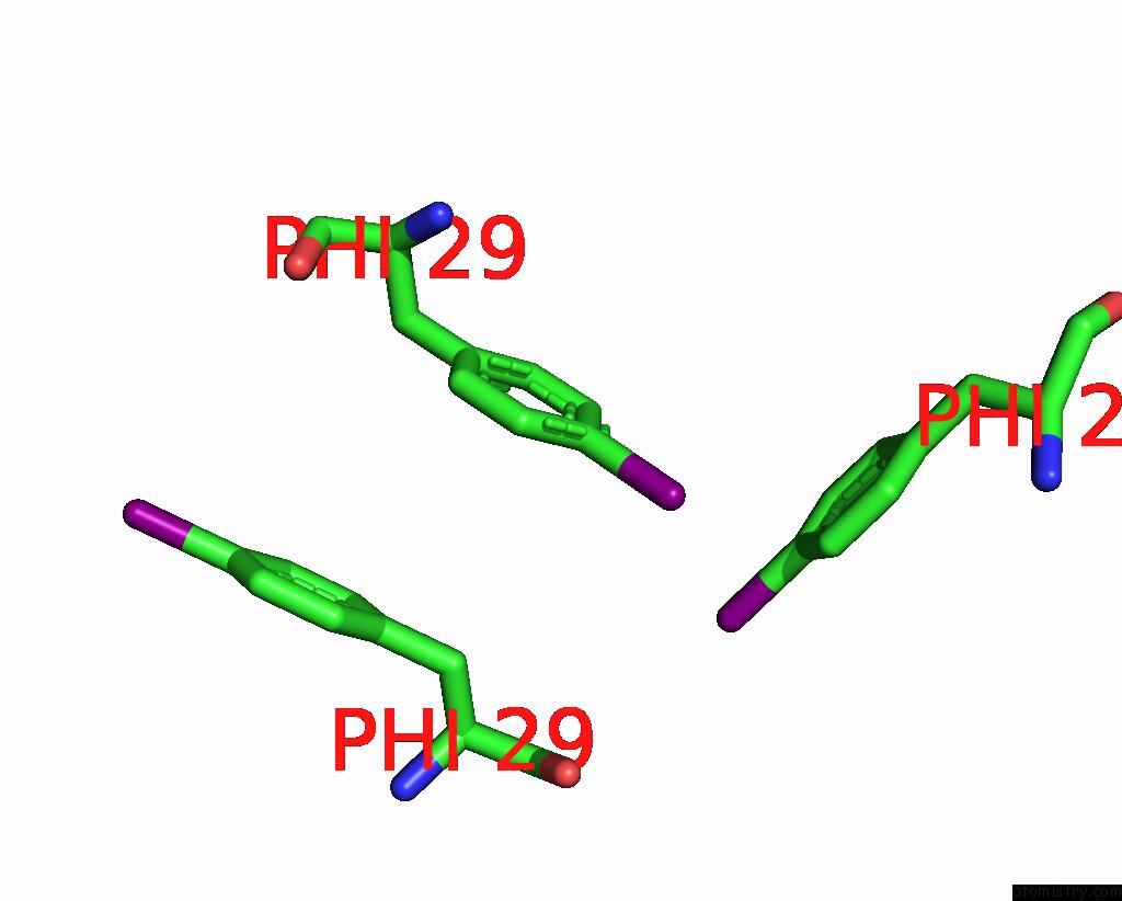
Mono view
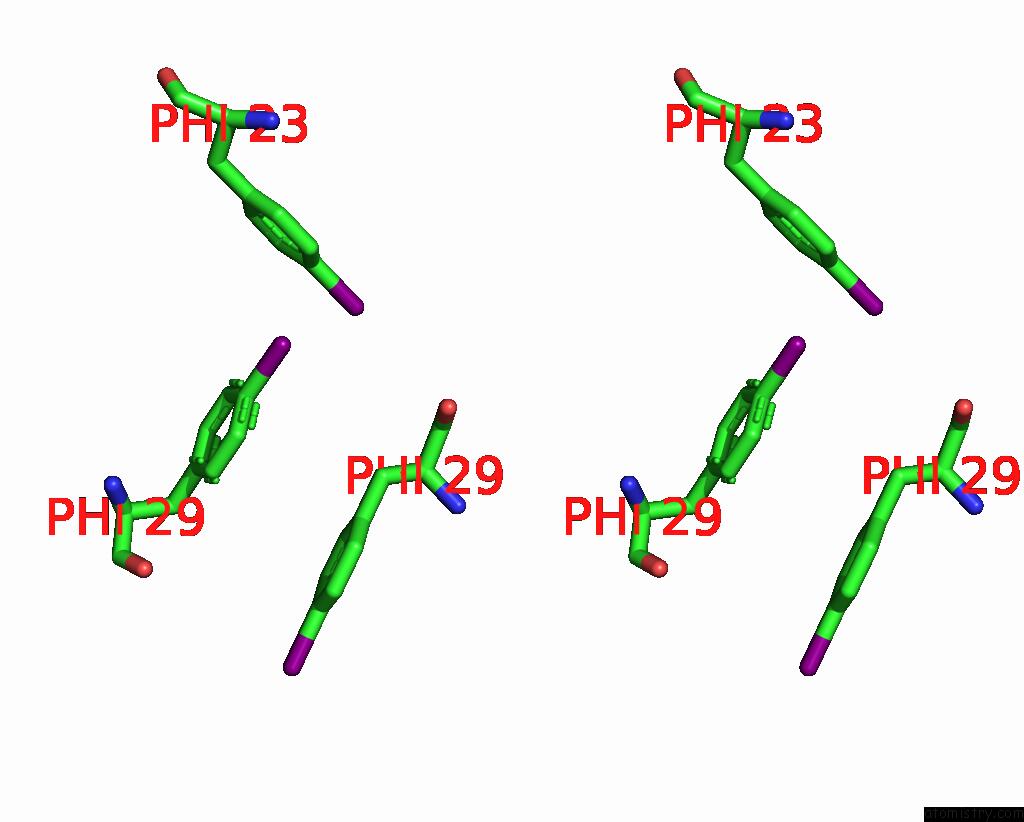
Stereo pair view
Mono view
Stereo pair view
A full contact list of Iodine with other atoms in the I binding
site number 2 of Cryo-Em Structure of Dip-2I8I Fibril POLYMORPH1 within 5.0Å range:
|
Iodine binding site 3 out of 48 in 8iy7
Go back to
Iodine binding site 3 out
of 48 in the Cryo-Em Structure of Dip-2I8I Fibril POLYMORPH1
Mono view
Stereo pair view
Mono view
Stereo pair view
A full contact list of Iodine with other atoms in the I binding
site number 3 of Cryo-Em Structure of Dip-2I8I Fibril POLYMORPH1 within 5.0Å range:
|
Iodine binding site 4 out of 48 in 8iy7
Go back to
Iodine binding site 4 out
of 48 in the Cryo-Em Structure of Dip-2I8I Fibril POLYMORPH1
Mono view
Stereo pair view
Mono view
Stereo pair view
A full contact list of Iodine with other atoms in the I binding
site number 4 of Cryo-Em Structure of Dip-2I8I Fibril POLYMORPH1 within 5.0Å range:
|
Iodine binding site 5 out of 48 in 8iy7
Go back to
Iodine binding site 5 out
of 48 in the Cryo-Em Structure of Dip-2I8I Fibril POLYMORPH1
Mono view
Stereo pair view
Mono view
Stereo pair view
A full contact list of Iodine with other atoms in the I binding
site number 5 of Cryo-Em Structure of Dip-2I8I Fibril POLYMORPH1 within 5.0Å range:
|
Iodine binding site 6 out of 48 in 8iy7
Go back to
Iodine binding site 6 out
of 48 in the Cryo-Em Structure of Dip-2I8I Fibril POLYMORPH1
Mono view
Stereo pair view
Mono view
Stereo pair view
A full contact list of Iodine with other atoms in the I binding
site number 6 of Cryo-Em Structure of Dip-2I8I Fibril POLYMORPH1 within 5.0Å range:
|
Iodine binding site 7 out of 48 in 8iy7
Go back to
Iodine binding site 7 out
of 48 in the Cryo-Em Structure of Dip-2I8I Fibril POLYMORPH1
Mono view
Stereo pair view
Mono view
Stereo pair view
A full contact list of Iodine with other atoms in the I binding
site number 7 of Cryo-Em Structure of Dip-2I8I Fibril POLYMORPH1 within 5.0Å range:
|
Iodine binding site 8 out of 48 in 8iy7
Go back to
Iodine binding site 8 out
of 48 in the Cryo-Em Structure of Dip-2I8I Fibril POLYMORPH1
Mono view
Stereo pair view
Mono view
Stereo pair view
A full contact list of Iodine with other atoms in the I binding
site number 8 of Cryo-Em Structure of Dip-2I8I Fibril POLYMORPH1 within 5.0Å range:
|
Iodine binding site 9 out of 48 in 8iy7
Go back to
Iodine binding site 9 out
of 48 in the Cryo-Em Structure of Dip-2I8I Fibril POLYMORPH1
Mono view
Stereo pair view
Mono view
Stereo pair view
A full contact list of Iodine with other atoms in the I binding
site number 9 of Cryo-Em Structure of Dip-2I8I Fibril POLYMORPH1 within 5.0Å range:
|
Iodine binding site 10 out of 48 in 8iy7
Go back to
Iodine binding site 10 out
of 48 in the Cryo-Em Structure of Dip-2I8I Fibril POLYMORPH1
Mono view
Stereo pair view
Mono view
Stereo pair view
A full contact list of Iodine with other atoms in the I binding
site number 10 of Cryo-Em Structure of Dip-2I8I Fibril POLYMORPH1 within 5.0Å range:
|
Reference:
D.N.Li,
Y.Y.Ma,
D.Li,
B.Dai,
C.Liu.
Cryo-Em Structure of Dip-2I8I Fibril POLYMORPH1 To Be Published.
Page generated: Mon Aug 12 02:49:08 2024
Last articles
Zn in 9MJ5Zn in 9HNW
Zn in 9G0L
Zn in 9FNE
Zn in 9DZN
Zn in 9E0I
Zn in 9D32
Zn in 9DAK
Zn in 8ZXC
Zn in 8ZUF